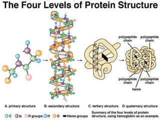
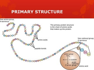
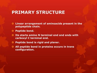
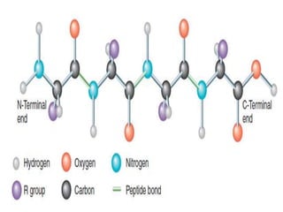
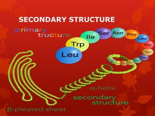
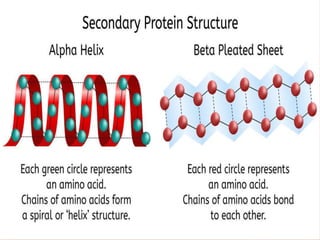
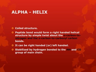
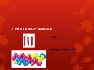
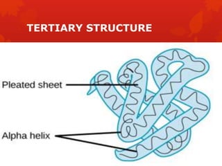
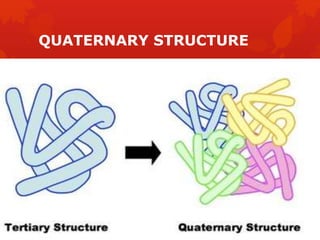
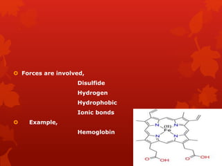
s
Structure of
protein
R.HARISHMA,
I MSC MICROBIOLOGY,
18PY07.
PROTEIN
 Derived from Greek word “ protos ” –
meaning primary.
 It plays a variety of activities.
 Each protein is a polymer of aminoacid.
 Amino acids linked by a peptide bond-
polypeptide.
 Each amino acid consists of a carbon atom,
an amino group, a carboxyl group as a side
chain.
STRUCTURE OF PROTEIN
 Four levels of protein structure,
Primary structure
Secondary structure
Tertiary structure
Quaternary structure
PRIMARY STRUCTURE
PRIMARY STRUCTURE
 Linear arrangement of aminoacids present in the
polypeptide chain.
 Peptide bond.
 Its starts amino N terminal end and ends with
carboxyl C terminal end.
 Peptide bond is rigid and planer.
 All peptide bond in proteins occurs in trans
configuration.
 The rotation is permitted about the bond between
nitrogen and alpha carbon atoms and between the
alpha carbon and carbonyl carbon atoms.
 Two rotatory angles,
phi (φ)
Psi (ψ)
 Phi and psi have a value between −180º to
+180º
RAMACHANDRAN PLOT
 To identify secondary structure of proteins
depending upon the rotation (or) degree of
freedom of dihedral angles.
CHARACTERISTICS OF RAMACHANDRAN PLOT:
 Four quadrants.
 Y axis – ψ and X axis – φ .
 Definite position in this plot indicate definite
secondary structure of protein.
RAMACHANDRAN PLOT
 Both allowed and disallowed region.
 Glysine – Most allowed region.
 Proline – Least allowed region.
 Two phi and psi angles are 180º .
s
SECONDARY STRUCTURE
SECONDARY STRUCTURE
 Regular folding of primary structure is called
secondary structure.
 Folding and hydrogen bonding between
neighbouring aminoacids – results to form rigid
and tubular structure called helix.
 Pauling and corey identified ,
Alpha helix
Beta pleated sheet
;.
ALPHA - HELIX
 Coiled structure.
 Peptide bond would form a right handed helical
structure by simple twist about the α-carbon-to-
nitrogen and the α-carbon-to-carboxyl carbon
bonds.
 It can be right handed (or) left handed.
 Stabilized by hydrogen bonded to the NH and CO
group of main chain.
 Pitch of 5.4 Å .
 Per turn of helix 3.6 aminoacids are present.
 One residue occupies 1.5 Å.
 Conformations with φ = − 60º and ψ = − 45º to −
50º.
 Alpha helix found in protein alpha keratin.
BETA PLEATED SHEET
 Pauling and corey identified a stable conformation
– Beta pleated sheet.
 Stabilized by hydrogen bond between NH and CO
groups in different polypeptide strands.
 One residue occupies 3.5 Å.
 Two types,
Parallel β pleated sheet
Antiparallel β pleated sheet
Parallel beta pleated sheet:
The polypeptide strands run in the same direction.
N-C terminus.
Antiparallel beta pleated sheet:
The polypeptide strands run in the opposite direction.
The N- is opposite to the C terminus.
Both parallel and antiparallel beta sheets have
similar structure.
Random coil:
It is the third type of secondary structure.
 Other secondary structures:
β turn
Collagen triple helix
TERTIARY STRUCTURE
TERTIARY STRUCTURE
 The tertiary structure of protein is the overall
three Dimensional shape that arises from all of the
secondary structures of its polypeptide chain.
 functions are depends…
 Globular (or) fibrous tertiary structure.
 Do not occur randomly.
 Forces involved in stabilizing tertiary structure,
including
Electrostatic forces
Vander Waals
Hydrogen bond
Disulfide bonds
 For example,
Myoglobin
 Myoglobin
Relatively small.
Compact macromolecule.
Oxygen binding hemi protein.
Found in muscle cells.
Molecules contain single polypeptide
chain of 153 aminoacid residues.
MYOGLOBIN
QUATERNARY STRUCTURE
 Connectivity between two (or) more polypeptide
chain.
 Complexity – very high
 Some globular proteins consists of two (or) more
interacting peptide chains.
 These chains may be identical (or) different in
primary structure.
 Forces are involved,
Disulfide
Hydrogen
Hydrophobic
Ionic bonds
 Example,
Hemoglobin
s

Structure of protein

  • 1.
  • 2.
    PROTEIN  Derived fromGreek word “ protos ” – meaning primary.  It plays a variety of activities.  Each protein is a polymer of aminoacid.  Amino acids linked by a peptide bond- polypeptide.  Each amino acid consists of a carbon atom, an amino group, a carboxyl group as a side chain.
  • 3.
    STRUCTURE OF PROTEIN Four levels of protein structure, Primary structure Secondary structure Tertiary structure Quaternary structure
  • 6.
  • 7.
    PRIMARY STRUCTURE  Lineararrangement of aminoacids present in the polypeptide chain.  Peptide bond.  Its starts amino N terminal end and ends with carboxyl C terminal end.  Peptide bond is rigid and planer.  All peptide bond in proteins occurs in trans configuration.
  • 9.
     The rotationis permitted about the bond between nitrogen and alpha carbon atoms and between the alpha carbon and carbonyl carbon atoms.  Two rotatory angles, phi (φ) Psi (ψ)  Phi and psi have a value between −180º to +180º
  • 10.
    RAMACHANDRAN PLOT  Toidentify secondary structure of proteins depending upon the rotation (or) degree of freedom of dihedral angles. CHARACTERISTICS OF RAMACHANDRAN PLOT:  Four quadrants.  Y axis – ψ and X axis – φ .  Definite position in this plot indicate definite secondary structure of protein.
  • 12.
  • 13.
     Both allowedand disallowed region.  Glysine – Most allowed region.  Proline – Least allowed region.  Two phi and psi angles are 180º .
  • 14.
  • 15.
  • 16.
    SECONDARY STRUCTURE  Regularfolding of primary structure is called secondary structure.  Folding and hydrogen bonding between neighbouring aminoacids – results to form rigid and tubular structure called helix.  Pauling and corey identified , Alpha helix Beta pleated sheet
  • 17.
  • 18.
    ALPHA - HELIX Coiled structure.  Peptide bond would form a right handed helical structure by simple twist about the α-carbon-to- nitrogen and the α-carbon-to-carboxyl carbon bonds.  It can be right handed (or) left handed.  Stabilized by hydrogen bonded to the NH and CO group of main chain.
  • 19.
     Pitch of5.4 Å .  Per turn of helix 3.6 aminoacids are present.  One residue occupies 1.5 Å.  Conformations with φ = − 60º and ψ = − 45º to − 50º.  Alpha helix found in protein alpha keratin.
  • 20.
    BETA PLEATED SHEET Pauling and corey identified a stable conformation – Beta pleated sheet.  Stabilized by hydrogen bond between NH and CO groups in different polypeptide strands.  One residue occupies 3.5 Å.  Two types, Parallel β pleated sheet Antiparallel β pleated sheet
  • 21.
    Parallel beta pleatedsheet: The polypeptide strands run in the same direction. N-C terminus.
  • 22.
    Antiparallel beta pleatedsheet: The polypeptide strands run in the opposite direction. The N- is opposite to the C terminus.
  • 23.
    Both parallel andantiparallel beta sheets have similar structure. Random coil: It is the third type of secondary structure.
  • 24.
     Other secondarystructures: β turn Collagen triple helix
  • 25.
  • 26.
    TERTIARY STRUCTURE  Thetertiary structure of protein is the overall three Dimensional shape that arises from all of the secondary structures of its polypeptide chain.  functions are depends…  Globular (or) fibrous tertiary structure.  Do not occur randomly.
  • 27.
     Forces involvedin stabilizing tertiary structure, including Electrostatic forces Vander Waals Hydrogen bond Disulfide bonds  For example, Myoglobin
  • 28.
     Myoglobin Relatively small. Compactmacromolecule. Oxygen binding hemi protein. Found in muscle cells. Molecules contain single polypeptide chain of 153 aminoacid residues.
  • 29.
  • 30.
  • 31.
    QUATERNARY STRUCTURE  Connectivitybetween two (or) more polypeptide chain.  Complexity – very high  Some globular proteins consists of two (or) more interacting peptide chains.  These chains may be identical (or) different in primary structure.
  • 32.
     Forces areinvolved, Disulfide Hydrogen Hydrophobic Ionic bonds  Example, Hemoglobin
  • 33.