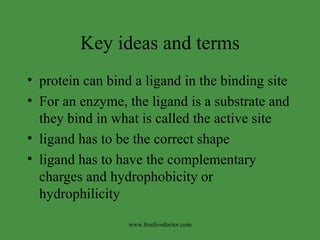
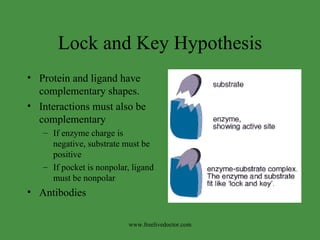
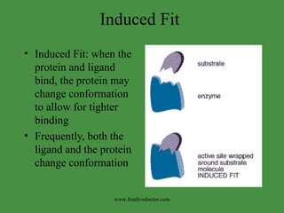
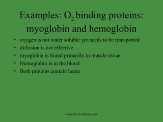
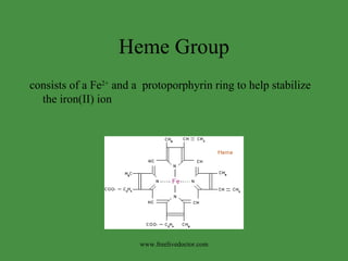
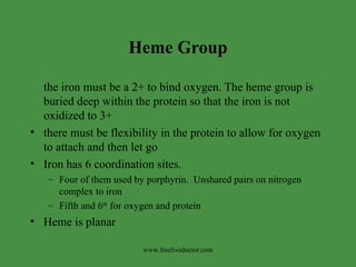
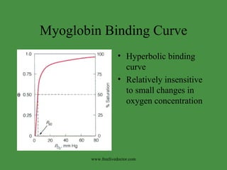
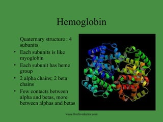
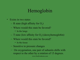
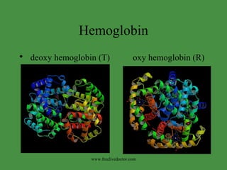
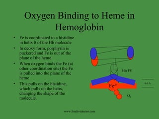
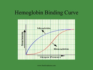
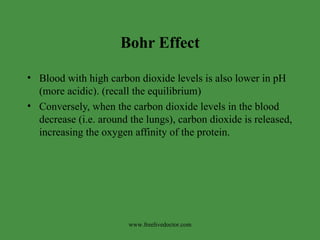
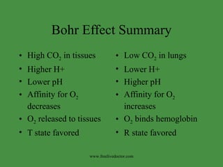
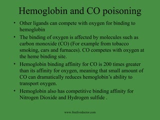
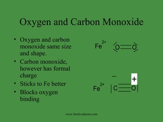
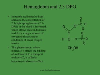
Protein Function Structure will determine the function of the protein www.freelivedoctor.com
Key ideas and terms protein can bind a ligand in the binding site  For an enzyme, the ligand is a substrate and they bind in what is called the active site ligand has to be the correct shape ligand has to have the complementary charges and hydrophobicity or hydrophilicity www.freelivedoctor.com
Lock and Key Hypothesis Protein and ligand have complementary shapes. Interactions must also be complementary If enzyme charge is negative, substrate must be positive If pocket is nonpolar, ligand must be nonpolar Antibodies www.freelivedoctor.com
Induced Fit Induced Fit: when the protein and ligand bind, the protein may change conformation to allow for tighter binding Frequently, both the ligand and the protein change conformation www.freelivedoctor.com
Examples: O 2  binding proteins: myoglobin and hemoglobin oxygen is not water soluble yet needs to be transported diffusion is not effective myoglobin is found primarily in muscle tissue Hemoglobin is in the blood Both proteins contain heme www.freelivedoctor.com
Heme Group consists of a Fe 2+  and a  protoporphyrin ring to help stabilize the iron(II) ion www.freelivedoctor.com
Heme Group the iron must be a 2+ to bind oxygen. The heme group is buried deep within the protein so that the iron is not oxidized to 3+ there must be flexibility in the protein to allow for oxygen to attach and then let go Iron has 6 coordination sites.  Four of them used by porphyrin.  Unshared pairs on nitrogen complex to iron Fifth and 6 th  for oxygen and protein Heme is planar www.freelivedoctor.com
Myoglobin has heme group eight alpha helical segments dense hydrophobic core all but two polar groups on outside room for only 4 water molecules flat heme in pocket iron coordinated to poryphorin and H As well as the heme www.freelivedoctor.com
Myoglobin Binding Curve Hyperbolic binding curve Relatively insensitive to small changes in oxygen concentration www.freelivedoctor.com
Myoglobin The P50 (oxygen partial pressure required for half saturation) for myoglobin is very low Myoglobin has a high affinity for oxygen-an important characteristic for a protein that must extract oxygen from the small amounts present in blood.  At the oxygen concentration existing in the capillaries, the myoglobin in adjacent tissues is nearly saturated.  When cells are metabolically active, their internal P O2  falls to levels where myoglobin will lose   (deliver) its oxygen . www.freelivedoctor.com
Hemoglobin Quaternary structure : 4 subunits Each subunits is like myoglobin Each subunit has heme group 2 alpha chains; 2 beta chains  Few contacts between alpha and betas, more between alphas and betas www.freelivedoctor.com
Hemoglobin Exists in two states  R state (high affinity for O 2 )  Where would this state be favored?  In the lungs T state (low affinity for O 2 ) (deoxyhemoglobin) Where would this state be favored?  In the tissue Sensitive to pressure changes On oxygenation, one pair of subunits shifts with respect to the other by a rotation of 15 degrees. www.freelivedoctor.com
Hemoglobin deoxy hemoglobin (T)  oxy hemoglobin (R) www.freelivedoctor.com
Oxygen Binding to Heme in Hemoglobin Fe is coordinated to a histidine in helix 8 of the Hb molecule In deoxy form, porphyrin is puckered and Fe is out of the plane of the heme When oxygen binds the Fe (at other coordination site) the Fe is pulled into the plane of the heme This pulls on the histidine, which pulls on the helix, changing the shape of the molecule. Fe 2+ His F8 0.6 A O 2 www.freelivedoctor.com
Hemoglobin Conformational changes in hemoglobin alter its binding ability Binding of oxygen in one subunit causes conformational changes in the next subunit This is called cooperative binding This can happen because it is composed of 4 independent subunits produces a different binding curve that is sigmoidal The modulation of the affinity of a site for a ligand by ligand binding at another site is called Allostery . www.freelivedoctor.com
Hemoglobin Binding Curve www.freelivedoctor.com
Bohr Effect Hemoglobin's affinity for oxygen is decreased in the presence of carbon dioxide and at lower pH.  Carbon dioxide reacts with water to give bicarbonate, carbonic acid free protons via the reaction:  CO 2   +  H 2 O ---> H 2 CO 3  ---> H +   +  HCO 3 -   Protons bind at various places along the protein and carbon dioxide binds at the alpha-amino group forming carbamate.  This causes a conformational change in the protein and facilitates the release of oxygen.  www.freelivedoctor.com
Bohr Effect Blood with high carbon dioxide levels is also lower in pH (more acidic). (recall the equilibrium) Conversely, when the carbon dioxide levels in the blood decrease (i.e. around the lungs), carbon dioxide is released, increasing the oxygen affinity of the protein.  www.freelivedoctor.com
Bohr Effect Summary High CO 2  in tissues  Higher H+ Lower pH Affinity for O 2  decreases O 2  released to tissues T state favored Low CO 2  in lungs  Lower H+ Higher pH Affinity for O 2  increases O 2  binds hemoglobin R state favored www.freelivedoctor.com
Hemoglobin and CO poisoning Other ligands can compete with oxygen for binding to hemoglobin The binding of oxygen is affected by molecules such as carbon monoxide (CO) (For example from tobacco smoking, cars and furnaces). CO competes with oxygen at the heme binding site.  Hemoglobin binding affinity for CO is 200 times greater than its affinity for oxygen, meaning that small amount of CO can dramatically reduces hemoglobin’s ability to transport oxygen. Hemoglobin also has competitive binding affinity for  Nitrogen Dioxide and Hydrogen sulfide . www.freelivedoctor.com
Oxygen and Carbon Monoxide Oxygen and carbon monoxide same size and shape. Carbon monoxide, however has formal charge  Sticks to Fe better Blocks oxygen binding www.freelivedoctor.com
Hemoglobin and 2,3 DPG In people acclimated to high altitudes, the concentration of 2,3-diphosphoglycerate (2,3-DPG) in the blood is increased, which allows these individuals to deliver a larger amount of oxygen to tissues under conditions of lower oxygen tension.  This phenomenon, where molecule Y affects the binding of molecule X to a transport molecule Z, is called a heterotropic allosteric effect . www.freelivedoctor.com
Sickle Cell Anemia Sickle cell disease is caused by an abnormal adult hemoglobin, called hemoglobin S. People with sickle cell disease make hemoglobin S instead of hemoglobin A. Single amino acid substitution  glutamate changed to valine To show condition, have to have mutation in both genes (Homozygous) www.freelivedoctor.com
Sickle vs normal hemoglobin first 9 amino acids of normal hemoglobin  beta chain v h l t p  e  e k s  first 9 amino acids of sickle hemoglobin  beta chain v h l t p  v  e k s Notice the single amino acid change? www.freelivedoctor.com
Sickle Cell Anemia Position 6 is on outside of molecule Glutamate is polar Valine substitution causes “sticky spot” on outside of hemoglobin Causes hemoglobin molecules to stick together Forms long chains which cause red blood cell to sickle www.freelivedoctor.com

Protien Metabolism

  • 1.
    Protein Function Structurewill determine the function of the protein www.freelivedoctor.com
  • 2.
    Key ideas andterms protein can bind a ligand in the binding site For an enzyme, the ligand is a substrate and they bind in what is called the active site ligand has to be the correct shape ligand has to have the complementary charges and hydrophobicity or hydrophilicity www.freelivedoctor.com
  • 3.
    Lock and KeyHypothesis Protein and ligand have complementary shapes. Interactions must also be complementary If enzyme charge is negative, substrate must be positive If pocket is nonpolar, ligand must be nonpolar Antibodies www.freelivedoctor.com
  • 4.
    Induced Fit InducedFit: when the protein and ligand bind, the protein may change conformation to allow for tighter binding Frequently, both the ligand and the protein change conformation www.freelivedoctor.com
  • 5.
    Examples: O 2 binding proteins: myoglobin and hemoglobin oxygen is not water soluble yet needs to be transported diffusion is not effective myoglobin is found primarily in muscle tissue Hemoglobin is in the blood Both proteins contain heme www.freelivedoctor.com
  • 6.
    Heme Group consistsof a Fe 2+ and a protoporphyrin ring to help stabilize the iron(II) ion www.freelivedoctor.com
  • 7.
    Heme Group theiron must be a 2+ to bind oxygen. The heme group is buried deep within the protein so that the iron is not oxidized to 3+ there must be flexibility in the protein to allow for oxygen to attach and then let go Iron has 6 coordination sites. Four of them used by porphyrin. Unshared pairs on nitrogen complex to iron Fifth and 6 th for oxygen and protein Heme is planar www.freelivedoctor.com
  • 8.
    Myoglobin has hemegroup eight alpha helical segments dense hydrophobic core all but two polar groups on outside room for only 4 water molecules flat heme in pocket iron coordinated to poryphorin and H As well as the heme www.freelivedoctor.com
  • 9.
    Myoglobin Binding CurveHyperbolic binding curve Relatively insensitive to small changes in oxygen concentration www.freelivedoctor.com
  • 10.
    Myoglobin The P50(oxygen partial pressure required for half saturation) for myoglobin is very low Myoglobin has a high affinity for oxygen-an important characteristic for a protein that must extract oxygen from the small amounts present in blood. At the oxygen concentration existing in the capillaries, the myoglobin in adjacent tissues is nearly saturated. When cells are metabolically active, their internal P O2 falls to levels where myoglobin will lose (deliver) its oxygen . www.freelivedoctor.com
  • 11.
    Hemoglobin Quaternary structure: 4 subunits Each subunits is like myoglobin Each subunit has heme group 2 alpha chains; 2 beta chains Few contacts between alpha and betas, more between alphas and betas www.freelivedoctor.com
  • 12.
    Hemoglobin Exists intwo states R state (high affinity for O 2 ) Where would this state be favored? In the lungs T state (low affinity for O 2 ) (deoxyhemoglobin) Where would this state be favored? In the tissue Sensitive to pressure changes On oxygenation, one pair of subunits shifts with respect to the other by a rotation of 15 degrees. www.freelivedoctor.com
  • 13.
    Hemoglobin deoxy hemoglobin(T) oxy hemoglobin (R) www.freelivedoctor.com
  • 14.
    Oxygen Binding toHeme in Hemoglobin Fe is coordinated to a histidine in helix 8 of the Hb molecule In deoxy form, porphyrin is puckered and Fe is out of the plane of the heme When oxygen binds the Fe (at other coordination site) the Fe is pulled into the plane of the heme This pulls on the histidine, which pulls on the helix, changing the shape of the molecule. Fe 2+ His F8 0.6 A O 2 www.freelivedoctor.com
  • 15.
    Hemoglobin Conformational changesin hemoglobin alter its binding ability Binding of oxygen in one subunit causes conformational changes in the next subunit This is called cooperative binding This can happen because it is composed of 4 independent subunits produces a different binding curve that is sigmoidal The modulation of the affinity of a site for a ligand by ligand binding at another site is called Allostery . www.freelivedoctor.com
  • 16.
    Hemoglobin Binding Curvewww.freelivedoctor.com
  • 17.
    Bohr Effect Hemoglobin'saffinity for oxygen is decreased in the presence of carbon dioxide and at lower pH. Carbon dioxide reacts with water to give bicarbonate, carbonic acid free protons via the reaction: CO 2 + H 2 O ---> H 2 CO 3 ---> H + + HCO 3 - Protons bind at various places along the protein and carbon dioxide binds at the alpha-amino group forming carbamate. This causes a conformational change in the protein and facilitates the release of oxygen. www.freelivedoctor.com
  • 18.
    Bohr Effect Bloodwith high carbon dioxide levels is also lower in pH (more acidic). (recall the equilibrium) Conversely, when the carbon dioxide levels in the blood decrease (i.e. around the lungs), carbon dioxide is released, increasing the oxygen affinity of the protein. www.freelivedoctor.com
  • 19.
    Bohr Effect SummaryHigh CO 2 in tissues Higher H+ Lower pH Affinity for O 2 decreases O 2 released to tissues T state favored Low CO 2 in lungs Lower H+ Higher pH Affinity for O 2 increases O 2 binds hemoglobin R state favored www.freelivedoctor.com
  • 20.
    Hemoglobin and COpoisoning Other ligands can compete with oxygen for binding to hemoglobin The binding of oxygen is affected by molecules such as carbon monoxide (CO) (For example from tobacco smoking, cars and furnaces). CO competes with oxygen at the heme binding site. Hemoglobin binding affinity for CO is 200 times greater than its affinity for oxygen, meaning that small amount of CO can dramatically reduces hemoglobin’s ability to transport oxygen. Hemoglobin also has competitive binding affinity for Nitrogen Dioxide and Hydrogen sulfide . www.freelivedoctor.com
  • 21.
    Oxygen and CarbonMonoxide Oxygen and carbon monoxide same size and shape. Carbon monoxide, however has formal charge Sticks to Fe better Blocks oxygen binding www.freelivedoctor.com
  • 22.
    Hemoglobin and 2,3DPG In people acclimated to high altitudes, the concentration of 2,3-diphosphoglycerate (2,3-DPG) in the blood is increased, which allows these individuals to deliver a larger amount of oxygen to tissues under conditions of lower oxygen tension. This phenomenon, where molecule Y affects the binding of molecule X to a transport molecule Z, is called a heterotropic allosteric effect . www.freelivedoctor.com
  • 23.
    Sickle Cell AnemiaSickle cell disease is caused by an abnormal adult hemoglobin, called hemoglobin S. People with sickle cell disease make hemoglobin S instead of hemoglobin A. Single amino acid substitution glutamate changed to valine To show condition, have to have mutation in both genes (Homozygous) www.freelivedoctor.com
  • 24.
    Sickle vs normalhemoglobin first 9 amino acids of normal hemoglobin beta chain v h l t p e e k s first 9 amino acids of sickle hemoglobin beta chain v h l t p v e k s Notice the single amino acid change? www.freelivedoctor.com
  • 25.
    Sickle Cell AnemiaPosition 6 is on outside of molecule Glutamate is polar Valine substitution causes “sticky spot” on outside of hemoglobin Causes hemoglobin molecules to stick together Forms long chains which cause red blood cell to sickle www.freelivedoctor.com