SlideShare a Scribd company logo
Radiological Imaging of Anal Carcinoma.
Dr/ ABD ALLAH NAZEER. MD.
Normal Anatomy:
The anal canal begins at the narrowing of the rectal ampulla at the
anorectal junction where the rectum enters the puborectalis sling at the
apex of the anal sphincter complex. It extends distally for approximately
4 cm and ends at the anal verge where the squamous mucosa blends with
the perianal skin. The anal margin is the circular rim of pigmented skin
with folds surrounding the anus, extending over an approximately 5-cm
radius away from the anal verge. The most significant landmark of the
anal canal is the dentate line, which lies 2.5–3 cm proximal to the anal
verge and is visible macroscopically but not on MRI. Its position can be
estimated either by measuring 2.5 cm above the anal verge or by dividing
the anal canal into thirds so that the dentate line lies at the junction of the
middle and upper thirds. The anal canal is divided by the dentate line into
an upper part, lined with transitional or rectal glandular mucosa, and a
lower part, lined by nonkeratinizing squamous epithelium, that merges
with the perianal skin. The anal transition zone is defined as the histologic
zone interposed between uniform rectal glandular mucosa above and
uninterrupted squamous epithelium below, and it often has a
heterogeneous histologic appearance.
Illustrative anatomy of anal canal.
A, Line diagram shows anal anatomy.
B, Healthy 67-year-old man (with treated prostate
cancer).Coronal T2-weighted image that
corresponds to A shows ischioanal fossa (IAF),
external sphincter (ES), and internal sphincter (IS).
C, Healthy 53-year-old woman (with treated
carcinoma cervix). Axial T2-weighted image through
upper anal canal shows urethra (Ur) and vagina (Va).
D, 67-year-old woman (post TAH and BSO for endometrial
sarcoma). Sagittal T2-weighted image shows pubococcygeal line
(long straight line), which is generally at level of anorectal junction.
Angled line shows reference plane that one should use when
reporting distance of epicenter of abnormality from anal verge.
Anal cancer is a relatively uncommon malignancy. It accounts for
less than 2% of large bowel malignancies and 1-6% of anorectal
tumours (~1.5% of all gastro-intestinal tract malignancies in the
Unites States).
Epidemiology
There may be a slight male predilection where its incidence has been
reported to be approximately 0.5 per 100 000 in men and 1.0 per
100 000 in women. Its incidence is thought to be rising over the
years.
Clinical presentation
Approximately 45% of patients may present with bleeding per
rectum. Around 30% of patients may have pain and/or a sensation
of a mass.
Pathology
Anal carcinoma typically originates between
the anorectal junction above and the anal verge below. The vast
majority of anal canal cancers are squamous cell cancers.
Risk factors
Both male and female:
HPV / HIV infection
immunosuppression
number of lifetime sexual partners, and receptive anal
intercourse
smoking
In females: previous in situ or invasive cervical, vulval or
vaginal cancer.
Lymphatic spread
tumour above dentate line: to pararectal and
paravertebral nodes
tumour below dentate line: to inguinal and femoral nodes
The accepted TNM staging of anal cancer is as follows:
Primary tumour (T)
TX: primary tumour cannot be assessed
T0: no evidence of primary tumour
Tis: carcinoma in situ
T1: tumour 2 cm or less in greatest dimension
T2: tumour >2 cm but <5 cm in greatest dimension
T3: tumour >5 cm in greatest dimension
T4: tumour of any size invades adjacent organ(s), e.g. vagina, urethra, bladder
Regional lymph nodes (N)
Nx: regional lymph nodes cannot be assessed
N0: no regional lymph node metastasis
N1: metastasis in perirectal lymph nodes(s)
N2: metastasis in unilateral internal iliac and/or inguinal lymph node(s)
N3: metastasis in perirectal and inguinal lymph nodes and/or bilateral internal
iliac lymph nodes
Distant metastasis (M)
Mx: distant metastasis cannot be assessed
M0: no distant metastasis
M1: distant metastasis
Radiologic staging is supplemented by clinical assessment of the
anal margin, verge, and canal. This assessment often involves an
examination with the patient under anesthesia and biopsy.
Biopsy or fine-needle aspiration cytology of any enlarged or
suspicious-appearing inguinal nodes is also essential.
Transanal Endoscopic Ultrasound Transanal endoscopic
ultrasound has results comparable to MRI for the detection of
local tumor spread in patients with anal carcinoma and may be
superior to MRI for the detection of small superficial tumors.
However, regional lymph nodes higher in the pelvis or groins are
outside the FOV of endosonography and supplementary MRI is
needed for N staging. A meta-analysis of staging rectal cancer
using endorectal ultrasound revealed other limitations including
operator dependency and inability to assess stenotic tumors ;
these limitations likely apply equally to anal cancer.
MRI
MRI has become the imaging modality of choice for locoregional staging and
assessment of tumor response after chemoradiotherapy. MRI provides high-resolution
multiplanar information about the location, size, circumferential and craniocaudal
extent of the primary tumor and information regarding the involvement of adjacent
structures.
There is no significant difference in diagnostic value between pelvic surface coils and
endoluminal coils. Moreover, endoluminal coil imaging is limited by a narrow FOV and
near-field artifact and may not be tolerated by patients with anal cancer. MRI with a
pelvic surface coil is easy, is more acceptable to the patient, and shows both local
spread of the disease as well as lymph node involvement.
We routinely perform MRI of anal cancers with a standard technique using a body coil;
we obtain transaxial T1-weighted images with a 5-mm slice thickness to cover the
abdomen and pelvis. High-resolution thin-section T2-weighted images are obtained
with a 3-mm slice thickness in three orthogonal planes through the anal canal using a
pelvic surface coil. In our experience, contrast-enhanced MRI offers limited advantages
over standard T2-weighted sequences in routine primary staging but can be helpful to
assess fistulation to other organs. Fat-suppressed imaging can help improve the
conspicuity of the primary tumor, and STIR sequences are also useful to show fistula
tracks. Diffusion-weighted imaging has an emerging role, particularly for detection of
recurrent tumor after therapy, and can aid in differentiation of suspected residual or
recurrent tumor from treatment-related change.
8F-FDG PET/CT
FDG PET/CT has an increasing role in staging and treatment
planning of anal carcinoma, particularly because up to 98% of anal
tumors are FDG-avid. At diagnosis, FDG PET/CT is used to evaluate
primary tumor size, lymph node status, and whether distant
metastases are present. FDG PET/CT can also be useful for planning
radiation therapy by clearly defining sites of metabolically active
tumor. Several studies have shown that FDG PET/CT (in comparison
with standard imaging) alters staging of anal carcinoma in
approximately 20% of cases and treatment intent in approximately
3–5% of cases. Although PET/CT has a lower sensitivity in detecting
perirectal nodes than MRI, this shortcoming does not affect
management because perirectal nodes are routinely irradiated.
The main impact of PET/CT on therapy stems from its superiority in
detecting involved pelvic or inguinal lymph nodes, provoking the
radiation oncologist to include them in the radiotherapy field.
T staging of rectal cancer according to depth of tumor invasion as seen on axial T2-weighted MR images. (a) T1 cancers invade
the submucosa (arrows) but do not involve the muscularis propria. (b) T2 cancers invade the muscularis propria but do not
reach the subserosa (arrow). (c) T3 cancers invade beyond the muscularis propria (arrow). (d) T4 cancers invade through the
serosal covering (T4a) or another fascial plane (T4b) and/or involve an adjacent organ (T4b) (arrows).
A 32-year-old HIV-positive woman with clinical diagnosis of anovaginal fistula. Axial T2-weighted
(a) and post-contrast fat-suppressed axial T1-weighted (b) images show inhomogeneous anal
tissue invading the left aspect of the vagina (arrowheads), with internal non-enhancing necrosis
and peripheral enhancement. Biopsy diagnosed SCAC with superimposed infection
Early anal tumour, MR image shows that normal low
signal of anal canal muscle has been replaced by
intermediate-signal tumor (arrow) that is less than 2 cm.
Early anal carcinoma, anterior T2 anal canal
carcinoma. MR image shows that lesion measures
more than 2 cm and hence achieves T2 status.
There is invasion of external sphincter (arrow).
A 40-year-old MSM with bioptic diagnosis of SCAC. Axial (a) and sagittal (b) T2-weighted images show
2-cm hyperintense nodule contained within the internal sphincter muscle, intensely enhancing as seen
on post-contrast fat-suppressed coronal T1-weighted image (c), consistent with T1 tumour (arrowheads).
A 62-year-old female with biopsy-proven SCAC. Axial T2-weighted (a), post-contrast fat-suppressed axial
(b) and coronal (c) T1-weighted images, and corresponding enhanced image from body CT (d) show a 5.5-
cm long (T3) enhancing tumour with infiltration of the left ischioanal fatty space (arrowheads).
A 57-year-old woman undergoing abdomino-pelvic MDCT for unrelated reasons. Post-contrast axial (a) and
coronal reformatted (b) detailed images of the anorectal region identify an unexpected 2-cm right-sided
enhancing anal nodule. Subsequent clinical and bioptic assessment confirmed poorly symptomatic ulcerated SCAC.
46-year-old man with T3 anal carcinoma. A, Coronal T2-
weighted image shows that neoplasm (bracket) extends from
anal margin into lower rectum and is more than 5 cm in
maximum dimension and therefore is T3. There is invasion
of external sphincter and of left levator ani muscle (arrow).
46-year-old man with T3 anal carcinoma. B, MR
image shows no evidence of adjacent organ invasion,
as evidenced by complete low-signal rim (arrowheads)
separating tumor from prostate anteriorly.
Axial contrast-enhanced MR images in a patient with stage IIIB (cT3N2M0) adenocarcinoma of the
rectum. (a) Image shows a low rectal mass with a small focus of extension into the adjacent mesorectal fat
(arrow) (T3 disease). (b, c) Multiple enhancing subcentimeter mesorectal lymph nodes (arrows in b) and a left
internal iliac lymph node (arrow in c) suggest N2 disease on the basis of the number of potentially involved lymph
nodes. The patient subsequently underwent neoadjuvant chemotherapy and radiation therapy and resection.
Stage IIIB (cT3N2M0) squamous cell carcinoma of the anus. (a, b) Coronal CT images obtained at different levels show a locally
extensive mass extending superiorly from the anorectal junction (arrows in a). Pathologic analysis demonstrated squamous cell
carcinoma consistent with anal cancer, despite the relatively superior location. A prominent left internal iliac lymph node is
also seen (arrow in b). (c, d) Coronal CT images obtained at different levels 3 months after completion of definitive
chemoradiation therapy show a partial radiographic response of both the primary mass and the left internal iliac lymph node.
An elderly, 92-year-old man with previous prostatectomy and kidney failure has unenhanced MRI.
Sagittal T2- (a) and axial T1-weighted (b) images show 5-cm long solid, inhomogeneous neoplastic
tissue (*) extending from the anus to encase the proximal urethra (note catheter in place).
A 65-year-old woman with history of previously treated small SCAC 3 years earlier. Axial T2- (a) and post-contrast
T1-weighted (b) images show roundish 1-cm left inguinal node (arrowheads) with internal fluid-like necrosis and
inhomogeneous enhancement, confirmed by ultrasound (c) as hypoechoic with loss of normal nodal structure.
Surgical exeresis (postoperative status as seen in d, follow-up MRI) confirmed metastatic node from SCAC.
Axial T2-weighted (a, b) and post-contrast fat-suppressed T1-weighted (c) images show inguinal nodal
metastases, larger on left side (arrowheads) plus bilateral enhancing perirectal adenopathies (arrows)
35-year-old woman with T4 anal cancer. MR image shows lobulated
tumor with central necrosis and invasion into posterior vagina (arrow).
Axial fused FDG PET/CT images of 45-
year-old man with upper anal cancer.
A, Bulky anal cancer shows increased
FDG uptake (arrow) and metabolically
active right inguinal node (arrowhead).
Axial fused FDG PET/CT images of
45-year-old man with upper anal
cancer. B, Increased uptake in pelvic
sidewall lymph node (arrowhead) is
consistent with disease involvement.
Axial fused FDG PET/CT images of 45-year-
old man with upper anal cancer.
C, Increased uptake in unsuspected liver
lesion (arrowhead) was subsequently
shown to be metastasis on liver MRI (not
shown). Ability of FDG PET/CT to stage
regional and metastatic disease accurately
in single examination is its great strength
A, Coronal T2-weighted MR image shows bulky
T3 tumor in upper anal canal (arrow) that was
subsequently treated with chemoradiotherapy.
Coronal T2-weighted image obtained 8 weeks after therapy
shows that tumor has diminished in size and there is tram-track
appearance of low-signal fibrotic tissue encasing anal canal wall
(arrowhead). Note that mass like pseudotumor of high signal in
rectal mucosa (arrow) is edema from radiotherapy.
37-year-old man with bulky T3 anal cancer. A, Axial
T2-weighted MR image obtained before treatment.
37-year-old man with bulky T3 anal cancer. B, Axial T2-
weighted image obtained 3 months after therapy depicts
low-signal fibrosis in place of tumor (arrow). Patient had
complete response clinically and radiologically.
A, T2-weighted MR image obtained
at time of initial staging shows
abnormal tumor tissue in lower
anal canal, which was treated with
chemoradiotherapy.
B, T2-weighted MR image obtained 8
weeks after therapy shows significant
size reduction and fibrotic reaction,
but residual nodule (arrow)
suspicious for remnant tumor is seen.
C, Repeat T2-weighted MR image obtained
immediately before resection shows
nodule (arrow) has enlarged compared to
initial post treatment (B). This nodule was
confirmed to be tumor at histology after
salvage abdominoperineal resection.
A, Coronal T2-weighted MR
image shows small tumor
involving lower anal canal and
anal margin (arrow) that was
treated with chemoradiotherapy
with good initial response
B, Coronal MR image obtained
for surveillance 1 year after
chemoradiotherapy shows
intermediate- to high-signal
abnormality (arrow) suspicious
for recurrent tumor at site of
original primary.
C, Axial T2-weighted MR image
from same examination as B shows
areas of small-volume, linear post-
therapy fibrosis (arrowhead) and
bulky intermediate- to high-signal
abnormality (arrow) suspicious for
recurrence.
D, Apparent diffusion
coefficient map from diffusion-
weighted imaging series
shows rim of restricted
diffusion (arrow) in
intermediate T2 signal areas
indicative of high cellular
content.
A 60-year-old man with AIDS and clinical finding of ulcero-fungating anal mass. Axial (a) and coronal reformatted (b)
CT images show moderately heterogeneous tissue (*) in its entire longitudinal extent from the anorectal junction to
below the anal verge, associated with large necrotic iliac adenopathies (* in c). Biopsy confirmed super infected SCAC.
An 83-year-old man with known SCAC. Initial MRI shows moderate circumferential thickening of the anus with
T2-hyperintense signal (a) and contrast enhancement (b) (arrowheads). Complete disappearance of the lesion is
observed on axial T2 (c) and post-contrast fat-suppressed T1-weighted (d) images following chemo-radiotherapy.
An elderly, 85-year-old lady with biopsy-proven SCAC and multiple comorbidities. Initial MRI (a, b) shows showed
circumferential anal wall thickening with abnormal solid signal (arrowheads) measuring 6 cm in length, plus a suspicious
centimetric left inguinal lymph node (arrow in a), findings consistent with T3N2 lesion. After reduced chemo-radiotherapy,
follow-up MRI (c, d) 4 months later disclosed progression and partial necrosis of both primary tumour (arrowhead) and
inguinal adenopathy (arrow), plus appearance of an exophytic tissue mass protruding from the external anal orifice (* in d)
A 40-year-old male patient with long-standing perianal Crohn’s disease, being treated with seton.
Contrast-enhanced MDCT images (a, b, c in cranio-caudal order) show right-sided levator ani abscess (*),
extensive perianal fistulization occupying the ischioanal space (arrow), and marked solid-appearing
circumferential anorectal thickening (arrowheads). Abdomino-perineal resection for SCAC was performed
Image shows an HIV-positive 41-year-old male with squamous cell carcinoma of the anus. Pretreatment FDG-PET/CT (left)
demonstrated increased uptake in the anal canal (arrows) and a left internal iliac lymph node. Post-treatment FDG-PET/CT (right)
demonstrated persistent uptake in the anal canal and new activity in the right lobe of the liver (arrows). The patient had biopsy-
proven local and distant disease and died from local and metastatic disease 12 months after completion of chemoradiation.
Image shows a 47-year-old female with squamous cell carcinoma of the anus. Pretreatment FDG-PET/CT (left)
demonstrated increased uptake in the right anal canal and two 5-mm perirectal lymph nodes. Post-treatment FDG-PET/CT
(right) showed complete metabolic response. She remains without evidence of disease 31 months after chemoradiation.
Thank You.

More Related Content

What's hot

54 DAVID SUTTON PICTURES NEURORADIOLOGY OF SPINE
54 DAVID SUTTON PICTURES NEURORADIOLOGY OF SPINE54 DAVID SUTTON PICTURES NEURORADIOLOGY OF SPINE
54 DAVID SUTTON PICTURES NEURORADIOLOGY OF SPINE
Dr. Muhammad Bin Zulfiqar
 
Bowel Wall Thickening CT Pattern
Bowel Wall Thickening CT PatternBowel Wall Thickening CT Pattern
Bowel Wall Thickening CT Pattern
DrBhishm Sevendra
 
Imaging Of Peritoneal Pathology
Imaging Of Peritoneal Pathology  Imaging Of Peritoneal Pathology
Imaging Of Peritoneal Pathology
Sakher Alkhaderi
 
Presentation1.pptx, radiological imaging of pediatric neck masses.
Presentation1.pptx, radiological imaging of pediatric neck masses.Presentation1.pptx, radiological imaging of pediatric neck masses.
Presentation1.pptx, radiological imaging of pediatric neck masses.Abdellah Nazeer
 
Imaging in Skull base
Imaging in Skull baseImaging in Skull base
Imaging in Skull base
Rakesh Ca
 
CT anatomy of Neck Spaces RV
CT anatomy of Neck Spaces RVCT anatomy of Neck Spaces RV
CT anatomy of Neck Spaces RV
Roshan Valentine
 
Presentation1.pptx, radiological imaging of extra nodal lymphoma.
Presentation1.pptx, radiological imaging of extra nodal lymphoma.Presentation1.pptx, radiological imaging of extra nodal lymphoma.
Presentation1.pptx, radiological imaging of extra nodal lymphoma.Abdellah Nazeer
 
Spots with keys
Spots with keysSpots with keys
Spots with keys
Anish Choudhary
 
Presentation2.pptx, radiological imaging of the rectal diseases.
Presentation2.pptx, radiological imaging of the rectal diseases.Presentation2.pptx, radiological imaging of the rectal diseases.
Presentation2.pptx, radiological imaging of the rectal diseases.Abdellah Nazeer
 
Presentation1.pptx, radilogical imaging of ovarian lesions.
Presentation1.pptx, radilogical imaging of ovarian lesions.Presentation1.pptx, radilogical imaging of ovarian lesions.
Presentation1.pptx, radilogical imaging of ovarian lesions.Abdellah Nazeer
 
Prostate carcinoma raiology
Prostate carcinoma raiologyProstate carcinoma raiology
Prostate carcinoma raiology
Dr. Mohit Goel
 
Radiology spotters
Radiology spottersRadiology spotters
Radiology spotters
priyanka rana
 
Retroperitoneal masses radiology
Retroperitoneal masses radiologyRetroperitoneal masses radiology
Retroperitoneal masses radiology
Dr. Mohit Goel
 
Orbital imaging
Orbital imagingOrbital imaging
Orbital imaging
Navni Garg
 
Presentation1.pptx, radiological imaging of adult neck masses.
Presentation1.pptx, radiological imaging of adult neck masses.Presentation1.pptx, radiological imaging of adult neck masses.
Presentation1.pptx, radiological imaging of adult neck masses.Abdellah Nazeer
 
Radiological approach for malignant breast lesions
Radiological approach for malignant breast lesionsRadiological approach for malignant breast lesions
Radiological approach for malignant breast lesions
Nazia Ashraf
 
Diagnostic Imaging of Cerebellopontine Angle Masses
Diagnostic Imaging of Cerebellopontine Angle MassesDiagnostic Imaging of Cerebellopontine Angle Masses
Diagnostic Imaging of Cerebellopontine Angle Masses
Mohamed M.A. Zaitoun
 
Presentation1, radiological imaging of gastro intestinal stromal tumour(gist).
Presentation1, radiological imaging of gastro intestinal stromal tumour(gist).Presentation1, radiological imaging of gastro intestinal stromal tumour(gist).
Presentation1, radiological imaging of gastro intestinal stromal tumour(gist).
Abdellah Nazeer
 
Presentation1, mri imaging of the prostate
Presentation1, mri imaging of the prostatePresentation1, mri imaging of the prostate
Presentation1, mri imaging of the prostate
Abdellah Nazeer
 
Presentation1.pptx, radiological anatomy of the thigh and leg.
Presentation1.pptx, radiological anatomy of the thigh and leg.Presentation1.pptx, radiological anatomy of the thigh and leg.
Presentation1.pptx, radiological anatomy of the thigh and leg.Abdellah Nazeer
 

What's hot (20)

54 DAVID SUTTON PICTURES NEURORADIOLOGY OF SPINE
54 DAVID SUTTON PICTURES NEURORADIOLOGY OF SPINE54 DAVID SUTTON PICTURES NEURORADIOLOGY OF SPINE
54 DAVID SUTTON PICTURES NEURORADIOLOGY OF SPINE
 
Bowel Wall Thickening CT Pattern
Bowel Wall Thickening CT PatternBowel Wall Thickening CT Pattern
Bowel Wall Thickening CT Pattern
 
Imaging Of Peritoneal Pathology
Imaging Of Peritoneal Pathology  Imaging Of Peritoneal Pathology
Imaging Of Peritoneal Pathology
 
Presentation1.pptx, radiological imaging of pediatric neck masses.
Presentation1.pptx, radiological imaging of pediatric neck masses.Presentation1.pptx, radiological imaging of pediatric neck masses.
Presentation1.pptx, radiological imaging of pediatric neck masses.
 
Imaging in Skull base
Imaging in Skull baseImaging in Skull base
Imaging in Skull base
 
CT anatomy of Neck Spaces RV
CT anatomy of Neck Spaces RVCT anatomy of Neck Spaces RV
CT anatomy of Neck Spaces RV
 
Presentation1.pptx, radiological imaging of extra nodal lymphoma.
Presentation1.pptx, radiological imaging of extra nodal lymphoma.Presentation1.pptx, radiological imaging of extra nodal lymphoma.
Presentation1.pptx, radiological imaging of extra nodal lymphoma.
 
Spots with keys
Spots with keysSpots with keys
Spots with keys
 
Presentation2.pptx, radiological imaging of the rectal diseases.
Presentation2.pptx, radiological imaging of the rectal diseases.Presentation2.pptx, radiological imaging of the rectal diseases.
Presentation2.pptx, radiological imaging of the rectal diseases.
 
Presentation1.pptx, radilogical imaging of ovarian lesions.
Presentation1.pptx, radilogical imaging of ovarian lesions.Presentation1.pptx, radilogical imaging of ovarian lesions.
Presentation1.pptx, radilogical imaging of ovarian lesions.
 
Prostate carcinoma raiology
Prostate carcinoma raiologyProstate carcinoma raiology
Prostate carcinoma raiology
 
Radiology spotters
Radiology spottersRadiology spotters
Radiology spotters
 
Retroperitoneal masses radiology
Retroperitoneal masses radiologyRetroperitoneal masses radiology
Retroperitoneal masses radiology
 
Orbital imaging
Orbital imagingOrbital imaging
Orbital imaging
 
Presentation1.pptx, radiological imaging of adult neck masses.
Presentation1.pptx, radiological imaging of adult neck masses.Presentation1.pptx, radiological imaging of adult neck masses.
Presentation1.pptx, radiological imaging of adult neck masses.
 
Radiological approach for malignant breast lesions
Radiological approach for malignant breast lesionsRadiological approach for malignant breast lesions
Radiological approach for malignant breast lesions
 
Diagnostic Imaging of Cerebellopontine Angle Masses
Diagnostic Imaging of Cerebellopontine Angle MassesDiagnostic Imaging of Cerebellopontine Angle Masses
Diagnostic Imaging of Cerebellopontine Angle Masses
 
Presentation1, radiological imaging of gastro intestinal stromal tumour(gist).
Presentation1, radiological imaging of gastro intestinal stromal tumour(gist).Presentation1, radiological imaging of gastro intestinal stromal tumour(gist).
Presentation1, radiological imaging of gastro intestinal stromal tumour(gist).
 
Presentation1, mri imaging of the prostate
Presentation1, mri imaging of the prostatePresentation1, mri imaging of the prostate
Presentation1, mri imaging of the prostate
 
Presentation1.pptx, radiological anatomy of the thigh and leg.
Presentation1.pptx, radiological anatomy of the thigh and leg.Presentation1.pptx, radiological anatomy of the thigh and leg.
Presentation1.pptx, radiological anatomy of the thigh and leg.
 

Similar to Presentation1, radiological imaging of anal carcinoma.

Ca Rectum Imaging
Ca Rectum ImagingCa Rectum Imaging
Ca Rectum Imaging
Dr. Manoj Krishnan Sarojam
 
esophageal cancer staging.pptx
esophageal cancer staging.pptxesophageal cancer staging.pptx
esophageal cancer staging.pptx
DrRohan NU
 
rectal cancer staging;Imaging for rectal cancer staging -Endoscopic ultrasoun...
rectal cancer staging;Imaging for rectal cancer staging -Endoscopic ultrasoun...rectal cancer staging;Imaging for rectal cancer staging -Endoscopic ultrasoun...
rectal cancer staging;Imaging for rectal cancer staging -Endoscopic ultrasoun...
jim kuok
 
CARCINOMA RECTUM MANAGEMENT
CARCINOMA RECTUM MANAGEMENTCARCINOMA RECTUM MANAGEMENT
CARCINOMA RECTUM MANAGEMENT
Nabeel Yahiya
 
Carcinoma oesophagus
Carcinoma oesophagusCarcinoma oesophagus
Carcinoma oesophagus
Dr Vandana Singh Kushwaha
 
Oesophageal carcinoma
Oesophageal carcinomaOesophageal carcinoma
Oesophageal carcinoma
Priyadarshan Konar
 
Treatment of Muscle Invasive Bladder Carcinoma
Treatment of Muscle Invasive Bladder Carcinoma Treatment of Muscle Invasive Bladder Carcinoma
Treatment of Muscle Invasive Bladder Carcinoma
Dr.Bhavin Vadodariya
 
Carcinoma rectum (Rectal Cancer)
Carcinoma rectum (Rectal Cancer)Carcinoma rectum (Rectal Cancer)
Carcinoma rectum (Rectal Cancer)
Dr Vandana Singh Kushwaha
 
Malignant lesions of uterus
Malignant lesions of uterusMalignant lesions of uterus
Malignant lesions of uterus
Gobardhan Thapa
 
Cancer of the esophagus
Cancer of the esophagusCancer of the esophagus
Cancer of the esophagus
krisshk1989
 
Cancer of the anal canal
Cancer of the anal canalCancer of the anal canal
Cancer of the anal canal
Nilesh Kucha
 
Rectal cancer MRI (for staging of CA rectum), Dr. Adnan Rashid, MD
Rectal cancer  MRI (for staging of CA rectum), Dr. Adnan Rashid, MDRectal cancer  MRI (for staging of CA rectum), Dr. Adnan Rashid, MD
Rectal cancer MRI (for staging of CA rectum), Dr. Adnan Rashid, MD
Adnan Rashid, MD
 
CANCER DE TIROIDES IMAGENOLOGIA
CANCER DE TIROIDES IMAGENOLOGIACANCER DE TIROIDES IMAGENOLOGIA
CANCER DE TIROIDES IMAGENOLOGIA
Pedro Proaño T
 
Breast Cancer by dr Isa Basuki
Breast Cancer by dr Isa BasukiBreast Cancer by dr Isa Basuki
Breast Cancer by dr Isa Basuki
Isa Basuki
 
zfEuQE_114610.ppt
zfEuQE_114610.pptzfEuQE_114610.ppt
zfEuQE_114610.ppt
TyronBn
 
nasopharyngeal carcinoma an impportant cancer
nasopharyngeal carcinoma an impportant cancernasopharyngeal carcinoma an impportant cancer
nasopharyngeal carcinoma an impportant cancer
Mubasharullahjan
 
Nasopharyngeal carcinoma an importamnt cancer of nasopharynx
Nasopharyngeal carcinoma an importamnt cancer of nasopharynxNasopharyngeal carcinoma an importamnt cancer of nasopharynx
Nasopharyngeal carcinoma an importamnt cancer of nasopharynx
Mubasharullahjan
 
Carcinomarectum 111113085726-phpapp01
Carcinomarectum 111113085726-phpapp01Carcinomarectum 111113085726-phpapp01
Carcinomarectum 111113085726-phpapp01
Drrajan Paliwal
 
Staging and Diagnostic approach of rectal cancer
 Staging and Diagnostic approach  of rectal cancer Staging and Diagnostic approach  of rectal cancer
Staging and Diagnostic approach of rectal cancer
Dr.Bhavin Vadodariya
 

Similar to Presentation1, radiological imaging of anal carcinoma. (20)

Ca Rectum Imaging
Ca Rectum ImagingCa Rectum Imaging
Ca Rectum Imaging
 
Rectal cancer
Rectal cancerRectal cancer
Rectal cancer
 
esophageal cancer staging.pptx
esophageal cancer staging.pptxesophageal cancer staging.pptx
esophageal cancer staging.pptx
 
rectal cancer staging;Imaging for rectal cancer staging -Endoscopic ultrasoun...
rectal cancer staging;Imaging for rectal cancer staging -Endoscopic ultrasoun...rectal cancer staging;Imaging for rectal cancer staging -Endoscopic ultrasoun...
rectal cancer staging;Imaging for rectal cancer staging -Endoscopic ultrasoun...
 
CARCINOMA RECTUM MANAGEMENT
CARCINOMA RECTUM MANAGEMENTCARCINOMA RECTUM MANAGEMENT
CARCINOMA RECTUM MANAGEMENT
 
Carcinoma oesophagus
Carcinoma oesophagusCarcinoma oesophagus
Carcinoma oesophagus
 
Oesophageal carcinoma
Oesophageal carcinomaOesophageal carcinoma
Oesophageal carcinoma
 
Treatment of Muscle Invasive Bladder Carcinoma
Treatment of Muscle Invasive Bladder Carcinoma Treatment of Muscle Invasive Bladder Carcinoma
Treatment of Muscle Invasive Bladder Carcinoma
 
Carcinoma rectum (Rectal Cancer)
Carcinoma rectum (Rectal Cancer)Carcinoma rectum (Rectal Cancer)
Carcinoma rectum (Rectal Cancer)
 
Malignant lesions of uterus
Malignant lesions of uterusMalignant lesions of uterus
Malignant lesions of uterus
 
Cancer of the esophagus
Cancer of the esophagusCancer of the esophagus
Cancer of the esophagus
 
Cancer of the anal canal
Cancer of the anal canalCancer of the anal canal
Cancer of the anal canal
 
Rectal cancer MRI (for staging of CA rectum), Dr. Adnan Rashid, MD
Rectal cancer  MRI (for staging of CA rectum), Dr. Adnan Rashid, MDRectal cancer  MRI (for staging of CA rectum), Dr. Adnan Rashid, MD
Rectal cancer MRI (for staging of CA rectum), Dr. Adnan Rashid, MD
 
CANCER DE TIROIDES IMAGENOLOGIA
CANCER DE TIROIDES IMAGENOLOGIACANCER DE TIROIDES IMAGENOLOGIA
CANCER DE TIROIDES IMAGENOLOGIA
 
Breast Cancer by dr Isa Basuki
Breast Cancer by dr Isa BasukiBreast Cancer by dr Isa Basuki
Breast Cancer by dr Isa Basuki
 
zfEuQE_114610.ppt
zfEuQE_114610.pptzfEuQE_114610.ppt
zfEuQE_114610.ppt
 
nasopharyngeal carcinoma an impportant cancer
nasopharyngeal carcinoma an impportant cancernasopharyngeal carcinoma an impportant cancer
nasopharyngeal carcinoma an impportant cancer
 
Nasopharyngeal carcinoma an importamnt cancer of nasopharynx
Nasopharyngeal carcinoma an importamnt cancer of nasopharynxNasopharyngeal carcinoma an importamnt cancer of nasopharynx
Nasopharyngeal carcinoma an importamnt cancer of nasopharynx
 
Carcinomarectum 111113085726-phpapp01
Carcinomarectum 111113085726-phpapp01Carcinomarectum 111113085726-phpapp01
Carcinomarectum 111113085726-phpapp01
 
Staging and Diagnostic approach of rectal cancer
 Staging and Diagnostic approach  of rectal cancer Staging and Diagnostic approach  of rectal cancer
Staging and Diagnostic approach of rectal cancer
 

More from Abdellah Nazeer

Muculoskeletal Pediatic Imaging..pptx
Muculoskeletal Pediatic Imaging..pptxMuculoskeletal Pediatic Imaging..pptx
Muculoskeletal Pediatic Imaging..pptx
Abdellah Nazeer
 
Presentation1, Ultrasound of the bowel loops and the lymph nodes..pptx
Presentation1, Ultrasound of the bowel loops and the lymph nodes..pptxPresentation1, Ultrasound of the bowel loops and the lymph nodes..pptx
Presentation1, Ultrasound of the bowel loops and the lymph nodes..pptx
Abdellah Nazeer
 
Presentation1 Short cases MD..pptx
Presentation1 Short cases MD..pptxPresentation1 Short cases MD..pptx
Presentation1 Short cases MD..pptx
Abdellah Nazeer
 
Presentation1, MD MCQ Cases..pptx
Presentation1, MD MCQ Cases..pptxPresentation1, MD MCQ Cases..pptx
Presentation1, MD MCQ Cases..pptx
Abdellah Nazeer
 
Presentation1, Short Cases Quiz..pptx
Presentation1, Short Cases Quiz..pptxPresentation1, Short Cases Quiz..pptx
Presentation1, Short Cases Quiz..pptx
Abdellah Nazeer
 
Presentation1, radiological imaging of lateral hindfoot impingement.
Presentation1, radiological imaging of lateral hindfoot impingement.Presentation1, radiological imaging of lateral hindfoot impingement.
Presentation1, radiological imaging of lateral hindfoot impingement.
Abdellah Nazeer
 
Presentation2, radiological anatomy of the liver and spleen.
Presentation2, radiological anatomy of the liver and spleen.Presentation2, radiological anatomy of the liver and spleen.
Presentation2, radiological anatomy of the liver and spleen.
Abdellah Nazeer
 
Presentation1, artifacts and pitfalls of the wrist and elbow joints.
Presentation1, artifacts and pitfalls of the wrist and elbow joints.Presentation1, artifacts and pitfalls of the wrist and elbow joints.
Presentation1, artifacts and pitfalls of the wrist and elbow joints.
Abdellah Nazeer
 
Presentation1, artifact and pitfalls of the knee, hip and ankle joints.
Presentation1, artifact and pitfalls of the knee, hip and ankle joints.Presentation1, artifact and pitfalls of the knee, hip and ankle joints.
Presentation1, artifact and pitfalls of the knee, hip and ankle joints.
Abdellah Nazeer
 
Presentation1, radiological imaging of artifact and pitfalls in shoulder join...
Presentation1, radiological imaging of artifact and pitfalls in shoulder join...Presentation1, radiological imaging of artifact and pitfalls in shoulder join...
Presentation1, radiological imaging of artifact and pitfalls in shoulder join...
Abdellah Nazeer
 
Presentation1, radiological imaging of internal abdominal hernia.
Presentation1, radiological imaging of internal abdominal hernia.Presentation1, radiological imaging of internal abdominal hernia.
Presentation1, radiological imaging of internal abdominal hernia.
Abdellah Nazeer
 
Presentation11, radiological imaging of ovarian torsion.
Presentation11, radiological imaging of ovarian torsion.Presentation11, radiological imaging of ovarian torsion.
Presentation11, radiological imaging of ovarian torsion.
Abdellah Nazeer
 
Presentation1, musculoskeletal anatomy.
Presentation1, musculoskeletal anatomy.Presentation1, musculoskeletal anatomy.
Presentation1, musculoskeletal anatomy.
Abdellah Nazeer
 
Presentation1, new mri techniques in the diagnosis and monitoring of multiple...
Presentation1, new mri techniques in the diagnosis and monitoring of multiple...Presentation1, new mri techniques in the diagnosis and monitoring of multiple...
Presentation1, new mri techniques in the diagnosis and monitoring of multiple...
Abdellah Nazeer
 
Presentation1, radiological application of diffusion weighted mri in neck mas...
Presentation1, radiological application of diffusion weighted mri in neck mas...Presentation1, radiological application of diffusion weighted mri in neck mas...
Presentation1, radiological application of diffusion weighted mri in neck mas...
Abdellah Nazeer
 
Presentation1, radiological application of diffusion weighted images in breas...
Presentation1, radiological application of diffusion weighted images in breas...Presentation1, radiological application of diffusion weighted images in breas...
Presentation1, radiological application of diffusion weighted images in breas...
Abdellah Nazeer
 
Presentation1, radiological application of diffusion weighted images in abdom...
Presentation1, radiological application of diffusion weighted images in abdom...Presentation1, radiological application of diffusion weighted images in abdom...
Presentation1, radiological application of diffusion weighted images in abdom...
Abdellah Nazeer
 
Presentation1, radiological application of diffusion weighted imges in neuror...
Presentation1, radiological application of diffusion weighted imges in neuror...Presentation1, radiological application of diffusion weighted imges in neuror...
Presentation1, radiological application of diffusion weighted imges in neuror...
Abdellah Nazeer
 
Presentation1, mr physics.
Presentation1, mr physics.Presentation1, mr physics.
Presentation1, mr physics.
Abdellah Nazeer
 
Presentation1. ct physics.
Presentation1. ct physics.Presentation1. ct physics.
Presentation1. ct physics.
Abdellah Nazeer
 

More from Abdellah Nazeer (20)

Muculoskeletal Pediatic Imaging..pptx
Muculoskeletal Pediatic Imaging..pptxMuculoskeletal Pediatic Imaging..pptx
Muculoskeletal Pediatic Imaging..pptx
 
Presentation1, Ultrasound of the bowel loops and the lymph nodes..pptx
Presentation1, Ultrasound of the bowel loops and the lymph nodes..pptxPresentation1, Ultrasound of the bowel loops and the lymph nodes..pptx
Presentation1, Ultrasound of the bowel loops and the lymph nodes..pptx
 
Presentation1 Short cases MD..pptx
Presentation1 Short cases MD..pptxPresentation1 Short cases MD..pptx
Presentation1 Short cases MD..pptx
 
Presentation1, MD MCQ Cases..pptx
Presentation1, MD MCQ Cases..pptxPresentation1, MD MCQ Cases..pptx
Presentation1, MD MCQ Cases..pptx
 
Presentation1, Short Cases Quiz..pptx
Presentation1, Short Cases Quiz..pptxPresentation1, Short Cases Quiz..pptx
Presentation1, Short Cases Quiz..pptx
 
Presentation1, radiological imaging of lateral hindfoot impingement.
Presentation1, radiological imaging of lateral hindfoot impingement.Presentation1, radiological imaging of lateral hindfoot impingement.
Presentation1, radiological imaging of lateral hindfoot impingement.
 
Presentation2, radiological anatomy of the liver and spleen.
Presentation2, radiological anatomy of the liver and spleen.Presentation2, radiological anatomy of the liver and spleen.
Presentation2, radiological anatomy of the liver and spleen.
 
Presentation1, artifacts and pitfalls of the wrist and elbow joints.
Presentation1, artifacts and pitfalls of the wrist and elbow joints.Presentation1, artifacts and pitfalls of the wrist and elbow joints.
Presentation1, artifacts and pitfalls of the wrist and elbow joints.
 
Presentation1, artifact and pitfalls of the knee, hip and ankle joints.
Presentation1, artifact and pitfalls of the knee, hip and ankle joints.Presentation1, artifact and pitfalls of the knee, hip and ankle joints.
Presentation1, artifact and pitfalls of the knee, hip and ankle joints.
 
Presentation1, radiological imaging of artifact and pitfalls in shoulder join...
Presentation1, radiological imaging of artifact and pitfalls in shoulder join...Presentation1, radiological imaging of artifact and pitfalls in shoulder join...
Presentation1, radiological imaging of artifact and pitfalls in shoulder join...
 
Presentation1, radiological imaging of internal abdominal hernia.
Presentation1, radiological imaging of internal abdominal hernia.Presentation1, radiological imaging of internal abdominal hernia.
Presentation1, radiological imaging of internal abdominal hernia.
 
Presentation11, radiological imaging of ovarian torsion.
Presentation11, radiological imaging of ovarian torsion.Presentation11, radiological imaging of ovarian torsion.
Presentation11, radiological imaging of ovarian torsion.
 
Presentation1, musculoskeletal anatomy.
Presentation1, musculoskeletal anatomy.Presentation1, musculoskeletal anatomy.
Presentation1, musculoskeletal anatomy.
 
Presentation1, new mri techniques in the diagnosis and monitoring of multiple...
Presentation1, new mri techniques in the diagnosis and monitoring of multiple...Presentation1, new mri techniques in the diagnosis and monitoring of multiple...
Presentation1, new mri techniques in the diagnosis and monitoring of multiple...
 
Presentation1, radiological application of diffusion weighted mri in neck mas...
Presentation1, radiological application of diffusion weighted mri in neck mas...Presentation1, radiological application of diffusion weighted mri in neck mas...
Presentation1, radiological application of diffusion weighted mri in neck mas...
 
Presentation1, radiological application of diffusion weighted images in breas...
Presentation1, radiological application of diffusion weighted images in breas...Presentation1, radiological application of diffusion weighted images in breas...
Presentation1, radiological application of diffusion weighted images in breas...
 
Presentation1, radiological application of diffusion weighted images in abdom...
Presentation1, radiological application of diffusion weighted images in abdom...Presentation1, radiological application of diffusion weighted images in abdom...
Presentation1, radiological application of diffusion weighted images in abdom...
 
Presentation1, radiological application of diffusion weighted imges in neuror...
Presentation1, radiological application of diffusion weighted imges in neuror...Presentation1, radiological application of diffusion weighted imges in neuror...
Presentation1, radiological application of diffusion weighted imges in neuror...
 
Presentation1, mr physics.
Presentation1, mr physics.Presentation1, mr physics.
Presentation1, mr physics.
 
Presentation1. ct physics.
Presentation1. ct physics.Presentation1. ct physics.
Presentation1. ct physics.
 

Recently uploaded

How STIs Influence the Development of Pelvic Inflammatory Disease.pptx
How STIs Influence the Development of Pelvic Inflammatory Disease.pptxHow STIs Influence the Development of Pelvic Inflammatory Disease.pptx
How STIs Influence the Development of Pelvic Inflammatory Disease.pptx
FFragrant
 
Non-respiratory Functions of the Lungs.pdf
Non-respiratory Functions of the Lungs.pdfNon-respiratory Functions of the Lungs.pdf
Non-respiratory Functions of the Lungs.pdf
MedicoseAcademics
 
Maxilla, Mandible & Hyoid Bone & Clinical Correlations by Dr. RIG.pptx
Maxilla, Mandible & Hyoid Bone & Clinical Correlations by Dr. RIG.pptxMaxilla, Mandible & Hyoid Bone & Clinical Correlations by Dr. RIG.pptx
Maxilla, Mandible & Hyoid Bone & Clinical Correlations by Dr. RIG.pptx
Dr. Rabia Inam Gandapore
 
Are There Any Natural Remedies To Treat Syphilis.pdf
Are There Any Natural Remedies To Treat Syphilis.pdfAre There Any Natural Remedies To Treat Syphilis.pdf
Are There Any Natural Remedies To Treat Syphilis.pdf
Little Cross Family Clinic
 
Flu Vaccine Alert in Bangalore Karnataka
Flu Vaccine Alert in Bangalore KarnatakaFlu Vaccine Alert in Bangalore Karnataka
Flu Vaccine Alert in Bangalore Karnataka
addon Scans
 
Physiology of Special Chemical Sensation of Taste
Physiology of Special Chemical Sensation of TastePhysiology of Special Chemical Sensation of Taste
Physiology of Special Chemical Sensation of Taste
MedicoseAcademics
 
micro teaching on communication m.sc nursing.pdf
micro teaching on communication m.sc nursing.pdfmicro teaching on communication m.sc nursing.pdf
micro teaching on communication m.sc nursing.pdf
Anurag Sharma
 
New Drug Discovery and Development .....
New Drug Discovery and Development .....New Drug Discovery and Development .....
New Drug Discovery and Development .....
NEHA GUPTA
 
Ocular injury ppt Upendra pal optometrist upums saifai etawah
Ocular injury  ppt  Upendra pal  optometrist upums saifai etawahOcular injury  ppt  Upendra pal  optometrist upums saifai etawah
Ocular injury ppt Upendra pal optometrist upums saifai etawah
pal078100
 
ARTHROLOGY PPT NCISM SYLLABUS AYURVEDA STUDENTS
ARTHROLOGY PPT NCISM SYLLABUS AYURVEDA STUDENTSARTHROLOGY PPT NCISM SYLLABUS AYURVEDA STUDENTS
ARTHROLOGY PPT NCISM SYLLABUS AYURVEDA STUDENTS
Dr. Vinay Pareek
 
Evaluation of antidepressant activity of clitoris ternatea in animals
Evaluation of antidepressant activity of clitoris ternatea in animalsEvaluation of antidepressant activity of clitoris ternatea in animals
Evaluation of antidepressant activity of clitoris ternatea in animals
Shweta
 
Ozempic: Preoperative Management of Patients on GLP-1 Receptor Agonists
Ozempic: Preoperative Management of Patients on GLP-1 Receptor Agonists  Ozempic: Preoperative Management of Patients on GLP-1 Receptor Agonists
Ozempic: Preoperative Management of Patients on GLP-1 Receptor Agonists
Saeid Safari
 
Alcohol_Dr. Jeenal Mistry MD Pharmacology.pdf
Alcohol_Dr. Jeenal Mistry MD Pharmacology.pdfAlcohol_Dr. Jeenal Mistry MD Pharmacology.pdf
Alcohol_Dr. Jeenal Mistry MD Pharmacology.pdf
Dr Jeenal Mistry
 
The Normal Electrocardiogram - Part I of II
The Normal Electrocardiogram - Part I of IIThe Normal Electrocardiogram - Part I of II
The Normal Electrocardiogram - Part I of II
MedicoseAcademics
 
Ophthalmology Clinical Tests for OSCE exam
Ophthalmology Clinical Tests for OSCE examOphthalmology Clinical Tests for OSCE exam
Ophthalmology Clinical Tests for OSCE exam
KafrELShiekh University
 
basicmodesofventilation2022-220313203758.pdf
basicmodesofventilation2022-220313203758.pdfbasicmodesofventilation2022-220313203758.pdf
basicmodesofventilation2022-220313203758.pdf
aljamhori teaching hospital
 
Novas diretrizes da OMS para os cuidados perinatais de mais qualidade
Novas diretrizes da OMS para os cuidados perinatais de mais qualidadeNovas diretrizes da OMS para os cuidados perinatais de mais qualidade
Novas diretrizes da OMS para os cuidados perinatais de mais qualidade
Prof. Marcus Renato de Carvalho
 
Cervical & Brachial Plexus By Dr. RIG.pptx
Cervical & Brachial Plexus By Dr. RIG.pptxCervical & Brachial Plexus By Dr. RIG.pptx
Cervical & Brachial Plexus By Dr. RIG.pptx
Dr. Rabia Inam Gandapore
 
Pharynx and Clinical Correlations BY Dr.Rabia Inam Gandapore.pptx
Pharynx and Clinical Correlations BY Dr.Rabia Inam Gandapore.pptxPharynx and Clinical Correlations BY Dr.Rabia Inam Gandapore.pptx
Pharynx and Clinical Correlations BY Dr.Rabia Inam Gandapore.pptx
Dr. Rabia Inam Gandapore
 
Couples presenting to the infertility clinic- Do they really have infertility...
Couples presenting to the infertility clinic- Do they really have infertility...Couples presenting to the infertility clinic- Do they really have infertility...
Couples presenting to the infertility clinic- Do they really have infertility...
Sujoy Dasgupta
 

Recently uploaded (20)

How STIs Influence the Development of Pelvic Inflammatory Disease.pptx
How STIs Influence the Development of Pelvic Inflammatory Disease.pptxHow STIs Influence the Development of Pelvic Inflammatory Disease.pptx
How STIs Influence the Development of Pelvic Inflammatory Disease.pptx
 
Non-respiratory Functions of the Lungs.pdf
Non-respiratory Functions of the Lungs.pdfNon-respiratory Functions of the Lungs.pdf
Non-respiratory Functions of the Lungs.pdf
 
Maxilla, Mandible & Hyoid Bone & Clinical Correlations by Dr. RIG.pptx
Maxilla, Mandible & Hyoid Bone & Clinical Correlations by Dr. RIG.pptxMaxilla, Mandible & Hyoid Bone & Clinical Correlations by Dr. RIG.pptx
Maxilla, Mandible & Hyoid Bone & Clinical Correlations by Dr. RIG.pptx
 
Are There Any Natural Remedies To Treat Syphilis.pdf
Are There Any Natural Remedies To Treat Syphilis.pdfAre There Any Natural Remedies To Treat Syphilis.pdf
Are There Any Natural Remedies To Treat Syphilis.pdf
 
Flu Vaccine Alert in Bangalore Karnataka
Flu Vaccine Alert in Bangalore KarnatakaFlu Vaccine Alert in Bangalore Karnataka
Flu Vaccine Alert in Bangalore Karnataka
 
Physiology of Special Chemical Sensation of Taste
Physiology of Special Chemical Sensation of TastePhysiology of Special Chemical Sensation of Taste
Physiology of Special Chemical Sensation of Taste
 
micro teaching on communication m.sc nursing.pdf
micro teaching on communication m.sc nursing.pdfmicro teaching on communication m.sc nursing.pdf
micro teaching on communication m.sc nursing.pdf
 
New Drug Discovery and Development .....
New Drug Discovery and Development .....New Drug Discovery and Development .....
New Drug Discovery and Development .....
 
Ocular injury ppt Upendra pal optometrist upums saifai etawah
Ocular injury  ppt  Upendra pal  optometrist upums saifai etawahOcular injury  ppt  Upendra pal  optometrist upums saifai etawah
Ocular injury ppt Upendra pal optometrist upums saifai etawah
 
ARTHROLOGY PPT NCISM SYLLABUS AYURVEDA STUDENTS
ARTHROLOGY PPT NCISM SYLLABUS AYURVEDA STUDENTSARTHROLOGY PPT NCISM SYLLABUS AYURVEDA STUDENTS
ARTHROLOGY PPT NCISM SYLLABUS AYURVEDA STUDENTS
 
Evaluation of antidepressant activity of clitoris ternatea in animals
Evaluation of antidepressant activity of clitoris ternatea in animalsEvaluation of antidepressant activity of clitoris ternatea in animals
Evaluation of antidepressant activity of clitoris ternatea in animals
 
Ozempic: Preoperative Management of Patients on GLP-1 Receptor Agonists
Ozempic: Preoperative Management of Patients on GLP-1 Receptor Agonists  Ozempic: Preoperative Management of Patients on GLP-1 Receptor Agonists
Ozempic: Preoperative Management of Patients on GLP-1 Receptor Agonists
 
Alcohol_Dr. Jeenal Mistry MD Pharmacology.pdf
Alcohol_Dr. Jeenal Mistry MD Pharmacology.pdfAlcohol_Dr. Jeenal Mistry MD Pharmacology.pdf
Alcohol_Dr. Jeenal Mistry MD Pharmacology.pdf
 
The Normal Electrocardiogram - Part I of II
The Normal Electrocardiogram - Part I of IIThe Normal Electrocardiogram - Part I of II
The Normal Electrocardiogram - Part I of II
 
Ophthalmology Clinical Tests for OSCE exam
Ophthalmology Clinical Tests for OSCE examOphthalmology Clinical Tests for OSCE exam
Ophthalmology Clinical Tests for OSCE exam
 
basicmodesofventilation2022-220313203758.pdf
basicmodesofventilation2022-220313203758.pdfbasicmodesofventilation2022-220313203758.pdf
basicmodesofventilation2022-220313203758.pdf
 
Novas diretrizes da OMS para os cuidados perinatais de mais qualidade
Novas diretrizes da OMS para os cuidados perinatais de mais qualidadeNovas diretrizes da OMS para os cuidados perinatais de mais qualidade
Novas diretrizes da OMS para os cuidados perinatais de mais qualidade
 
Cervical & Brachial Plexus By Dr. RIG.pptx
Cervical & Brachial Plexus By Dr. RIG.pptxCervical & Brachial Plexus By Dr. RIG.pptx
Cervical & Brachial Plexus By Dr. RIG.pptx
 
Pharynx and Clinical Correlations BY Dr.Rabia Inam Gandapore.pptx
Pharynx and Clinical Correlations BY Dr.Rabia Inam Gandapore.pptxPharynx and Clinical Correlations BY Dr.Rabia Inam Gandapore.pptx
Pharynx and Clinical Correlations BY Dr.Rabia Inam Gandapore.pptx
 
Couples presenting to the infertility clinic- Do they really have infertility...
Couples presenting to the infertility clinic- Do they really have infertility...Couples presenting to the infertility clinic- Do they really have infertility...
Couples presenting to the infertility clinic- Do they really have infertility...
 

Presentation1, radiological imaging of anal carcinoma.

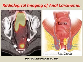
  • 1. Radiological Imaging of Anal Carcinoma. Dr/ ABD ALLAH NAZEER. MD.
  • 2. Normal Anatomy: The anal canal begins at the narrowing of the rectal ampulla at the anorectal junction where the rectum enters the puborectalis sling at the apex of the anal sphincter complex. It extends distally for approximately 4 cm and ends at the anal verge where the squamous mucosa blends with the perianal skin. The anal margin is the circular rim of pigmented skin with folds surrounding the anus, extending over an approximately 5-cm radius away from the anal verge. The most significant landmark of the anal canal is the dentate line, which lies 2.5–3 cm proximal to the anal verge and is visible macroscopically but not on MRI. Its position can be estimated either by measuring 2.5 cm above the anal verge or by dividing the anal canal into thirds so that the dentate line lies at the junction of the middle and upper thirds. The anal canal is divided by the dentate line into an upper part, lined with transitional or rectal glandular mucosa, and a lower part, lined by nonkeratinizing squamous epithelium, that merges with the perianal skin. The anal transition zone is defined as the histologic zone interposed between uniform rectal glandular mucosa above and uninterrupted squamous epithelium below, and it often has a heterogeneous histologic appearance.
  • 3. Illustrative anatomy of anal canal. A, Line diagram shows anal anatomy. B, Healthy 67-year-old man (with treated prostate cancer).Coronal T2-weighted image that corresponds to A shows ischioanal fossa (IAF), external sphincter (ES), and internal sphincter (IS).
  • 4. C, Healthy 53-year-old woman (with treated carcinoma cervix). Axial T2-weighted image through upper anal canal shows urethra (Ur) and vagina (Va). D, 67-year-old woman (post TAH and BSO for endometrial sarcoma). Sagittal T2-weighted image shows pubococcygeal line (long straight line), which is generally at level of anorectal junction. Angled line shows reference plane that one should use when reporting distance of epicenter of abnormality from anal verge.
  • 5. Anal cancer is a relatively uncommon malignancy. It accounts for less than 2% of large bowel malignancies and 1-6% of anorectal tumours (~1.5% of all gastro-intestinal tract malignancies in the Unites States). Epidemiology There may be a slight male predilection where its incidence has been reported to be approximately 0.5 per 100 000 in men and 1.0 per 100 000 in women. Its incidence is thought to be rising over the years. Clinical presentation Approximately 45% of patients may present with bleeding per rectum. Around 30% of patients may have pain and/or a sensation of a mass. Pathology Anal carcinoma typically originates between the anorectal junction above and the anal verge below. The vast majority of anal canal cancers are squamous cell cancers.
  • 6. Risk factors Both male and female: HPV / HIV infection immunosuppression number of lifetime sexual partners, and receptive anal intercourse smoking In females: previous in situ or invasive cervical, vulval or vaginal cancer. Lymphatic spread tumour above dentate line: to pararectal and paravertebral nodes tumour below dentate line: to inguinal and femoral nodes
  • 7. The accepted TNM staging of anal cancer is as follows: Primary tumour (T) TX: primary tumour cannot be assessed T0: no evidence of primary tumour Tis: carcinoma in situ T1: tumour 2 cm or less in greatest dimension T2: tumour >2 cm but <5 cm in greatest dimension T3: tumour >5 cm in greatest dimension T4: tumour of any size invades adjacent organ(s), e.g. vagina, urethra, bladder Regional lymph nodes (N) Nx: regional lymph nodes cannot be assessed N0: no regional lymph node metastasis N1: metastasis in perirectal lymph nodes(s) N2: metastasis in unilateral internal iliac and/or inguinal lymph node(s) N3: metastasis in perirectal and inguinal lymph nodes and/or bilateral internal iliac lymph nodes Distant metastasis (M) Mx: distant metastasis cannot be assessed M0: no distant metastasis M1: distant metastasis
  • 8. Radiologic staging is supplemented by clinical assessment of the anal margin, verge, and canal. This assessment often involves an examination with the patient under anesthesia and biopsy. Biopsy or fine-needle aspiration cytology of any enlarged or suspicious-appearing inguinal nodes is also essential. Transanal Endoscopic Ultrasound Transanal endoscopic ultrasound has results comparable to MRI for the detection of local tumor spread in patients with anal carcinoma and may be superior to MRI for the detection of small superficial tumors. However, regional lymph nodes higher in the pelvis or groins are outside the FOV of endosonography and supplementary MRI is needed for N staging. A meta-analysis of staging rectal cancer using endorectal ultrasound revealed other limitations including operator dependency and inability to assess stenotic tumors ; these limitations likely apply equally to anal cancer.
  • 9. MRI MRI has become the imaging modality of choice for locoregional staging and assessment of tumor response after chemoradiotherapy. MRI provides high-resolution multiplanar information about the location, size, circumferential and craniocaudal extent of the primary tumor and information regarding the involvement of adjacent structures. There is no significant difference in diagnostic value between pelvic surface coils and endoluminal coils. Moreover, endoluminal coil imaging is limited by a narrow FOV and near-field artifact and may not be tolerated by patients with anal cancer. MRI with a pelvic surface coil is easy, is more acceptable to the patient, and shows both local spread of the disease as well as lymph node involvement. We routinely perform MRI of anal cancers with a standard technique using a body coil; we obtain transaxial T1-weighted images with a 5-mm slice thickness to cover the abdomen and pelvis. High-resolution thin-section T2-weighted images are obtained with a 3-mm slice thickness in three orthogonal planes through the anal canal using a pelvic surface coil. In our experience, contrast-enhanced MRI offers limited advantages over standard T2-weighted sequences in routine primary staging but can be helpful to assess fistulation to other organs. Fat-suppressed imaging can help improve the conspicuity of the primary tumor, and STIR sequences are also useful to show fistula tracks. Diffusion-weighted imaging has an emerging role, particularly for detection of recurrent tumor after therapy, and can aid in differentiation of suspected residual or recurrent tumor from treatment-related change.
  • 10. 8F-FDG PET/CT FDG PET/CT has an increasing role in staging and treatment planning of anal carcinoma, particularly because up to 98% of anal tumors are FDG-avid. At diagnosis, FDG PET/CT is used to evaluate primary tumor size, lymph node status, and whether distant metastases are present. FDG PET/CT can also be useful for planning radiation therapy by clearly defining sites of metabolically active tumor. Several studies have shown that FDG PET/CT (in comparison with standard imaging) alters staging of anal carcinoma in approximately 20% of cases and treatment intent in approximately 3–5% of cases. Although PET/CT has a lower sensitivity in detecting perirectal nodes than MRI, this shortcoming does not affect management because perirectal nodes are routinely irradiated. The main impact of PET/CT on therapy stems from its superiority in detecting involved pelvic or inguinal lymph nodes, provoking the radiation oncologist to include them in the radiotherapy field.
  • 11. T staging of rectal cancer according to depth of tumor invasion as seen on axial T2-weighted MR images. (a) T1 cancers invade the submucosa (arrows) but do not involve the muscularis propria. (b) T2 cancers invade the muscularis propria but do not reach the subserosa (arrow). (c) T3 cancers invade beyond the muscularis propria (arrow). (d) T4 cancers invade through the serosal covering (T4a) or another fascial plane (T4b) and/or involve an adjacent organ (T4b) (arrows).
  • 12. A 32-year-old HIV-positive woman with clinical diagnosis of anovaginal fistula. Axial T2-weighted (a) and post-contrast fat-suppressed axial T1-weighted (b) images show inhomogeneous anal tissue invading the left aspect of the vagina (arrowheads), with internal non-enhancing necrosis and peripheral enhancement. Biopsy diagnosed SCAC with superimposed infection
  • 13. Early anal tumour, MR image shows that normal low signal of anal canal muscle has been replaced by intermediate-signal tumor (arrow) that is less than 2 cm. Early anal carcinoma, anterior T2 anal canal carcinoma. MR image shows that lesion measures more than 2 cm and hence achieves T2 status. There is invasion of external sphincter (arrow).
  • 14. A 40-year-old MSM with bioptic diagnosis of SCAC. Axial (a) and sagittal (b) T2-weighted images show 2-cm hyperintense nodule contained within the internal sphincter muscle, intensely enhancing as seen on post-contrast fat-suppressed coronal T1-weighted image (c), consistent with T1 tumour (arrowheads).
  • 15. A 62-year-old female with biopsy-proven SCAC. Axial T2-weighted (a), post-contrast fat-suppressed axial (b) and coronal (c) T1-weighted images, and corresponding enhanced image from body CT (d) show a 5.5- cm long (T3) enhancing tumour with infiltration of the left ischioanal fatty space (arrowheads).
  • 16. A 57-year-old woman undergoing abdomino-pelvic MDCT for unrelated reasons. Post-contrast axial (a) and coronal reformatted (b) detailed images of the anorectal region identify an unexpected 2-cm right-sided enhancing anal nodule. Subsequent clinical and bioptic assessment confirmed poorly symptomatic ulcerated SCAC.
  • 17. 46-year-old man with T3 anal carcinoma. A, Coronal T2- weighted image shows that neoplasm (bracket) extends from anal margin into lower rectum and is more than 5 cm in maximum dimension and therefore is T3. There is invasion of external sphincter and of left levator ani muscle (arrow). 46-year-old man with T3 anal carcinoma. B, MR image shows no evidence of adjacent organ invasion, as evidenced by complete low-signal rim (arrowheads) separating tumor from prostate anteriorly.
  • 18. Axial contrast-enhanced MR images in a patient with stage IIIB (cT3N2M0) adenocarcinoma of the rectum. (a) Image shows a low rectal mass with a small focus of extension into the adjacent mesorectal fat (arrow) (T3 disease). (b, c) Multiple enhancing subcentimeter mesorectal lymph nodes (arrows in b) and a left internal iliac lymph node (arrow in c) suggest N2 disease on the basis of the number of potentially involved lymph nodes. The patient subsequently underwent neoadjuvant chemotherapy and radiation therapy and resection.
  • 19. Stage IIIB (cT3N2M0) squamous cell carcinoma of the anus. (a, b) Coronal CT images obtained at different levels show a locally extensive mass extending superiorly from the anorectal junction (arrows in a). Pathologic analysis demonstrated squamous cell carcinoma consistent with anal cancer, despite the relatively superior location. A prominent left internal iliac lymph node is also seen (arrow in b). (c, d) Coronal CT images obtained at different levels 3 months after completion of definitive chemoradiation therapy show a partial radiographic response of both the primary mass and the left internal iliac lymph node.
  • 20. An elderly, 92-year-old man with previous prostatectomy and kidney failure has unenhanced MRI. Sagittal T2- (a) and axial T1-weighted (b) images show 5-cm long solid, inhomogeneous neoplastic tissue (*) extending from the anus to encase the proximal urethra (note catheter in place).
  • 21. A 65-year-old woman with history of previously treated small SCAC 3 years earlier. Axial T2- (a) and post-contrast T1-weighted (b) images show roundish 1-cm left inguinal node (arrowheads) with internal fluid-like necrosis and inhomogeneous enhancement, confirmed by ultrasound (c) as hypoechoic with loss of normal nodal structure. Surgical exeresis (postoperative status as seen in d, follow-up MRI) confirmed metastatic node from SCAC.
  • 22. Axial T2-weighted (a, b) and post-contrast fat-suppressed T1-weighted (c) images show inguinal nodal metastases, larger on left side (arrowheads) plus bilateral enhancing perirectal adenopathies (arrows)
  • 23. 35-year-old woman with T4 anal cancer. MR image shows lobulated tumor with central necrosis and invasion into posterior vagina (arrow).
  • 24. Axial fused FDG PET/CT images of 45- year-old man with upper anal cancer. A, Bulky anal cancer shows increased FDG uptake (arrow) and metabolically active right inguinal node (arrowhead). Axial fused FDG PET/CT images of 45-year-old man with upper anal cancer. B, Increased uptake in pelvic sidewall lymph node (arrowhead) is consistent with disease involvement. Axial fused FDG PET/CT images of 45-year- old man with upper anal cancer. C, Increased uptake in unsuspected liver lesion (arrowhead) was subsequently shown to be metastasis on liver MRI (not shown). Ability of FDG PET/CT to stage regional and metastatic disease accurately in single examination is its great strength
  • 25. A, Coronal T2-weighted MR image shows bulky T3 tumor in upper anal canal (arrow) that was subsequently treated with chemoradiotherapy. Coronal T2-weighted image obtained 8 weeks after therapy shows that tumor has diminished in size and there is tram-track appearance of low-signal fibrotic tissue encasing anal canal wall (arrowhead). Note that mass like pseudotumor of high signal in rectal mucosa (arrow) is edema from radiotherapy.
  • 26. 37-year-old man with bulky T3 anal cancer. A, Axial T2-weighted MR image obtained before treatment. 37-year-old man with bulky T3 anal cancer. B, Axial T2- weighted image obtained 3 months after therapy depicts low-signal fibrosis in place of tumor (arrow). Patient had complete response clinically and radiologically.
  • 27. A, T2-weighted MR image obtained at time of initial staging shows abnormal tumor tissue in lower anal canal, which was treated with chemoradiotherapy. B, T2-weighted MR image obtained 8 weeks after therapy shows significant size reduction and fibrotic reaction, but residual nodule (arrow) suspicious for remnant tumor is seen. C, Repeat T2-weighted MR image obtained immediately before resection shows nodule (arrow) has enlarged compared to initial post treatment (B). This nodule was confirmed to be tumor at histology after salvage abdominoperineal resection.
  • 28. A, Coronal T2-weighted MR image shows small tumor involving lower anal canal and anal margin (arrow) that was treated with chemoradiotherapy with good initial response B, Coronal MR image obtained for surveillance 1 year after chemoradiotherapy shows intermediate- to high-signal abnormality (arrow) suspicious for recurrent tumor at site of original primary. C, Axial T2-weighted MR image from same examination as B shows areas of small-volume, linear post- therapy fibrosis (arrowhead) and bulky intermediate- to high-signal abnormality (arrow) suspicious for recurrence. D, Apparent diffusion coefficient map from diffusion- weighted imaging series shows rim of restricted diffusion (arrow) in intermediate T2 signal areas indicative of high cellular content.
  • 29. A 60-year-old man with AIDS and clinical finding of ulcero-fungating anal mass. Axial (a) and coronal reformatted (b) CT images show moderately heterogeneous tissue (*) in its entire longitudinal extent from the anorectal junction to below the anal verge, associated with large necrotic iliac adenopathies (* in c). Biopsy confirmed super infected SCAC.
  • 30. An 83-year-old man with known SCAC. Initial MRI shows moderate circumferential thickening of the anus with T2-hyperintense signal (a) and contrast enhancement (b) (arrowheads). Complete disappearance of the lesion is observed on axial T2 (c) and post-contrast fat-suppressed T1-weighted (d) images following chemo-radiotherapy.
  • 31. An elderly, 85-year-old lady with biopsy-proven SCAC and multiple comorbidities. Initial MRI (a, b) shows showed circumferential anal wall thickening with abnormal solid signal (arrowheads) measuring 6 cm in length, plus a suspicious centimetric left inguinal lymph node (arrow in a), findings consistent with T3N2 lesion. After reduced chemo-radiotherapy, follow-up MRI (c, d) 4 months later disclosed progression and partial necrosis of both primary tumour (arrowhead) and inguinal adenopathy (arrow), plus appearance of an exophytic tissue mass protruding from the external anal orifice (* in d)
  • 32. A 40-year-old male patient with long-standing perianal Crohn’s disease, being treated with seton. Contrast-enhanced MDCT images (a, b, c in cranio-caudal order) show right-sided levator ani abscess (*), extensive perianal fistulization occupying the ischioanal space (arrow), and marked solid-appearing circumferential anorectal thickening (arrowheads). Abdomino-perineal resection for SCAC was performed
  • 33. Image shows an HIV-positive 41-year-old male with squamous cell carcinoma of the anus. Pretreatment FDG-PET/CT (left) demonstrated increased uptake in the anal canal (arrows) and a left internal iliac lymph node. Post-treatment FDG-PET/CT (right) demonstrated persistent uptake in the anal canal and new activity in the right lobe of the liver (arrows). The patient had biopsy- proven local and distant disease and died from local and metastatic disease 12 months after completion of chemoradiation.
  • 34. Image shows a 47-year-old female with squamous cell carcinoma of the anus. Pretreatment FDG-PET/CT (left) demonstrated increased uptake in the right anal canal and two 5-mm perirectal lymph nodes. Post-treatment FDG-PET/CT (right) showed complete metabolic response. She remains without evidence of disease 31 months after chemoradiation.