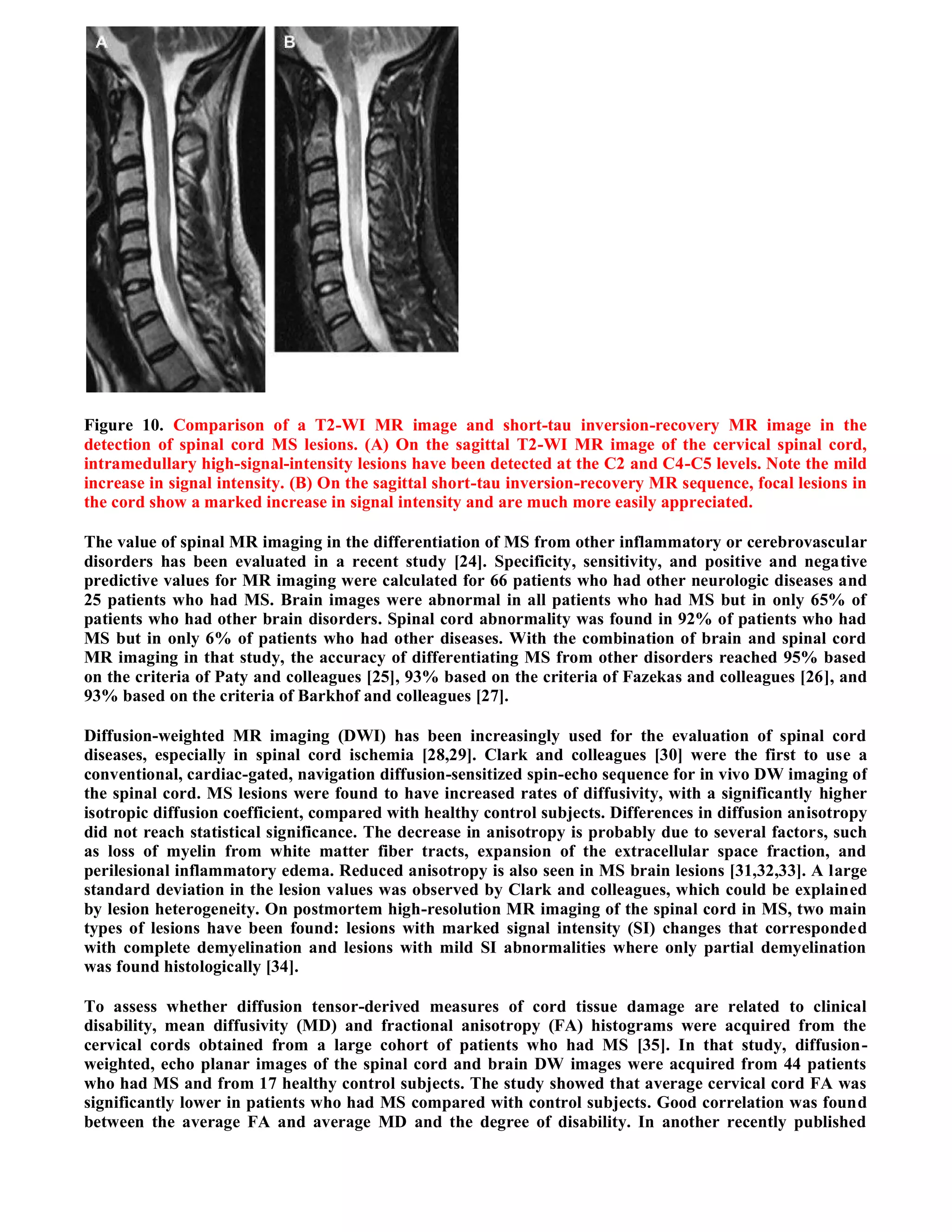
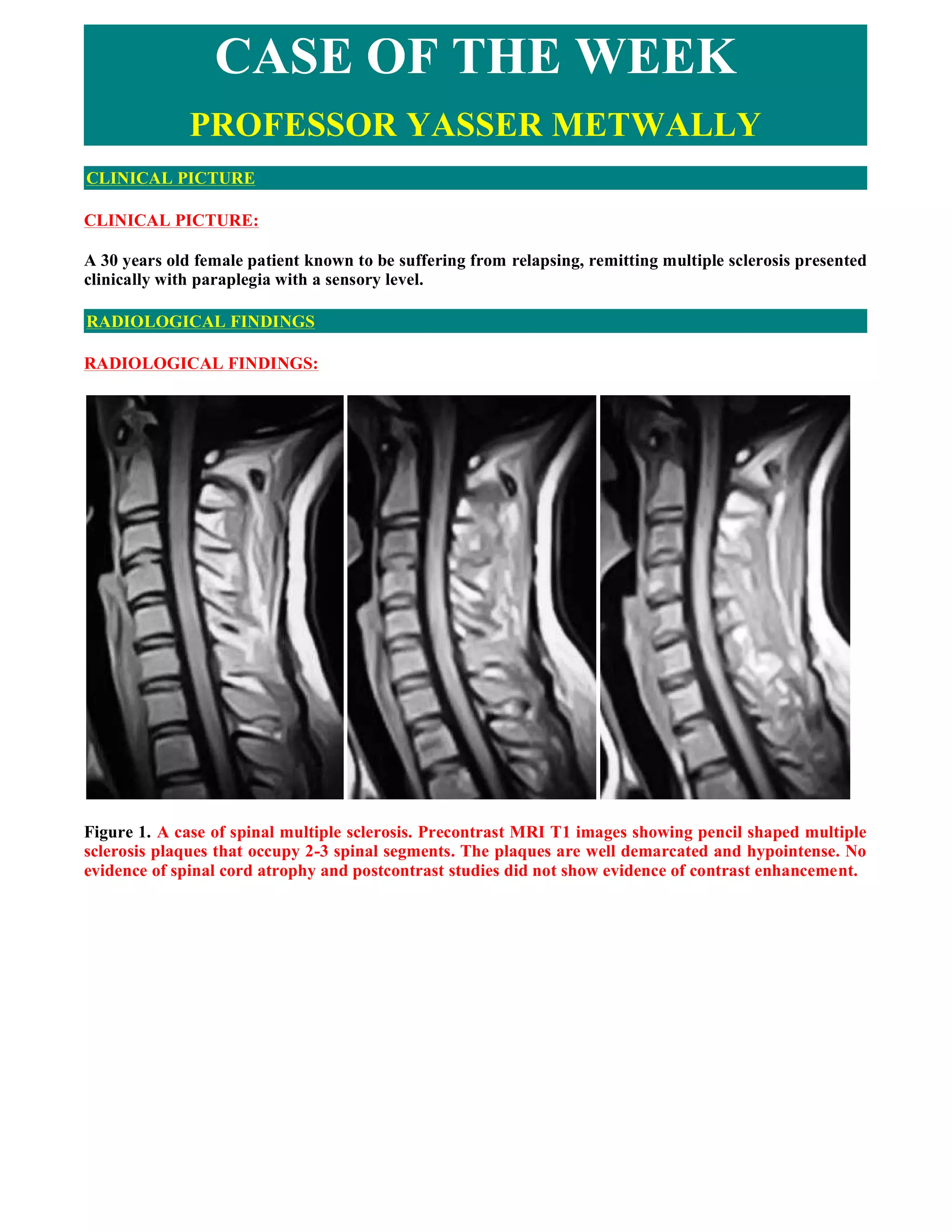
This document summarizes a case study of spinal multiple sclerosis seen on MRI imaging. It includes the following key points:
1) MRI images of the patient show multiple well-defined pencil-shaped lesions occupying 2-3 spinal segments that appear hypointense on T1-weighted images and hyperintense on T2-weighted images.
2) The lesions are characteristic of multiple sclerosis and located peripherally within the spinal cord.
3) The imaging and clinical presentation lead to a diagnosis of spinal multiple sclerosis.
1. Paraplegia refers to weakness or paralysis of both legs, which is most often caused by diseases of the thoracic spinal cord, cauda equina, or peripheral nerves.
2. Spastic paraplegia involves increased muscle tone and weakness due to upper motor neuron lesions, while flaccid paraplegia presents with decreased muscle tone and weakness from lower motor neuron lesions.
3. The key distinguishing features between the two types are muscle tone (increased in spastic vs decreased in flaccid) and deep tendon reflexes (exaggerated in spastic vs absent in flaccid).
Applied cross sectional anatomy of spinal cordTanat Tabtieang
The document provides an overview of the anatomy and imaging features of the spine and spinal cord. It describes the basic anatomy of the vertebrae and spinal segments. Common spinal pathologies are summarized, including degenerative changes, trauma, infection, tumors and congenital abnormalities. For each condition, the document explains the imaging appearance and features to evaluate on radiographs, CT and MRI scans. Key anatomical structures and imaging signs are illustrated with examples.
The document discusses the anatomy and classification of brachial plexus injuries. It notes that the brachial plexus extends from the spinal cord to the axilla, supplying the upper extremity and shoulder. It is susceptible to trauma due to its size, location, and position between mobile structures. The brachial plexus contains over 100,000 axons and is composed of five roots, three trunks, six divisions, three cords, and five terminal nerves. Injuries are classified as supraclavicular, retroclavicular, or infraclavicular depending on the location of the lesion. Supraclavicular injuries tend to be more severe due to the forces required to cause them and often result from
The document discusses anatomy of the spine, including identifying vertebrae and describing their features. It covers the roles of intervertebral discs, ligaments, and muscles in load bearing and spinal movement. Common spinal abnormalities and causes of back pain are outlined. Procedures like lumbar puncture and considerations for spinal injury management are also summarized.
spinal cord and applied aspects of spinemrinal joshi
The document provides information on the gross appearance, meninges, organization of nerves, and mobility of the spinal cord. It describes the structure of the spinal cord including the gray and white matter. The gray matter contains nerve cell groups that innervate muscles and receive sensory information. The white matter surrounds the gray matter and contains ascending and descending tracts for transmitting signals to and from the brain. The ascending tracts convey sensory information for pain, temperature, touch and proprioception. The descending tracts control skeletal muscles through pathways from the cortex, reticular formation, tectum, red nucleus and vestibular system.
The spinal cord runs through the vertebral canal and extends from the foramen magnum to the L2 vertebra. It gives rise to 31 pairs of spinal nerves and has cervical, thoracic, lumbar, sacral and coccygeal regions. The meninges that surround it include the dura mater, arachnoid mater and pia mater. The spinal cord has gray matter containing neuron cell bodies and white matter containing myelinated axons. Descending motor pathways control voluntary movement while ascending sensory pathways transmit proprioceptive and discriminative touch information to the brain.
The document discusses brachial plexopathies, including the anatomy of the brachial plexus, classifications of brachial plexopathies, assessment of the brachial plexus, and selected site-specific disorders of the brachial plexus. It describes how brachial plexopathies can be classified by the region involved, such as supraclavicular, retroclavicular, and infraclavicular. Supraclavicular plexopathies are more common, severe, and associated with a worse prognosis compared to other regions. Selected disorders that are discussed include burner syndrome, rucksack paralysis, classic postoperative paralysis, and obstetric brachial plexopathy.
This document describes the structure and function of the spinal cord. It discusses the external and internal anatomy, including the protective meninges, enlargements in the cervical and lumbar regions, conus medullaris, cauda equina, and filum terminale. Internally, it describes the gray matter, white matter, tracts, and roots. The spinal cord acts as a pathway for sensory and motor impulses and is responsible for reflexes. Ascending tracts conduct sensory information to the brain and descending tracts carry motor commands from the brain.
The spinal cord begins at the foramen magnum and extends down to the L1-L2 vertebrae. It has 31 segments and is enlarged in the cervical and lumbar regions. It is protected by vertebrae, meninges, and cerebrospinal fluid. The spinal cord contains gray matter with dorsal and ventral horns and white matter tracts. It transmits motor and sensory signals between the brain and body.
The spinal cord is approximately 42-45 cm long in adults and extends from the medulla oblongata down to the L1-L2 vertebrae. It is divided into 31 segments that correspond to nerve root attachments. The spinal cord receives its blood supply from the anterior and posterior spinal arteries as well as radicular arteries. Key arteries include the anterior spinal artery, posterior spinal arteries, and the artery of Adamkiewicz, which supplies the lumbar segments. Disruptions to the anterior spinal artery can cause ischemia to the anterior two-thirds of the spinal cord.
The brachial plexus is formed by the ventral rami of spinal nerves C5-T1, which supply the upper limb. It is a network of nerves that branches out to form the five main nerves of the arm - the median, ulnar, radial, musculocutaneous and axillary nerves. Injuries to different parts of the brachial plexus cause weakness and sensory loss in specific areas innervated by the affected nerves. Erb's palsy is a type of brachial plexus birth injury that results in weakness of shoulder and arm muscles on the affected side.
A 19-year-old college student fell 30 feet while rock climbing and was found unable to move his extremities with neck pain. X-rays showed a fractured fifth cervical vertebra. The spinal cord begins at the foramen magnum and extends down, giving off 31 pairs of spinal nerves. It is protected by three meninges and has gray matter in an H-shape containing motor, sensory and interneurons. Injuries can result in quadriplegia or paraplegia depending on the level and extent of damage.
This document provides an overview of the anatomy of the spinal cord including its structure, blood supply, sensory and motor systems, and approach to spinal cord diseases. Key points include:
- The spinal cord has grey matter containing nuclei and white matter containing tracts that transmit sensory and motor signals.
- It receives blood supply from the anterior and posterior spinal arteries as well as segmental radicular arteries like the artery of Adamkiewicz.
- Sensory systems assess proprioception, pain, temperature and touch. Motor systems involve upper and lower motor neurons controlling voluntary movement.
- Clinical examination assesses sensory levels and motor function to localize spinal cord lesions, while investigations include CSF analysis, imaging
anatomy,physiology of spinal cord 7CSFAbebe Assaye
The document provides information on the anatomy and physiology of the spinal cord, ventricles, and cerebrospinal fluid (CSF). It describes the anatomy of the spinal cord including its meninges, blood supply, internal organization into gray and white matter, and ascending and descending nerve pathways. It also discusses the anatomy and physiology of the ventricles, including their structure and how CSF is produced. The objectives are to review the basic anatomy of the spinal cord and ventricles and learn about the position of nerve pathways in the spinal cord and how CSF is produced.
The document describes the anatomy and structure of the spinal cord. It details the external structure including its cylindrical shape and location within the vertebral canal surrounded by meninges. Internally, it describes the gray and white matter, including the ascending and descending tracts that transmit sensory and motor signals. It also discusses the blood supply, clinical correlations, and applications related to injuries and diseases of the spinal cord.
The spinal cord extends from the skull to the lower back, and has sensory and motor functions. It contains gray matter in an H-shape containing nuclei, and white matter containing ascending and descending tracts. The spinal cord is surrounded by meninges and has 31 pairs of spinal nerves that connect it to the body. It carries sensory information from the body to the brain via ascending tracts, and carries motor commands from the brain to the body via descending tracts.
Plexopathy is a disorder affecting nerve networks like the brachial or lumbosacral plexus. Symptoms include pain, motor control loss, and sensory deficits. It is usually caused by localized trauma or compression. Brachial plexopathy specifically affects the network of nerves from the cervical spine to the shoulder, arm, and hand. Lumbosacral plexopathy affects the network of nerves from the lumbar spine and sacral spine. Diabetic plexopathy commonly affects the lumbosacral plexus and causes anterior thigh pain and proximal leg muscle weakness.
The spinal cord extends from the foramen magnum to the L1-L2 vertebral level. It has 31 spinal segments and contains gray matter in an H-shaped cross-section. The spinal cord enlarges at the cervical and lumbar regions, corresponding to the brachial and lumbosacral plexuses. White matter tracts in the spinal cord include the posterior columns (gracilis and cuneatus), spinothalamic tracts, corticospinal tracts and spinocerebellar tracts. The meninges surrounding the spinal cord are the dura, arachnoid and pia mater. The cauda equina is formed from spinal nerve roots distal to the conus
The spinal cord extends from the brainstem down to the lumbar region. It contains gray matter surrounded by white matter and is segmented into cervical, thoracic, lumbar, and sacral regions. The spinal cord transmits sensory information from the body to the brain via dorsal roots and carries motor commands from the brain to the body via ventral roots. Each segment innervates a specific dermatome of skin and muscles. The meninges protect the spinal cord and CSF circulates in the subarachnoid space.
The spinal cord has 31 pairs of spinal nerves that emerge from its sides. It occupies the upper two-thirds of the vertebral canal and is protected by bony vertebrae and meninges. The spinal cord receives its blood supply from the anterior and posterior spinal arteries as well as segmental arteries. It has an anterior median fissure and posterior median sulcus that contain the arteries supplying the cord. Lesions of the spinal cord can result in deficits depending on the location and structures involved, and the cord can be surgically approached through laminectomy.
The spinal cord ends at the lower border of the first lumbar vertebra in adults. During fetal development, the spinal cord is initially as long as the vertebral column but becomes shorter as the column elongates faster than the cord after the third month. Diseases of the vertebrae are a common cause of spinal cord pathology in adults from fractures, infections, or secondary deposits. The spinal cord has 31 segments including 8 cervical, 12 thoracic, 5 lumbar, and 5 sacral segments formed by fusion of vertebrae.
The spinal cord extends from the foramen magnum to the L1-L2 vertebrae in adults and functions to transmit information between the brain and body. It has 31 pairs of spinal nerves and cervical and lumbar enlargements corresponding to the brachial and lumbar plexuses. The spinal cord has grey matter containing nuclei and white matter tracts. Blood supply comes from the anterior and posterior spinal arteries along with segmental arteries from the vertebrae.
The spinal cord is a long, thin bundle of nervous tissue located along the back and neck that connects to the brain. It receives and transmits electric signals throughout the body and back to the brain. The spinal cord is protected by vertebrae bones and cerebral spinal fluid. It has gray matter containing nerve cell bodies and white matter containing nerve fibers. The spinal cord is divided into cervical, thoracic, lumbar, and sacral sections that each control certain muscles. Blood supply comes from anterior and posterior spinal arteries as well as radicular arteries.
The document discusses the anatomy and functions of the spinal cord. It describes the spinal cord's location and segments. The spinal cord contains gray matter containing neuron cell bodies and white matter containing myelinated axons. The white matter is divided into tracts that transmit sensory information ascending to the brain and motor signals descending from the brain. The document outlines various spinal cord syndromes and compressive myelopathies, their clinical presentations and causes.
The document provides information on the anatomy and structure of the spinal cord. It discusses the following key points:
- The spinal cord is surrounded by three meningeal coverings - dura mater, arachnoid mater, and pia mater. It has cervical and lumbar enlargements.
- Spinal nerves arise in pairs from the spinal cord. Each nerve has a dorsal root containing a ganglion and a ventral root.
- The spinal cord has gray matter on the outside containing nuclei and white matter on the inside. It also contains ascending and descending tracts that connect to the brain.
- Ascending tracts like the anterior and lateral spinothalamic tracts transmit sensory information from
This document summarizes a case report of a 22-year-old male patient diagnosed with Friedreich's ataxia. The patient presented with bilateral cerebellar ataxia, pyramidal signs, loss of tendon reflexes, peripheral neuropathy, enlarged nerves, kyphoscoliosis, and pes cavus. Nerve biopsy revealed demyelinating neuropathy. MRI showed cervical spinal cord atrophy. The clinical diagnosis was Friedreich's ataxia based on the progressive nature of symptoms starting at age 12. The document provides figures of MRI images and references for further information on the case.
This document summarizes a case study of a 37-year-old female patient who presented with Parinaud syndrome. She had first noticed eye movement abnormalities at age 12 but did not seek medical care until age 37. An MRI revealed a tectal plate glioma (focal midbrain glioma). The tumor enhanced with contrast and appeared hyperintense on T2-weighted MRI. The patient's condition remained stable with follow-up MRIs every two years.
The document describes a study that classified 21 patients with brain stem gliomas into 5 groups based on clinical features and radiological findings. Group 1 had diffuse gliomas and a relatively better prognosis. Group 2 also had diffuse gliomas but a worse prognosis. Group 3 consisted of patients with focal pontine or midbrain gliomas. Group 4 contained patients with cervicomedullary gliomas. The study analyzed each group's clinical features, treatment responses, and outcomes to better understand patterns in brain stem gliomas.
This document provides an overview of how to conduct a neurological examination, including taking a thorough patient history and performing a physical exam. Some key points:
1. Taking a thorough history is important for localizing lesions and making a differential diagnosis. Leading questions should be asked about symptoms, onset/progression, relieving/precipitating factors, and associated symptoms.
2. Common complaints warranting detailed history include headache, dizziness/vertigo, sensory symptoms, cognitive decline, speech disorders, weakness, and visual abnormalities.
3. The physical exam follows a standardized pattern but can be tailored based on pertinent findings. It includes tests of consciousness, cognition, cranial nerves, motor function, sensory function
A 66-year-old male patient presented with non-specific lower back pain. MRI images showed signs of lumbar spondylosis including disc degeneration and bulging in the lower lumbar spine. The document provides figures showing disc herniation and details on accessing additional materials on the author's website, including updates versions of the case publication.
This document discusses neurological complications that can occur during hematopoietic cell transplantation (HCT). It describes complications that occur during different stages of HCT: 1) During conditioning regimens, complications include encephalopathy, seizures, and cerebral infarction caused by chemotherapy drugs or medical procedures. 2) During bone marrow depletion, complications include encephalopathy, seizures, cerebral infarctions, and hemorrhages due to metabolic issues, drugs, or infections. 3) During chronic immunosuppression in allogeneic HCT, complications include infections by viruses and opportunistic organisms. 4) Late complications include relapse of the original disease, neurological graft-versus-host disease, and second neoplasms.
This document summarizes the pathology of pituitary tumors, including:
1) Pituitary adenomas are benign neoplasms originating from pituitary cells and are the most common tumors of the sella region.
2) Pituitary tumors are currently classified based on their hormonal content and ultrastructural morphology, with 14 recognized subtypes.
3) Growth hormone-secreting adenomas are associated with acromegaly or gigantism and can have various histologic appearances, most commonly densely or sparsely granulated somatotroph adenomas.
Issues in brainmapping...EEG and brainmap spectral profiles in cortical lesio...Professor Yasser Metwally
Issues in brainmapping...EEG and brainmap spectral profiles in cortical lesions, subcortical white matter lesions and subcortical gray matter (diencephalic) lesions
This document is a case publication from January 2008 edited by Professor Yasser Metwally about acute disseminated encephalomyelitis (ADEM). It describes an 18-year-old female patient who presented with disturbed consciousness, seizures, optic neuritis and meningism. MRI images show lesions in the brain consistent with ADEM. The publication discusses differentiating ADEM from other conditions like multiple sclerosis, infections and metabolic disorders based on clinical features and test results. It provides guidance on the diagnosis and management of ADEM cases.
A 40-year-old patient presented with relapsing-remitting spinal multiple sclerosis. MRI revealed asymptomatic brain involvement and recent progression of myelopathy. The document discusses the diagnosis of spinal MS and differentiating it from transverse myelitis using MRI characteristics such as lesion location and contrast enhancement. Figures show examples of MS plaques and diffuse abnormalities on MRI of the spinal cord.
A 40-year-old female patient presented with a history of migraines. MRI scans revealed two meningioma tumors, one in the parasellar region and one in the cerebellopontine angle. The tumors appeared hyperintense on T2-weighted MRI and showed dense contrast enhancement. Based on the MRI characteristics and location of the tumors, they were diagnosed as multiple syncytial meningiomas. Syncytial meningiomas tend to appear hyperintense on T2-weighted MRI due to high cell count and microcysts.
This document is a short case publication about a 60-year-old male patient presenting with symptoms of cerebellopontine angle syndrome affecting the 7th, 5th, and bulbar cranial nerves, as well as cerebellar deficits and right ear tinnitus. Imaging revealed a fusiform aneurysm of the vertebrobasilar system with mural thrombosis. Fusiform aneurysms commonly involve the vertebrobasilar system, causing arteries to be diffusely dilated, tortuous, and prolonged with frequent mural thrombosis. They rarely rupture but can cause ischemic manifestations or pressure effects from mass effect. The publication is periodically updated on the author's website.
Hepatic encephalopathy is a syndrome characterized by personality changes, impaired intellect, and depressed consciousness that occurs in patients with cirrhosis of the liver. An important prerequisite is the diversion of portal blood into systemic circulation through portosystemic shunts. Subtle signs are seen in 70% of cirrhosis patients, with symptoms debilitating in 24-53% of those with portosystemic shunt surgery. Triphasic waves seen on EEGs are nonspecific but classic for hepatic encephalopathy. While commonly caused by hepatic or renal failure, triphasic waves can result from various toxic, metabolic, or structural abnormalities.
Issues in brainmapping...The role of EEG in epileptic syndromes associated wi...Professor Yasser Metwally
1. EEG is essential for evaluating epilepsy and can provide information about background activity, epileptiform discharges, and help diagnose specific epilepsy syndromes.
2. The document discusses various epilepsy syndromes like symptomatic, cryptogenic, and idiopathic epilepsy and how EEG characteristics help differentiate these.
3. Specific syndromes discussed include infantile spasms and West syndrome, characterized by hypsarrhythmia on EEG, and Lennox-Gastaut syndrome, characterized by slow spike and wave activity on EEG.
This document summarizes the classification and treatment of headaches. Headaches are classified as either primary/idiopathic or secondary/symptomatic. Primary headaches include tension headaches, migraines, and cluster headaches and have no underlying cause, while secondary headaches are a symptom of another condition like hypertension or meningitis. The document outlines diagnostic criteria and treatment approaches for different headache types, including acute treatment with medications and preventative treatment.
This document discusses a case of spinal multiple sclerosis in a 40-year-old patient. MRI images show well-defined pencil-shaped lesions occupying 2-3 spinal segments that are hypointense on T1-weighted images and hyperintense on T2-weighted images. Diffuse abnormalities are also seen as poorly demarcated hyperintense regions on T2-weighted images. The diagnosis is spinal multiple sclerosis. The document then discusses features of spinal MS lesions seen on MRI such as focal lesions, diffuse abnormalities, and spinal cord atrophy, and compares features of MS to other conditions like neuromyelitis optica.
A spinal cord syndrome may develop from extramedullary or intramedullary pathological processes. Initial symptoms can be gradual or acute, including pain, numbness, or weakness. Radiographic imaging like MRI is used to identify intrinsic or extrinsic cord pathology. Patients with acute spinal cord injury after trauma can experience early or late changes in motor and sensory function. Differential diagnoses for diseases affecting the spinal cord include spinal shock, Brown-Séquard syndrome, central cord syndrome, and anterior spinal artery syndrome. Lesions in different regions of the spinal cord produce varying neurological deficits.
MRI of Spine and very easy details of spssuserc66686
The document discusses various neurological conditions that can be seen on CT and MRI imaging of the brain and spine. It provides examples of imaging findings for tumors such as meningiomas, medulloblastomas, ependymomas, glioblastomas, and metastases. It also reviews imaging appearances of conditions like cerebral abscesses, multiple sclerosis, cavernomas, spinal tuberculosis, and spondylitis. Key distinguishing radiological features of different lesions are highlighted.
Tips, Pearls and Pitfalls of Spinal Cord MRIWafik Bahnasy
- Many neurological disorders simultaneously or consecutively affect the brain and spinal cord, however most neurologist find their comfort zone in attending the diagnosis via the brain access.
- This concept resulted in lagging of spinal cord imaging researches compared to brain ones and consecutive underestimation of the opportunity of an important tool sometimes essential to reach a definite diagnosis.
This document discusses magnetic resonance imaging (MRI) for peripheral nerves. It begins by providing background on how MRI has become a standard imaging method for the central nervous system due to its soft tissue contrast. It then discusses some limitations of using MRI for peripheral nerves, including technical limitations of older scanners. The document outlines several clinical applications of peripheral nerve MRI, such as evaluating carpal tunnel syndrome, neural tumors, and traumatic nerve injuries. It describes the ability of MRI sequences like diffusion imaging to assess properties of neural tissue. The document concludes by discussing technical considerations for peripheral nerve MRI protocols and sequences.
This document provides guidance on how to read MRI scans by describing the key steps: check patient information and image details; examine the different MRI planes and sequences to analyze fat vs water content; look for abnormalities and determine their nature, location, size and shape; correlate MRI findings with prior imaging; and recognize signs of common musculoskeletal conditions like spinal stenosis, tumors, infections and arthritis. Example MRI images are included to illustrate moderate and severe spinal stenosis, different tumor types, and infections like spondylitis and septic arthritis.
Imaging in Neurovascular conflicts [Neurovascular compression syndrome ]Nija Panchal
- Neurovascular compression syndrome (NVCS) refers to nerve compression by aberrant or tortuous blood vessels, which can cause cranial nerve dysfunction including trigeminal neuralgia.
- Trigeminal neuralgia is characterized by abrupt, unilateral facial pain and is most often caused by neurovascular compression of the trigeminal nerve at the root entry/exit zone from the brainstem.
- MRI with techniques like CISS and MRA-TOF are effective in evaluating neurovascular relationships and compressions, aiding surgical planning for microvascular decompression to treat refractory trigeminal neuralgia.
This case report describes a 22-year-old male patient who presented with quadriplegia and sensory loss following an influenza infection. MRI imaging showed a central area of hyperintensity on the cervical spinal cord extending over multiple segments, consistent with transverse myelitis. Peripheral contrast enhancement was observed, a characteristic feature of transverse myelitis that helps differentiate it from multiple sclerosis. The patient was diagnosed with acute idiopathic transverse myelitis based on the clinical and radiological findings. Over the past decade, research has provided new insights into demyelinating diseases of the spinal cord. MRI has become particularly useful for distinguishing between transverse myelitis and multiple sclerosis based on the location and extent of
A 22-year-old male presented with symptoms of Friedreich's ataxia including bilateral cerebellar ataxia and peripheral neuropathy. MRI showed marked atrophy of the upper cervical spinal cord, a characteristic finding in Friedreich's ataxia. Pathological examination found diffuse demyelinating neuropathy with onion bulb formations. The patient was diagnosed with Friedreich's ataxia based on his clinical signs and supportive radiological and pathological findings.
- A 60-year-old male presented with persistent lumbar pain and was diagnosed with spinal metastasis from prostatic carcinoma. MRI showed an osteolytic lesion in his L2 vertebra extending into the epidural space.
- Spinal metastases are common manifestations of cancer that can be effectively treated with surgery alongside radiotherapy and chemotherapy. Evaluation and treatment of spinal metastases requires a multidisciplinary approach.
- This case report discusses the presentation, diagnosis, and management of spinal metastases as well as the mechanisms of spread to the spine.
1) The study analyzed MRI findings of Wallerian degeneration in the spinal cords of 11 patients with traumatic spinal injuries.
2) The most common pattern observed was degeneration in both the posterior and lateral tracts of the spinal cord.
3) The signal changes observed on MRI, including hyperintensity on T1 and T2 weighted images, likely correspond to later stages (3 and 4) of Wallerian degeneration as described in the brain.
multiple level spondylodiscitis in neurobrucllosis: int jr of medicineSachin Adukia
Multiple level spondylodiscitis with presacral abscess in spinal brucellosis: a rare presentation
A 56-year-old male farmer in India presented with lower back pain and weakness in his legs for 9 weeks. MRI revealed multi-level infective spondylodiscitis and a prevertebral sacral abscess. Blood culture grew Brucella melitensis after 8 days. Treatment was modified to a 12-month triple antibiotic therapy. Spinal brucellosis should be considered for those with back pain in endemic areas, as timely diagnosis and appropriate treatment is important.
The document discusses several radiology cases involving the brain and spine. Question 291 presents CT scans of a woman with headaches and asks for the most likely diagnosis of a fat-containing extraconal orbital mass. Question 292 shows MR images of a woman with extremity weakness and asks for the most likely diagnosis of multiple brain and spine lesions. Question 293 presents MR images of a man with back pain and asks for the diagnosis of a posterior spinal mass seen on the images.
Presentation2, radiological imaging of phakomatosis.Abdellah Nazeer
Von Hippel-Lindau disease is characterized by the development of numerous benign and malignant tumors in different organs due to mutations in the VHL tumor suppressor gene. Common manifestations include renal cell carcinomas, renal cysts, pheochromocytomas, pancreatic cysts and tumors, retinal hemangioblastomas, and cerebellar hemangioblastomas. Patients may develop tumors in the kidneys, pancreas, liver, adrenal glands, and brain. The variety of lesions that can occur has led to the mnemonic "HIPPEL" to remember the key features of VHL disease.
Presentation1 radiological imaging of carpal tunnel syndrome.Abdellah Nazeer
- Carpal tunnel syndrome results from compression of the median nerve as it passes through the carpal tunnel in the wrist. It commonly occurs between ages 36-60 and is more frequent in women. Symptoms include pain, numbness, and tingling in the hand.
- Ultrasound and MRI are useful imaging modalities. Ultrasound can show bowing of the flexor retinaculum, flattening and swelling of the median nerve. MRI also demonstrates these findings and can detect additional causes like masses or arthritic changes.
- Various pathologies can cause carpal tunnel syndrome by decreasing the size of the tunnel or enlarging its contents, compressing the median nerve. Imaging allows visualization
1. MRI plays a key role in evaluating suspected myelopathy by identifying the cause and extent of spinal cord abnormalities.
2. Common artifacts that can mimic cord abnormalities include Gibbs artifacts and pulsation artifacts. Extrinsic compression is also considered.
3. For intrinsic cord lesions, the differential depends on acute vs nonacute onset. Acute causes include demyelination, ischemia, infection while nonacute includes neoplasm, metabolic, neurodegenerative, and inflammatory diseases.
This patient presents with knee pain. Imaging shows a lesion within the epiphysis of the knee. On MRI, the lesion has low signal on T1 and T2 weighted images with a low signal margin and no aggressive features. The most likely diagnosis is chondroblastoma, which is a rare benign epiphyseal tumor seen in children before growth plate closure that appears as a well-defined lytic lesion on radiographs and MRI.
A 35-year-old female patient presented with paraplegia and acute onset sensory loss following a rabies vaccination. An MRI showed edema in her spinal cord occupying more than 2/3 of the cross-section over 8 spinal segments. She was diagnosed with acute idiopathic transverse myelitis. The document discusses the characteristic MRI findings of transverse myelitis and provides figures comparing it to other conditions like multiple sclerosis. It also provides information on accessing other publications by the author on neurological cases and imaging.
The document discusses various presacral lesions that can be seen on imaging. It describes the anatomy of the presacral space and then covers conditions with osteochondral origin like giant cell tumor and Ewing sarcoma. Neurogenic conditions such as neurofibromas, schwannomas, and perineural cysts are also discussed. Other lesions mentioned include dural ectasia and anterior myelomeningoceles. For each condition, the document provides details on clinical features, imaging appearance on modalities like CT and MRI, and examples of imaging findings.
Similar to Case record...Spinal multiple sclerosis (20)
The document discusses the benefits of exercise for mental health. It states that regular physical activity can help reduce anxiety and depression and improve mood and cognitive function. Exercise causes chemical changes in the brain that may help alleviate symptoms of mental illness.
The document discusses the benefits of exercise for mental health. Regular physical activity can help reduce anxiety and depression and improve mood and cognitive functioning. Exercise causes chemical changes in the brain that may help protect against mental illness and improve symptoms.
The document discusses the benefits of exercise for mental health. Regular physical activity can help reduce anxiety and depression and improve mood and cognitive function. Exercise causes chemical changes in the brain that may help protect against mental illness and improve symptoms.
The document discusses the benefits of exercise for mental health. Regular physical activity can help reduce anxiety and depression and improve mood and cognitive function. Exercise causes chemical changes in the brain that may help protect against mental illness and improve symptoms for those who already suffer from conditions like depression and anxiety.
The document discusses the benefits of exercise for mental health. Regular physical activity can help reduce anxiety and depression and improve mood and cognitive function. Exercise causes chemical changes in the brain that may help protect against mental illness and improve symptoms.
This document discusses the radiological pathology of seizure disorders. It describes various developmental anomalies, neoplasms, infections, immune-mediated disorders, cerebrovascular diseases, and trauma that can cause seizures. Specific conditions mentioned include cortical dysplasia, tuberous sclerosis, Sturge-Weber syndrome, neuronal migration disorders, vascular malformations, infections, and immune-mediated Rasmussen's encephalitis. The document provides detailed descriptions of the histopathological findings and MRI/CT appearances of different lesions that can underlie seizure disorders.
This document discusses cerebral haemorrhage (ICH), which accounts for 10-15% of strokes. ICH can result from several mechanisms, including hypertension (47-66% of cases), cerebral amyloid angiopathy (CAA), and vascular malformations. CAA typically affects the elderly and causes lobar ICH that is often recurrent or involves multiple simultaneous haemorrhages. Vascular malformations like arteriovenous malformations (AVMs) and cavernous angiomas are a common cause of ICH in young, non-hypertensive patients. Imaging techniques like CT and MRI can identify vascular malformations and help determine the underlying cause of ICH.
Cerebral amyloid angiopathy (CAA) refers to the deposition of β-amyloid in the arteries of the cerebral cortex. It is commonly seen in Alzheimer's disease but can also occur in healthy elderly individuals. CAA can cause intracerebral hemorrhage, dementia, or transient neurological symptoms. The deposition damages blood vessels and increases the risk of hemorrhage. Imaging such as CT scans can detect hemorrhages characteristic of CAA, which are often lobar and cortical. Genetic factors like the ApoE genotype can influence the severity and presentation of CAA.
Cerebral microbleeds are small brain hemorrhages detected by MRI that are caused by leakage of blood from damaged small vessel walls. They are increasingly recognized in patients with cerebrovascular disease, Alzheimer's disease, vascular cognitive impairment, and normal elderly populations. Microbleeds in lobar regions may indicate cerebral amyloid angiopathy and link vascular and amyloid neuropathologies, while deep or infratentorial microbleeds often reflect hypertensive vasculopathy. Detection of microbleeds provides insight into cerebral small vessel disease and its relationship to cognitive impairment and dementia.
The document discusses the benefits of exercise for mental health. Regular physical activity can help reduce anxiety and depression and improve mood and cognitive functioning. Exercise causes chemical changes in the brain that may help protect against mental illness and improve symptoms.
A 57-year-old male patient presented with left lower limb weakness that had progressed over three months. MRI images showed bilateral, symmetrical lesions in the posterior parieto-occipital white matter, which had scalloped margins and did not enhance or cause mass effect. Based on the clinical presentation and MRI findings, the patient was diagnosed with progressive multifocal leukoencephalopathy (PML), a demyelinating disease caused by JC virus reactivation that predominantly affects immunocompromised individuals. PML lesions are typically multifocal and located in the white matter of the brain, most often in the parieto-occipital region.
Issues in radiological pathology: Radiological pathology of watershed infarct...Professor Yasser Metwally
The document discusses border zone or watershed infarcts, which occur at the junction between two main arterial territories and constitute approximately 10% of all brain infarcts. There are two types - external (cortical) and internal (subcortical). External infarcts are often embolic in nature while internal infarcts are mainly caused by hemodynamic compromise. Advanced imaging can help identify areas of low perfusion and distinguish the two types. The document then examines the classification, imaging appearance, causal mechanisms, and clinical course of both external and internal border zone infarcts in more detail.
ISO/IEC 27001, ISO/IEC 42001, and GDPR: Best Practices for Implementation and...PECB
Denis is a dynamic and results-driven Chief Information Officer (CIO) with a distinguished career spanning information systems analysis and technical project management. With a proven track record of spearheading the design and delivery of cutting-edge Information Management solutions, he has consistently elevated business operations, streamlined reporting functions, and maximized process efficiency.
Certified as an ISO/IEC 27001: Information Security Management Systems (ISMS) Lead Implementer, Data Protection Officer, and Cyber Risks Analyst, Denis brings a heightened focus on data security, privacy, and cyber resilience to every endeavor.
His expertise extends across a diverse spectrum of reporting, database, and web development applications, underpinned by an exceptional grasp of data storage and virtualization technologies. His proficiency in application testing, database administration, and data cleansing ensures seamless execution of complex projects.
What sets Denis apart is his comprehensive understanding of Business and Systems Analysis technologies, honed through involvement in all phases of the Software Development Lifecycle (SDLC). From meticulous requirements gathering to precise analysis, innovative design, rigorous development, thorough testing, and successful implementation, he has consistently delivered exceptional results.
Throughout his career, he has taken on multifaceted roles, from leading technical project management teams to owning solutions that drive operational excellence. His conscientious and proactive approach is unwavering, whether he is working independently or collaboratively within a team. His ability to connect with colleagues on a personal level underscores his commitment to fostering a harmonious and productive workplace environment.
Date: May 29, 2024
Tags: Information Security, ISO/IEC 27001, ISO/IEC 42001, Artificial Intelligence, GDPR
-------------------------------------------------------------------------------
Find out more about ISO training and certification services
Training: ISO/IEC 27001 Information Security Management System - EN | PECB
ISO/IEC 42001 Artificial Intelligence Management System - EN | PECB
General Data Protection Regulation (GDPR) - Training Courses - EN | PECB
Webinars: https://pecb.com/webinars
Article: https://pecb.com/article
-------------------------------------------------------------------------------
For more information about PECB:
Website: https://pecb.com/
LinkedIn: https://www.linkedin.com/company/pecb/
Facebook: https://www.facebook.com/PECBInternational/
Slideshare: http://www.slideshare.net/PECBCERTIFICATION
Strategies for Effective Upskilling is a presentation by Chinwendu Peace in a Your Skill Boost Masterclass organisation by the Excellence Foundation for South Sudan on 08th and 09th June 2024 from 1 PM to 3 PM on each day.
LAND USE LAND COVER AND NDVI OF MIRZAPUR DISTRICT, UPRAHUL
This Dissertation explores the particular circumstances of Mirzapur, a region located in the
core of India. Mirzapur, with its varied terrains and abundant biodiversity, offers an optimal
environment for investigating the changes in vegetation cover dynamics. Our study utilizes
advanced technologies such as GIS (Geographic Information Systems) and Remote sensing to
analyze the transformations that have taken place over the course of a decade.
The complex relationship between human activities and the environment has been the focus
of extensive research and worry. As the global community grapples with swift urbanization,
population expansion, and economic progress, the effects on natural ecosystems are becoming
more evident. A crucial element of this impact is the alteration of vegetation cover, which plays a
significant role in maintaining the ecological equilibrium of our planet.Land serves as the foundation for all human activities and provides the necessary materials for
these activities. As the most crucial natural resource, its utilization by humans results in different
'Land uses,' which are determined by both human activities and the physical characteristics of the
land.
The utilization of land is impacted by human needs and environmental factors. In countries
like India, rapid population growth and the emphasis on extensive resource exploitation can lead
to significant land degradation, adversely affecting the region's land cover.
Therefore, human intervention has significantly influenced land use patterns over many
centuries, evolving its structure over time and space. In the present era, these changes have
accelerated due to factors such as agriculture and urbanization. Information regarding land use and
cover is essential for various planning and management tasks related to the Earth's surface,
providing crucial environmental data for scientific, resource management, policy purposes, and
diverse human activities.
Accurate understanding of land use and cover is imperative for the development planning
of any area. Consequently, a wide range of professionals, including earth system scientists, land
and water managers, and urban planners, are interested in obtaining data on land use and cover
changes, conversion trends, and other related patterns. The spatial dimensions of land use and
cover support policymakers and scientists in making well-informed decisions, as alterations in
these patterns indicate shifts in economic and social conditions. Monitoring such changes with the
help of Advanced technologies like Remote Sensing and Geographic Information Systems is
crucial for coordinated efforts across different administrative levels. Advanced technologies like
Remote Sensing and Geographic Information Systems
9
Changes in vegetation cover refer to variations in the distribution, composition, and overall
structure of plant communities across different temporal and spatial scales. These changes can
occur natural.
How to Manage Your Lost Opportunities in Odoo 17 CRMCeline George
Odoo 17 CRM allows us to track why we lose sales opportunities with "Lost Reasons." This helps analyze our sales process and identify areas for improvement. Here's how to configure lost reasons in Odoo 17 CRM
This presentation includes basic of PCOS their pathology and treatment and also Ayurveda correlation of PCOS and Ayurvedic line of treatment mentioned in classics.
it describes the bony anatomy including the femoral head , acetabulum, labrum . also discusses the capsule , ligaments . muscle that act on the hip joint and the range of motion are outlined. factors affecting hip joint stability and weight transmission through the joint are summarized.
This slide is special for master students (MIBS & MIFB) in UUM. Also useful for readers who are interested in the topic of contemporary Islamic banking.
Walmart Business+ and Spark Good for Nonprofits.pdfTechSoup
"Learn about all the ways Walmart supports nonprofit organizations.
You will hear from Liz Willett, the Head of Nonprofits, and hear about what Walmart is doing to help nonprofits, including Walmart Business and Spark Good. Walmart Business+ is a new offer for nonprofits that offers discounts and also streamlines nonprofits order and expense tracking, saving time and money.
The webinar may also give some examples on how nonprofits can best leverage Walmart Business+.
The event will cover the following::
Walmart Business + (https://business.walmart.com/plus) is a new shopping experience for nonprofits, schools, and local business customers that connects an exclusive online shopping experience to stores. Benefits include free delivery and shipping, a 'Spend Analytics” feature, special discounts, deals and tax-exempt shopping.
Special TechSoup offer for a free 180 days membership, and up to $150 in discounts on eligible orders.
Spark Good (walmart.com/sparkgood) is a charitable platform that enables nonprofits to receive donations directly from customers and associates.
Answers about how you can do more with Walmart!"
हिंदी वर्णमाला पीपीटी, hindi alphabet PPT presentation, hindi varnamala PPT, Hindi Varnamala pdf, हिंदी स्वर, हिंदी व्यंजन, sikhiye hindi varnmala, dr. mulla adam ali, hindi language and literature, hindi alphabet with drawing, hindi alphabet pdf, hindi varnamala for childrens, hindi language, hindi varnamala practice for kids, https://www.drmullaadamali.com
This presentation was provided by Steph Pollock of The American Psychological Association’s Journals Program, and Damita Snow, of The American Society of Civil Engineers (ASCE), for the initial session of NISO's 2024 Training Series "DEIA in the Scholarly Landscape." Session One: 'Setting Expectations: a DEIA Primer,' was held June 6, 2024.
A workshop hosted by the South African Journal of Science aimed at postgraduate students and early career researchers with little or no experience in writing and publishing journal articles.
1. CASE OF THE WEEK
PROFESSOR YASSER METWALLY
CLINICAL PICTURE
CLINICAL PICTURE:
A 30 years old female patient known to be suffering from relapsing, remitting multiple sclerosis presented
clinically with paraplegia with a sensory level.
RADIOLOGICAL FINDINGS
RADIOLOGICAL FINDINGS:
Figure 1. A case of spinal multiple sclerosis. Precontrast MRI T1 images showing pencil shaped multiple
sclerosis plaques that occupy 2-3 spinal segments. The plaques are well demarcated and hypointense. No
evidence of spinal cord atrophy and postcontrast studies did not show evidence of contrast enhancement.
2. Figure 2. A case of spinal multiple sclerosis. MRI T2 images showing pencil shaped multiple sclerosis
plaques that occupy 2-3 spinal segments The plaques are orientated along the longitudinal axis of the
spinal cord on sagittal sections. The plaques are short (less than two spinal segments), pencil shaped,
multiple and well demarcated. No evidence of spinal cord atrophy. The spinal cord parenchyma is
asymmetrically involved. Diffuse abnormalities seen as poorly demarcated areas of increased signal
intensity on MRI T2 images are also seen in this study. Diffuse abnormalities are more common in
primary progressive MS and secondary progressive MS.
Figure 3. A case of spinal multiple sclerosis. MRI T2 images showing pencil shaped multiple sclerosis
plaques that occupy 2-3 spinal segments The plaques are orientated along the longitudinal axis of the
spinal cord on sagittal sections. The plaques are short (less than two spinal segments), pencil shaped,
multiple and well demarcated. No evidence of spinal cord atrophy. The spinal cord parenchyma is
asymmetrically involved. Diffuse abnormalities seen as poorly demarcated areas of increased signal
3. intensity on MRI T2 images are also seen in this study. Diffuse abnormalities are more common in
primary progressive MS and secondary progressive MS.
Figure 4. MRI T2 images in a patient with spinal multiple sclerosis showing a peripherally located
hyperintense MS plaque. The plaque is surrounded by a hypointense incomplete ring that could be due to
the presence of paramagnetic free radicals within the phagocytosing macrophages which are
heterogeneously distributed in the periphery of the inflammatory lesion, paramagnetic free radicals
induce T2 hypointensity. Spinal cord demyelinating plaques present as well circumscribed foci of
increased T2 signal that asymmetrically involve the spinal cord parenchyma. They are characteristically
peripherally located, are less than two vertebral segments in length, and occupy less than half the cross-
sectional area of the cord. On axial MR images, the lesions located in the lateral segments have a wedge
shape with the basis at the cord surface or a round shape if there is no contact with the cord surface. The
distribution of MS lesions in the spinal cord closely corresponds to venous drainage areas.
DIAGNOSIS:
DIAGNOSIS: SPINAL MULTIPLE SCLEROSIS
DISCUSSION
DISCUSSION:
Over the past decade, researchers and clinicians have gained new insights into the core of demyelinating
diseases of the spinal cord, and much progress has been made in the management of these diseases. In the
following discussion we will concentrate on spinal multiple sclerosis, neuromyelitis optica, transverse
myelitis and spinal cord involvement in acute disseminated encephalomyelitis as typical examples of
demyelinating diseases of the spinal cord.
Multiple sclerosis
Multiple sclerosis (MS) is a chronic inflammatory demyelinating disease of the central nervous system
(CNS). Recent data suggest that MS is a T-cell–mediated disease with secondary macrophage activation.
The pathologic hallmark of MS is inflammatory demyelination, which can lead to irreversible tissue loss
or partial demyelination in cases where reparative processes occur with subsequent remyelination. Three
4. mechanisms of tissue injury in MS have been proposed: immunologic, excitotoxic, and metabolic [1]. The
spinal cord is frequently involved in MS, with cord lesions found in up to 99% of autopsy cases [2,3]. The
first pathologic descriptions of the macroscopic distribution of MS lesions in the spinal cord were by
Carswell in 1838 [4] and Cruveilhier in 1841 [5]. In 70% to 80% of patients who have MS, cord
abnormalities are detected on T2-weighted MR images [6]. MS spinal cord abnormalities can be divided
into three main types: (1) focal, well demarcated areas of high signal intensity on T2-WI; (2) diffuse
abnormalities seen as poorly demarcated areas of increased signal intensity on T2-WI; and (3) spinal cord
atrophy and axonal loss.
Focal lesions
Macroscopically, spinal cord lesions appear elongated in the direction of the long axis of the cord and
vary in length from a few millimeters to lesions that extend over multiple segments [7]. MR imaging is the
most sensitive technique for detecting MS lesions in the brain and spinal cord. Its role as a tool in the
diagnosis and longitudinal monitoring of patients who have MS has been well established in numerous
studies [8,9,10,11,12]. The recent introduction of the McDonald [11] criteria has further strengthened the
role of MR imaging in the diagnosis of MS. MS plaques are best seen with T2-weighted MR sequences
and are hyperintense on T2-WI and iso-hypointense on T1-weighted MR images. Spinal cord
demyelinating plaques present as well circumscribed foci of increased T2 signal that asymmetrically
involve the spinal cord parenchyma. They are characteristically peripherally located, are less than two
vertebral segments in length, and occupy less than half the cross-sectional area of the cord. On sagittal
sections, plaques have a cigar shape and may be located centrally, anteriorly, and dorsally. On axial MR
images, the lesions located in the lateral segments have a wedge shape with the basis at the cord surface or
a round shape if there is no contact with the cord surface (Fig. 5). The distribution of MS lesions in the
spinal cord closely corresponds to venous drainage areas. Cord swelling is usually found only in the
relapsing-remitting form of MS [12,13,14]. Because acute lesions are associated with transient breakdown
of the blood–brain barrier, enhancement may be seen on postcontrast images (Fig. 6, Fig. 7). The
incidence of enhancing lesions is significantly lower than in the brain [7]. Sixty-two percent of the plaques
occur in the cervical spinal cord. Chronic foci of hypointensity on T1-WI images, known in the brain as
“black holes,” are not present in the spinal cord [15].
5. Figure 5. Typical MS lesion in the cervical spinal cord. (A) Sagittal T2-weighted MR image showing
hyperintense, dorsally located spinal cord lesion at the C2 level. (B) On axial T2-weighted MR image, a
hyperintense, wedge-shaped lesion is located in the dorsal aspect of the spinal cord lesion, occupying less
than half the cross-sectional area of the cord. (C) Axial fluid-attenuated, inversion-recovery–weighted
MR image of the brain in the same patient showing hyperintense periventricular white matter lesions
consistent with MS lesions.
6. Figure 6. Multiple spinal cord focal lesions in a patient who has MS. (A) Sagittal T2-weighted MR image
showing several high-signal-intensity lesions in the spinal cord at levels C1/C2, T3, and from T6 to T8,
consistent with spinal MS manifestation. (B) A sagittal gadolinium-enhanced, T1-weighted MR image
demonstrating enhancement of the lesion at the T7 level, consistent with acute inflamed MS plaque. (C)
Ring enhancement of the lesions located at the C2 level is observed on a sagittal postcontrast, T1-
weighted MR image. (D) Axial T1-weighted, contrast-enhanced MR image of the brain showing a ring-
like enhancing lesion in the left occipital white matter lesions.
7. Figure 7. Active (enhancing) focal spinal cord lesion in two patients who have MS. (A) Sagittal T2-
weighted MR image of the thoracic spine showing a focal hyperintense spinal cord lesion consistent with a
focal MS lesion in the spinal cord. (B) On sagittal postcontrast, T1-weighted MR image, subtle nodular
enhancement of the MS lesion is observed. (C, D) In another patient, a wedge-shaped, high-signal-
intensity lesion located in the lateral aspect of the cord (D) extending from C2 to C4 with mild cord
expansion is demonstrated on a sagittal T2-weighted MR image (C). (E) Peripheral enhancement of the
lesion is demonstrated on an axial postcontrast T1-weighted MR image with fat suppression.
Diffuse abnormalities
Diffuse abnormalities are more common in primary progressive MS and secondary progressive MS.
Diffuse signal changes of the spinal cord are recognized on images as mild intramedullary
hyperintensities on T2-weighted MR images (Figure 8).
8. Figure 8. Diffuse abnormalities in the cervical spinal cord in a patient who has primary progressive
multiple sclerosis. (A) Sagittal T2-weighted MR-image showing increased signal intensity on T2-WI in the
cervical spinal cord, extending to multiple segments with cord enlargement. (B) Sagittal T1-weighted,
gadolinium-enhanced MR images showing diffuse, poorly demarcated enhancement of the spinal cord
lesions. (C) On axial T2-weighted MR image, the lesion involves almost the whole area of the spinal cord.
Spinal cord atrophy
In addition to plaques and diffuse spinal cord abnormalities, spinal cord atrophy has been recognized for
many years (Fig. 9). Axonal degeneration, or an alternative atrophic process, may be responsible for
spinal cord atrophy in MS. One recent study has shown that the degree of atrophy varies in different
parts of the cord, being more prominent in upper parts of the cord [16]. Studies have also shown that
spinal cord atrophy correlates with clinical disability [16]. Analysis of the amount of atrophy revealed a
correlation between upper cervical cord and cerebral white matter atrophy and an expanded disability
status scale [17]. Significant cerebral and spinal cord volume reductions have been found in all patient
subgroups of MS compared with control subjects [17]. Higher rates of atrophy have been reported in
relapsing-remitting MS than in secondary progressive forms of the disease [17]. Plaques associated with
cord atrophy are more likely to occur with the relapsing-progressive form of MS.
9. Figure 9. A marked decrease of the spinal cord diameter is demonstrated on a sagittal T2-WI MR image
in a patient who has MS.
Axonal loss
Postmortem studies have shown convincingly that cord damage is not limited to lesions visible on T2-WI
[18]. According to the neuropathologic studies about MS of the spinal cord, axonal loss can be found in
60% to 70% of chronic MS lesions. Magnetic Resonance Spectroscopy studies have shown reduced N-
acetyl aspartate in areas of the cord that were normal on conventional MR images. Significant
abnormalities in normal-appearing spinal cord have also been observed [19]. Decreased small fiber
density was found in one study in the lateral column of the cervical spinal cord of patients who have MS
compared with control subjects [20]. Data from recent neuropathologic studies suggest that extensive
axonal damage occurs during plaque formation soon after the onset of demyelination [21]. Furthermore,
during that process, significant axonal injury is found in the normal white matter. Ongoing, low -burning
axonal destruction has also been found in inactive demyelinated lesions in the brain [21].
The entire spinal cord should be imaged in patients who have spinal symptoms and who have a known or
presumptive diagnosis of MS. Slice thickness should not exceed 3 mm, with a maximum interslice gap of
10% [10]. The imaging protocol should include the following sequences: sagittal T2-WI, T1-WI, axial T2-
WI for exact anatomic location of the lesion, and contrast-enhanced T1-WI. Studies have shown the
superiority of short-tau inversion-recovery sequences to Fast Spin Echo sequences in the detection of MS
lesions in the spinal cord (Fig. 10) [22,23]. Fast fluid inversion recovery was rated unsatisfactory [22].
10. Figure 10. Comparison of a T2-WI MR image and short-tau inversion-recovery MR image in the
detection of spinal cord MS lesions. (A) On the sagittal T2-WI MR image of the cervical spinal cord,
intramedullary high-signal-intensity lesions have been detected at the C2 and C4-C5 levels. Note the mild
increase in signal intensity. (B) On the sagittal short-tau inversion-recovery MR sequence, focal lesions in
the cord show a marked increase in signal intensity and are much more easily appreciated.
The value of spinal MR imaging in the differentiation of MS from other inflammatory or cerebrovascular
disorders has been evaluated in a recent study [24]. Specificity, sensitivity, and positive and negative
predictive values for MR imaging were calculated for 66 patients who had other neurologic diseases and
25 patients who had MS. Brain images were abnormal in all patients who had MS but in only 65% of
patients who had other brain disorders. Spinal cord abnormality was found in 92% of patients who had
MS but in only 6% of patients who had other diseases. With the combination of brain and spinal cord
MR imaging in that study, the accuracy of differentiating MS from other disorders reached 95% based
on the criteria of Paty and colleagues [25], 93% based on the criteria of Fazekas and colleagues [26], and
93% based on the criteria of Barkhof and colleagues [27].
Diffusion-weighted MR imaging (DWI) has been increasingly used for the evaluation of spinal cord
diseases, especially in spinal cord ischemia [28,29]. Clark and colleagues [30] were the first to use a
conventional, cardiac-gated, navigation diffusion-sensitized spin-echo sequence for in vivo DW imaging of
the spinal cord. MS lesions were found to have increased rates of diffusivity, with a significantly higher
isotropic diffusion coefficient, compared with healthy control subjects. Differences in diffusion anisotropy
did not reach statistical significance. The decrease in anisotropy is probably due to several factors, such
as loss of myelin from white matter fiber tracts, expansion of the extracellular space fraction, and
perilesional inflammatory edema. Reduced anisotropy is also seen in MS brain lesions [31,32,33]. A large
standard deviation in the lesion values was observed by Clark and colleagues, which could be explained
by lesion heterogeneity. On postmortem high-resolution MR imaging of the spinal cord in MS, two main
types of lesions have been found: lesions with marked signal intensity (SI) changes that corresponded
with complete demyelination and lesions with mild SI abnormalities where only partial demyelination
was found histologically [34].
To assess whether diffusion tensor-derived measures of cord tissue damage are related to clinical
disability, mean diffusivity (MD) and fractional anisotropy (FA) histograms were acquired from the
cervical cords obtained from a large cohort of patients who had MS [35]. In that study, diffusion-
weighted, echo planar images of the spinal cord and brain DW images were acquired from 44 patients
who had MS and from 17 healthy control subjects. The study showed that average cervical cord FA was
significantly lower in patients who had MS compared with control subjects. Good correlation was found
between the average FA and average MD and the degree of disability. In another recently published
11. study, axial diffusion tensor MR imaging (DTI) was performed in 24 patients who had relapsing-
remitting MS and 24 age- and sex-matched control subjects [36]. FA and MD were calculated in the
anterior, lateral, and posterior spinal cord bilaterally and in the central spinal cord at the C2-C3 level.
Significantly lower FA values were found in the lateral, dorsal, and central parts of the normal-appearing
white matter in patients who had MS. The results of this study show that significant changes in DTI
metrics are present in the cervical spinal cord of patients who have MS in the absence of spinal cord
signal abnormality at conventional MR examination [36]. The exact value of DW imaginging and DTI in
MS of the spinal cord has not been completely evaluated [37].
Studies have been performed to evaluate the usefulness of T1 relaxation time and magnetization transfer
ratio [38,39,40]. In one study of 90 patients who had MS and 20 control subjects, reduced histogram
magnetization transfer ratio values were found in patients who had MS [39]. Although the results were
encouraging, the long acquisition times are clinically questionable.
Table 1. MRI criteria for relapsing remitting multiple sclerosis and progressive multiple sclerosis
MRI criteria MS type
Focal spinal cord lesions Relapsing-progressive form of MS
Plaques associated with cord atrophy Relapsing-progressive form of MS
Diffuse abnormalities (mild intramedullary Primary progressive MS and secondary
hyperintensities on T2-weighted MR images) progressive MS
Devic's neuromyelitis optica
Devic's neuromyelitis optica (DNMO) is a demyelinating disease characterized by bilateral visual
disturbance and transverse myelopathy. It was first described in 1894 by Eugene Devic [41] in a woman
who suffered from a bilateral optic neuritis and acute transverse myelitis. Pathologically, lesions are
restricted to the optic nerves and spinal cord, with areas of necrosis of gray and white matter, cavitations,
lack of inflammatory infiltrate, vascular hyalinization, and fibrosis [42]. Clinically, the disease may have
a mono- or multiphasic course [43]. The nosology is not clear, and the reports from the literature are
confusing. Historically, the disease was defined as a monophasic disorder consisting of fulminant bilateral
optic neuritis and myelitis, occurring in close temporal association. Cases of DNMO that followed in the
literature described more extensive findings, with a relapsing course, which raised the question of
whether DNMO represents a separate syndrome or a variant of MS. One of the largest series published
by a group from the Mayo Clinic included 71 patients who had DNMO [44]. Based on their findings, the
initial definition was revised. Clinical characteristics, course, and prognosis have been evaluated further
on 46 patients from 15 Italian MS centers [45]. Compared with patients who had MS, patients who had
DNMO had a poor prognosis, higher age at onset, and a more sever clinical course. DNMO was most like
to affect female patients. Corticosteroids are not helpful in DNMO, and the prognosis is poor. Cerebral
spinal fluid (CSF) abnormalities include pleocytosis, high protein, and high albumin ratio levels [45]. The
most common abnormality observed on MR images of the spinal cord is longitudinal, confluent lesions
extending across five or more vertebral segments, with a hyperintensity on T2-weighted images (Fig. 11)
[46,47]. Cord swelling and enhancement were present in 24 of 100 MR scans evaluated in one study [45].
In one study with nine patients diagnosed with possible DNMO in French hospitals, the authors observed
that cord atrophy was associated with complete para- or quadriplegia, whereas cord swelling was
associated with possible neurologic improvement [46]. Short inversion time inversion recovery techniques
depict the lesions of the optic nerves in cases of presumed neuromyelitis optica [48]. MR imaging findings
can be used to differentiate between DNMO and MS: In DNMO, no cerebral white matter lesions are
present; spinal cord lesions are confluent and extend to multiple segments in DNMO, which is uncommon
in MS; spinal cord atrophy is present in MS but is often described as part of the course of DNMO; and
cranial nerves or cerebellar involvement are common in MS but are not present in DNMO [43,44,45,46].
The discovery of a novel serum autoantibody, NMO-IgG, with high sensitivity and specificity for DNMO,
has significantly improved the early diagnosis of this severe demyelinating syndrome [49]. Clinical
findings that favor DNMO are higher age at onset and severe course [45]. Results from the recently
published study challenge the classic belief of a sparing of the brain tissue in DNMO; compared with
healthy control subjects, patients who had DNMO showed a reduced magnetization transfer ratio and
12. increased mean diffusivity of the normal-appearing gray matter of the brain [50].
Figure 11. Two patients who have DNMO. (A) Sagittal T2-weighted MR image of the cervico-thoracic
spine showing longitudinal, confluent lesions extending across several vertebral segments with cord
swelling. Note the cystic-appearing lesions at the T3–T6 level. (B) Patchy, confluent enhancement is
observed on a postcontrast T1-WI MR image. (C, D) Sagittal (C) and axial (D) T1–weighted, gadolinium-
enhanced MR images in another patient showing patchy enhancement of spinal cord abnormalities
extending to multiple segments of the cervical cord. (E) On an axial-enhanced, T1-weighted MR image of
the brain, marked enhancement of the optic nerves is demonstrated, consistent with bilateral optic
neuritis in DNMO.
Transverse myelitis
The first cases of acute transverse myelitis (ATM) were described in 1882 by Bastian [51]. In 1922 and
1923, 200 cases of so-called “post-vaccination encephalomyelitis” were reported in Holland and England.
It was in 1948 that the term ATM was used in reporting a case of severe myelopathy after pneumonia
[52].
Transverse myelitis is a clinical syndrome characterized by bilateral motor, sensory, and autonomic
disturbances [53]. About 50% of patients have paraparesis; 80% to 94% have numbness, paresthesias,
and band-like dysesthesias; and all have bladder dysfunction [53]. The histopathologic features of TM
include perivascular monocytic and lymphocytic infiltration, demyelination, and axonal injury [54]. TM
may exist as part of a multifocal CNS disease; as a multi-systemic disease; or as an isolated, idiopathic
13. entity. The immunopathogenesis of disease-associated TM is varied and includes vasculitis
neurosarcoidosis, MS, and lupus. Several reports of TM after vaccination have been published [55,56].
Recently, the term “parainfectious TM” has been introduced for TM cases with antecedent respiratory,
gastrointestinal, or systemic illness [54]. A variety of immune stimuli (eg, molecular mimicry,
superantigen-mediated immune activation) may trigger the immune system to injure the nervous system
[54]. In a retrospective study of 288 patients who had TM, 45 (15.6%) met the criteria for idiopathic TM
[57]. According to the published series, approximately one third of patients recover with little or no
sequelae, one third are left with a moderate degree of permanent disability, and one third develop severe
disability [58]. In 2002, the Transverse Myelitis Consortium Working Group proposed criteria for
idiopathic ATM, with incorporation of CSF testing and MR imaging findings [58]. The criteria include
(1) bilateral sensory, motor, or autonomic spinal cord dysfunction; (2) defined sensory level and bilateral
signs and symptoms; (3) proof of inflammation within the spinal cord by MR imaging or CSF
examination; (4) symptoms from onset to reach maximal deficit between few hours and 21 days; and (5)
exclusion of extra-axial compressive etiology [58]. The thoracic spine is most commonly involved, and
middle-aged adults are usually affected. MR imaging findings include focal, centrally located increased
signal on T2-weighted MR images, usually occupying more than two thirds of the cross-sectional area of
the cord (Fig. 12) [59]. This was observed in 88% of patients in a series of 17 patients who had idiopathic
TM [60]. Usually, the signal abnormality extends more than three to four vertebral segments in length.
Cord expansion may or may not be present; it was found in 47% in published series [59]. Enhancement is
usually absent; when enhancement was present, two patterns have been described: moderate patchy
enhancement or diffuse abnormal enhancement (Fig. 13, Fig. 14) [57,60,61,62]. Enhancement was found
in only 38% of cases of idiopathic TM in one series and in 47% and 53% in the two other series [57,59].
About 40% of TM cases display a normal MR imaging study [63]. MS is the most important differential
diagnosis of TM. Signal abnormality located peripherally in the spinal cord that is less than two vertebral
segments in length and occupying less than half the cross-sectional area of the cord favors a diagnosis of
MS rather than TM [9].
14. Figure 12. ATM. (A) Sagittal T2-weighted MR image showing high-signal-intensity abnormality in the
spinal cord lesion extending over several segments of the upper thoracic spine. (B) A focal, centrally
located increased signal occupying more than two thirds of the cross-sectional area of the cord is
demonstrated on the axial T2-weighted MR image. (C) On a sagittal, diffusion-weighted MR image
performed using navigated interleaved multishot echo planar imaging (5-mm slice thickness, b max = 700
s/mm2), high signal indicates increased diffusion in the area of increased signal on T2-WI. (D) High signal
was observed on the apparent diffusion coefficient map, suggesting a T2 shine-through effect rather than
restricted diffusion in spinal cord areas affected by myelitis.
15. Figure 13. Idiopathic ATM. (A) Sagittal T2-weighted MR image of the thoracic spine showing signal
abnormality extending from T7 to the L2 vertebral segment. (B) The lesion is isointense to the spinal cord
on sagittal T-weighted MR image. (C) Sagittal image showing focal enhancement in the cord.
Figure 14. A case of acute transverse myelitis in a patient who presented with sensory level. (A, B) T2-
weighted (A) and sagittal short-tau inversion-recovery (B) MR images show high-signal-intensity
abnormality in the cervical spinal cord extending from the C3 to the T1 level with cord swelling. (C)
Sagittal gadolinium-enhanced, T1-weighted MR image showing moderate patchy enhancement.
There is growing evidence that the length of the lesion is likely important from a pathogenic and a
prognostic standpoint. Patients who have acute partial transverse myelitis have signal abnormalities
extending less than two segments on MR imaging, and patients who have complete longitudinally
extensive transverse myelitis have abnormalities that extend to multiple segments (see Fig. 8). Patients in
16. the first group are at higher risk for developing MS compared with those in the second group, where the
risk is low [64].
DTI was recently used to characterize inflammatory processes of the spinal cord [65]. In cases of
inflammatory myelitis, decreased FA values have been found in the region of a T2-weighted lesion and
increased FA values in the lesion's boundaries. This pattern is different from that seen in invasive tumors,
in which FA is low in peripheral regions of edema.
Novel biomarkers, such as cytokine interleukin-6 and collapsin response-mediator protein–5 are
potentially useful prognostic indicators and markers of disease severity. The “idiopathic” form of ATM is
rarely seen [66].
The MRI picture characteristic of idiopathic transverse myelitis
1. A centrally located multisegmental (3 to 8 spinal segments) MRI T2 hyperintensity that
occupies more than two thirds of the cross-sectional area of the cord is characteristic of
transverse myelitis. The MRI T2 hyperintensity commonly shows a slow regression with
clinical improvement. The central spinal cord MRI T2 hyperintensity represents evenly
distributed central cord edema. MRI T1 Hypointensity might be present in the same spinal
segments that show T2 hyperintensity although to a lesser extent. The MRI T2
hyperintensity is central, bilateral, more or less symmetrical and multisegmental.
2. MRI T2 central isointensity, or dot (within and in the core of the MRI T2 hyperintensity)
might be present and is believed to represent central gray matter squeezed by the uniform,
evenly distributed edematous changes of the cord. (central dot sign). It might not be of any
clinical significance.
3. Contrast enhancement is commonly focal or peripheral and maximal at or near the
segmental MRI T2 hyperintensity. In idiopathic transverse myelitis enhancement is
peripheral to the centrally located area of high T2 signal intensity rather than in the very
same area. The prevalence of cord enhancement is significantly higher in patients with cord
expansion.
4. Spinal cord expansion might or might not be present and when present is usually
multisegmental and better appreciated on the sagittal MRI T1 images. Spinal cord
expansion tapers smoothly to the normal cord, and is of lesser extent than the high T2 signal
abnormality.
5. Multiple sclerosis plaques (and subsequent T2 hyperintensity) are located peripherally, are
less than 2 vertebral segments in length, and occupies less than half the cross-sectional area
of the cord. In contrast to transverse myelitis, enhancement in MS occurs in the same
location of high-signal-intensity lesions seen on T2-weighted images.
Table 2. Differences between idiopathic transverse myelitis and spinal multiple sclerosis
Number
T2 of
Disease entity Contrast element Pathology
hyperintensity segments
involved
Central, 4-8 In transverse myelitis Nonspecific necrosis that
multisegmental enhancement is peripheral affects gray and white
Idiopathic to the centrally located area matter indiscriminately
transverse myelitis of high T2 signal intensity and destroys axons and cell
rather than in the very same bodies as well as myelin.
area.
17. Peripheral 1-2 In contrast to transverse White matter
myelitis, enhancement in demyelination only.
Spinal multiple MS occurs in the same
sclerosis location of high-signal-
intensity lesions seen on T2-
weighted images.
Acute disseminated encephalomyelitis
Acute disseminated encephalomyelitis (ADEM) is an acute demyelinating disorder of the CNS, usually
occurring after infections and vaccinations. The most probable pathophysiology is an autoimmune
response to myelin basic protein, triggered by infection or immunization. Although ADEM is usually
considered a monophasic disease with a good prognosis, recurrent or multiphasic forms have been
described [67,68,69]. ADEM seems to occur more frequently in children and young adults. The most
frequent clinical symptoms include motor deficits, sensory deficits, brain stem signs, and ataxia [9,64,65].
CSF findings are nonspecific, with oligoclonal bands detected in up to 65% of patients [67]. On MR
imaging, ill-defined hyperintense lesions on T2-WI and hypointense lesions on T1-WI in the spinal cord
can be recognized (Fig. 15, Fig. 16) [70]. Lesions are usually large and extend over a long segment of the
spinal cord with cord expansion (see Fig. 15, Fig. 16). The thoracic cord is most commonly affected.
Spinal cord involvement was reported in 71% of patients in one series [71]. All patients who had spinal
involvement had cerebral lesions and signs of myelopathy [71]. The MR imaging appearance of ADEM is
nonspecific and indistinguishable from other inflammatory lesions, particularly MS plaques. Many
patients initially diagnosed with ADEM develop clinically definite MS upon long-term follow-up. In one
clinical study, 35% of all adult patients initially diagnosed with ADEM developed MS over a mean
observation period of 38 months [67]. Similar results have been reported in children, with 17 of 121
children initially diagnosed with ADEM later developing MS [71,72].
18. Figure 15. ADEM in the spinal cord and brain in a child who presented with symptoms 2 weeks after
respiratory illness. (A) On sagittal T2-WI MR image of the cervical spine, a high-signal-intensity lesion
extending to multiple segments is observed in the cervical spinal cord. Note expansion of the cord. (B, C)
No signal abnormality is noted on precontrast T1-WI MR image (B), and no enhancement is noted on
postcontrast image (C). (D, E) Multiple high-signal-intensity lesions are present in the brain (pons,
subcortical regions, basal ganglia) on axial T2-weighted MR images, consistent with ADEM. (F) Follow-
up sagittal T2-weighted image a few weeks later shows complete resolution of MR imaging abnormalities.
19. Figure 16 ADEM in a 7-year-old child and involvement of the brain and cervical spinal cord. (A) Sagittal
T2-weighted MR image showing a hyperintense cervical spinal cord lesion extending over multiple
segments. (B) The lesion is hypointense on sagittal T1-weighted MR image. (C) Hyperintense lesions are
present on axial fast fluid inversion recovery MR image in the subcortical regions, bilaterally in the
parietal lobes, representing ADEM lesions.
Some typical signs of ADEM have been described in the literature, such as involvement of the basal
ganglia and thalamus, cortical lesions, and brainstem involvement. Combined clinical and radiologic
studies failed to define reliable diagnostic criteria for the differentiation of a first episode of MS from
monophasic ADEM [67,70]. As long as accurate diagnostic criteria have not been established, it is wise to
use the term ADEM as a description of a clinical syndrome and not as a distinct entity, and the diagnosis
of monophasic ADEM should be made with caution in all cases, especially in patients who have an onset
in adulthood. Recently, magnetization transfer and diffusion tensor imaging have been used to
characterize normal-appearing brain tissue and cervical spinal cord in patients who have ADEM and to
compare these images with images from control subjects and patients who had MS [73]. Normal-
appearing brain tissue and cervical cord were spared except in the acute phase in patients who had
ADEM, which was not true for patients who had MS.
20. SUMMARY
SUMMARY
Over the past decade, researchers and clinicians have gained new insights into the core of demyelinating
diseases of the spinal cord, and much progress has been made in the management of these diseases.
Although we are starting to uncover some of the structural and physiologic substrates of demyelination of
the CNS, we are far from understanding what causes many of these demyelinating disorders and how to
prevent their progression. With further development of new techniques, such as DTI and more potent
MR units, spinal cord diseases may be distinguished from each other, and effective therapeutic strategies
may be initiated before any cord damage occurs (Fig. 17).
In particular MRI is very helpful in differentiation between Spinal multiple sclerosis and transverse
myelitis In the series reported by Choi et al, [59] the centrally located MRI T2 high signal intensity
occupied more than two thirds of the cross-sectional area of the cord in transverse myelitis. In multiple
sclerosis, plaques are usually located peripherally and occupy less than half the cross-sectional area of the
cord. The central isointensity, or dot (commonly seen in transverse myelitis), represents central gray
matter squeezed by the uniform, evenly distributed oedematous changes of the cord. Choi and colleagues
[59] have demonstrated the role of contrast media in differentiating transverse myelitis from multiple
sclerosis. In transverse myelitis, enhancement is in the periphery of a centrally located area of high T2
weighted images. In multiple sclerosis, the lesions show enhancement in the central zone of peripherally
located high signal intensity on T2 weighted images. [74]
In conclusion, certain MRI characteristics help in differentiating acute transverse myelitis from spinal
form of multiple sclerosis. These include: 1) centrally located high intensity signal extending over 3 to 4
segments and occupying more than two thirds of the cord cross-sectional area and 2) peripheral contrast
enhancement of high intensity signal.
21. Figure 17. Differential diagnoses of intramedullary lesions based on their location at the cross-sectional
area of the cord. (A) MS: Dorsally located wedge-shaped lesion involving less then two thirds of the cross-
sectional area of the spinal cord seen on axial T2-Wi MR image. (B) Poliomyelitis: Bilateral enhancing
anterior nerve roots demonstrated on postcontrast T1-Wi MR image. (C) Vacuolar myelopathy:
Bilateral, symmetrical, high-signal-intensity abnormality located dorsally in the spinal cord in an HIV-
positive patient. DD: Subacute combined degeneration. (D) ATM: On axial T2-Wi, a high-signal-intensity
lesion involving more than two thirds of cross-sectional area of the spinal cord is observed. (E) Herpes-
simplex-virus myelitis: Postcontrast T1-Wi axial MR image showing nodular enhancing lesion located in
the lateral part of the cervical spinal cord. DD: active MS plaque. (F) Spinal cord infarction: Swelling of
the anterior parts of the spinal cord is shown on axial T2-Wi MR images, indicating vulnerability of the
anterior portions of the spinal cord to ischemia.
Addendum
22. A new version of this PDF file (with a new case) is uploaded in my web site every week (every
Saturday and remains available till Friday.)
To download the current version follow the link quot;http://pdf.yassermetwally.com/case.pdfquot;.
You can also download the current version from my web site at quot;http://yassermetwally.comquot;.
To download the software version of the publication (crow.exe) follow the link:
http://neurology.yassermetwally.com/crow.zip
The case is also presented as a short case in PDF format, to download the short case follow the link:
http://pdf.yassermetwally.com/short.pdf
At the end of each year, all the publications are compiled on a single CD-ROM, please contact the
author to know more details.
Screen resolution is better set at 1024*768 pixel screen area for optimum display.
For an archive of the previously reported cases go to www.yassermetwally.net, then under pages in
the right panel, scroll down and click on the text entry quot;downloadable case records in PDF formatquot;
Also to view a list of the previously published case records follow the following link
(http://wordpress.com/tag/case-record/) or click on it if it appears as a link in your PDF reader
REFERENCES
References
[1] [1]Kappos L, Kuhle J, Gass A, et al.. Alternatives to current disease-modifying treatment in MS: what
do we need and what can we expect in the future?. J Neurol. 2004;251:57–64.
[2] [2]Ikuta F, Zimmerman HM. Distribution of plaques in seventy autopsy cases of multiple sclerosis in
the United States. Neurology. 1976;26:26–28.
[3] [3]Toussaint D, Perier O, Verstappen A, et al.. Clinicopathological study of the visual pathways, eyes,
and cerebral hemispheres in 32 cases of disseminated sclerosis. J Clin Neuroophthalmol. 1983;3:211–220.
[4] [4]Carswell R. Pathological anatomy: illustrations of the elementary forms of disease. London:
Longman; 1838;.
[5] [5]Cruveilhier J. Anatomie pathologique du corps humain; descriptions avec figures lithographiees et
calories: des diverses alterations morbides dont le corps humain est susceptible. Paris: Baillier; 1841;.
[6] [6]Lycklama G, Thompson A, Filippi M, et al.. Spinal cord MRI in multiple sclerosis. Lancet Neurol.
2003;2:555–562.
[7] [7]Tench CR, Morgan PS, Jaspan T, et al.. Spinal cord imaging in multiple sclerosis. J Neuroimaging.
2005;15:94S–102S.
[8] [8]Thielen KR, Miller GM. Multiple sclerosis of the spinal cord: magnetic resonance appearance. J
Comput Assist Tomogr. 1996;20(3):434–438.
[9] [9]Tartaglino LM, Friedman DP, Flanders AE, et al.. Multiple sclerosis in the spinal cord: MR
appearance and correlation with clinical parameters. Radiology. 1995;195(3):725–732.
[10] [10]Fazekas F, Barkhof F, Filippi M, et al.. The contribution of magnetic resonance imaging to the
diagnosis of multiple sclerosis. Neurology. 1999;53:448–456.
[11] [11]McDonald WI, Compston A, Edan G, et al.. Recommended diagnostic criteria for multiple
sclerosis: guidelines from the international panel on the diagnosis of multiple sclerosis. Ann Neurol.
2001;50:121–127.
23. [12] [12]Bachmann S, Kesselring J. Multiple sclerosis and infectious childhood diseases.
Neuroepidemiology. 1998;17(3):154–160.
[13] [13]Dietmann JL, Thibaut-Menard A, Warter JM, et al.. MRI in multiple sclerosis of the spinal cord:
evaluation of fast short-tan inversion-recovery and spin-echo sequences. Neuroradiology. 2000;42:810–
813.
[14] [14]Bot JCJ, Blezer ELA, Kamphorst W, et al.. The spinal cord in multiple sclerosis: relationship of
high-spatial-resolution quantitative MR imaging findings to histopathological results. Radiology.
2004;233:531–540.
[15] [15]Gass A, Filippi M, Rodegher ME, et al.. Characteristics of chronic MS lesions in the cerebrum,
brainstem, spinal cord, and optic nerve on T1-weighted MRI. Neurology. 1998;50:548–550.
[16] [16]Evangelou N, DeLuca GC, Owens T, et al.. Pathological study of spinal cord atrophy in multiple
sclerosis suggests limited role of local lesions. Brain. 2005;128:29–34.
[17] [17]Liu C, Edwards S, Gong Q, et al.. Three dimensional MRI estimates of brain and spinal cord
atrophy in multiple sclerosis. J Neurol Neurosurg Psychiatry. 1999;66(3):323–330.
[18] [18]Bergers E, Bot JCJ, de Groot CJA, et al.. Axonal damage in spinal cord of MS patients occurs
largely independently of T2 MRI lesions. Neurology. 2002;59:1766–1771.
[19] [19]Rovaris M, Bozzali M, Santuccio G, et al.. Relative contributions of brain and cervical cord
pathology to multiple sclerosis disability: a study with magnetisation transfer ratio histogram analysis. J
Neurol Neurosurg Psychiatry. 2000;69:723–727.
[20] [20]Ganter P, Prince C, Esiri MM. Spinal cord axonal loss in multiple sclerosis: a post-mortem study.
Neuropathol Appl Neurobiol. 1999;25(6):459–467.
[21] [21]Kornek B, Storch MK, Weissert R, et al.. Multiple sclerosis and chronic autoimmune
encephalomyelitis: a comparative quantitative study of axonal injury in active, inactive, and remyelinated
lesions. Am J Pathol. 2000;157(1):267–276.
[22] [22]Hittmair K, Mallek R, Prayer D, et al.. Spinal cord lesions in patients with multiple sclerosis:
comparison of MR pulse sequences. AJNR Am J Neuroradiol. 1996;17:1555–1565.
[23] [23]Rocca MA, Mastronardo G, Horsfield MA, et al.. Comparison of three MR sequences for the
detection of cervical cord lesions in patients with multiple sclerosis. AJNR Am J Neuroradiol.
1999;244:119–124.
[24] [24]Bot JCJ, Barkhof F, Lyclama a Nijeholt G, et al.. Differentiation of multiple sclerosis from other
inflammatory disorders and cerebrovascular disease: value of spinal MR imaging. Radiology.
2002;233:46–56.
[25] [25]Paty DW, Oger JJ, Kastrukoff LF, et al.. MRI in the diagnosis of MS: a prospective study with
comparison of clinical evaluation, evoked potentials, oligoclonal banding, and CT. Neurology.
1988;38:180–185.
[26] [26]Fazekas F, Offenbacher H, Fuchs S, et al.. Criteria for an increased specificity of MRI
interpretation in elderly subjects with suspected multiple sclerosis. Neurology. 1988;38:1822–1825.
[27] [27]Barkhof F, Filippi M, Miller DH, et al.. Comparison of MRI criteria at first presentation to
predict conversion to clinically definite multiple sclerosis. Brain. 1997;120:2059–2069.
[28] [28]Bammer R. Basic principles of diffusion-weighted imaging. Eur J Radiol. 2003;43:169–184.
24. [29] [29]Thurnher MM, Bammer R. Diffusion-weighted MR imaging (DWI) in spinal cord ischemia.
Neuroradiology. 2006;48(11):795–801.
[30] [30]Clark CA, Werring DJ, Miller DH. Diffusion imaging of the spinal cord in vivo: estimation of the
principal diffusivities and application to multiple sclerosis. Magn Reson Med. 2000;43:133–138.
[31] [31]Larsson HBW, Thomsen C, Fredriksen J, et al.. In vivo magnetic resonance diffusion
measurements in the brain of patients with multiple sclerosis. Magn Reson Imaging. 1992;10:7–12.
[32] [32]Christensen P, Gideon P, Thomsen C, et al.. Increased water self-diffusion in chronic plaques and
in apparently normal white matter in patients with multiple sclerosis. Acta Neurol Scand. 1993;87:195–
197.
[33] [33]Werring DJ, Clark CA, Barker GJ, et al.. Diffusion tensor imaging of lesions and normal
appearing white matter in multiple sclerosis. Neurology. 1999;52:1626–1632.
[34] [34]Lycklama a Nijeholt GJ, Bergers E, Kamphorst W, et al.. Post-mortem high-resolution MRI of
the spinal cord in multiple sclerosis: a correlative study with conventional MRI, histopathology and
clinical phenotype. Brain. 2001;124:154–166.
[35] [35]Valsasina P, Rocca MA, Agosta F, et al.. Mean diffusivity and fractional anisotropy histogram
analysis of the cervical cord in MS patients. Neuroimage. 2005;26:822–828.
[36] [36]Hasseltine SM, Law M, Babb J, et al.. Diffusion tensor imaging in multiple sclerosis: assessment
of regional differences in the axial plane within normal-appearing cervical spinal cord. AJNR Am J
Neuroradiol. 2006;27(6):1189–1193.
[37] [37]Agosta F, Benedetti B, Rocca MA, et al.. Quantification of cervical cord pathology in primary
progressive MS using diffusion tensor MRI. Neurology. 2005;22:631–635.
[38] [38]Hickmann SJ, Hadjiprocopis A, Coulon O, et al.. Cervical spinal cord MTR histogram analysis
in multiple sclerosis using 3D acquisition and a B-spline active surface segmentation technique. Magn
Reson Imaging. 2004;22:891–895.
[39] [39]Bozalli M, Rocca MA, Iannucci G, et al.. Magnetization-transfer histogram analysis of the
cervical cord in patients with multiple sclerosis. AJNR Am J Neuroradiol. 1999;20:1803–1808.
[40] [40]Filippi M, Bozzali M, Horsfield MA, et al.. A conventional and magnetization transfer MRI study
of the cervical spinal cord in patients with MS. Neurology. 2000;54:207–213.
[41] [41]Devic E. Myelite subaiguë compliquée de névrite optique. Bull Med. 1894;8:1033–1034.
[42] [42]Mandler RN, Gambarelli D, Gayraud D, et al.. Devic's neuromyelitis optica: a clinicopathological
study of 8 patients. Ann Neurol. 1993;34:162–168.
[43] [43]Filippi M, Rocca MA. MR imaging of Devic's neuromyelitis optica. Neurol Sci. 2004;25:S371–
S373.
[44] [44]Wingerchuk DM, Weinshenker BG. Neuromyelitis optica: clinical predictors of a relapsing
course and survival. Neurology. 2003;60:848–853.
[45] [45]Ghezzi A, Bergamaschi R, Martinelli V, et al.. Clinical characteristics, course and prognosis of
relapsing Devic's neuromyelitis optica. J neurol. 2004;251:47–52.
[46] [46]Fardet L, Genereau T, Mikaeloff Y, et al.. Devic's neuromyelitis optica: study of nine cases. Acta
Neurol Scand. 2003;108:193–200.
25. [47] [47]Tashiro K, Ito K, Maruo Y, et al.. MR imaging in spinal cord in Devic disease. J Comput Assist
Tomogr. 1987;11:516–517.
[48] [48]Barkhof F, Scheltens P, Valk J, et al.. Serial quantitative MR assessment of optic neuritis in a
case of neuromyelitis optica, using Gadolinium-“enhanced” STIR imaging. Neuroradiology. 1991;33:70–
71.
[49] [49]Lennon VA, Wingerchik DM, Kryzer TJ, et al.. A serum autoantibody marker of neuromyelitis
optica: distinction from multiple sclerosis. Lancet. 2004;364(9451):2106–2112.
[50] [50]Rocca MA, Agosta F, Mezzapesa DM, et al.. Magnetization transfer and diffusion tensor MRI
show gray matter damage in neuromyelitis optica. Neurology. 2004;10(62):476–478.
[51] [51]Bastian HC. Special diseases of the spinal cord. In: Quain R, editor. A dictionary of medicine:
including general pathology, general therapeutics, hygiene, and the diseases peculiar to women and
children. London: Longmans, Green; 1882. p. 1479–83.
[52] [52]Suchett-Kaye AL. Acute transverse myelitis complicating pneumonia. Lancet. 1948;255:417.
[53] [53]Krishnan C, Kaplin AI, Pardo CA, et al.. Demyelinating disorders: update on transverse myelitis.
Curr Neurol Neurosci Rep. 2006;6(3):236–243.
[54] [54]Kerr DA, Ayetey H. Immunopathogenesis of acute transverse myelitis. Curr Opin Neurol.
2002;15:339–347.
[55] [55]Larner AJ, Farmer SF. Myelopathy following influenza vaccination in inflammatory CNS
disorder treated with chronic immunosuppression. Eur J Neurol. 2000;7:731–733.
[56] [56]Bakshi R, Mazziotta JC. Acute transverse myelitis after influenza vaccination: magnetic
resonance imaging findings. J Neuroimaging. 1996;6(4):248–250.
[57] [57]De Seze J, Lanctin C, Lebrun C, et al.. Idiopathic acute transverse myelitis: application of the
recent diagnostic criteria. Neurology. 2005;65:1950–1953.
[58] [58]Transverse Myelitis Consortium Working Group (TMCWG) . Proposed diagnostic criteria and
nosology of acute transverse myelitis. Neurology. 2002;59:499–505.
[59] [59]Choi KH, Lee KS, Chung SO, et al.. Idiopathic transverse myelitis: MR characteristics. AJNR
Am J Neuroradiol. 1996;17:1151–1160.
[60] [60]Kim KK. Idiopathic reccurent transverse myelitis. Arch Neurol. 2003;60:1290–1294.
[61] [61]Brinar VV, Habek M, Brinar M, et al.. The differential diagnosis of acute transverse myelitis.
Clin Neurol Neurosurg. 2006;108:278–283.
[62] [62]Holtas S, Basibüyük N, Fredriksson K. MRI in acute transverse myelopathy. Neuroradiology.
1993;35:221–226.
[63] [63]Scotti G, Gerevini S. Diagnosis and differential diagnosis of acute transverse myelopathy: the
role of neuroradiological investigations and review of the literature. Neurol Sci. 2001;22(2):S69–S73.
[64] [64]Pittock SJ, Lucchinetti CF. Inflammatory transverse myelitis: evolving concepts. Current Opin
Neurol. 2006;19:362–368.
[65] [65]Renoux J, Facon D, Fillard P, et al.. MR diffusion tensor imaging and fiber tracking in
inflammatory diseases of the spinal cord. AJNR Am J Neuroradiol. 2006;27:1947–1951.
26. [66] [66]Cree BA, Wingerchuk DM. Acute transverse myelitis: is the “idiopathic” form vanishing?.
Neurology. 2005;65(12):1857–1858.
[67] [67]Schwarz S, Mohr A, Knauth M, et al.. Acute disseminated encephalomyelitis: a follow-up study
of 40 adult patients. Neurology. 2001;56:1313–1318.
[68] [68]Poser CM. Magnetic resonance imaging in asymptomatic disseminated vasculinomyelopathy. J
Neurol Sci. 1989;94:69–77.
[69] [69]Spieker S, Petersen D, Rolfs A, et al.. Acute disseminated encephalomyelitis following Pontiac
fever. Eur Neurol. 1998;40:169–172.
[70] [70]Singh S, Prabhakar S, Korah IP, et al.. Acute disseminated encephalomyelitis and multiple
sclerosis: magnetic resonance imaging differentiation. Australas Radiol. 2000;44:404–411.
[71] [71]Khong PL, Ho HK, Cheng PW, et al.. Childhood acute disseminated encephalomyelitis: the role
of brain and spinal cord MRI. Pediatr Radiol. 2002;32:59–66.
[72] [72]Rust RS. Multiple sclerosis, acute disseminated encephalomyelitis, and related conditions. Semin
Pediatr Neurol. 2000;7(2):66–90.
[73] [73]Inglese M, Salvi F, Iannucco G, et al.. Magnetization transfer and diffusion tensor MR imaging
of acute disseminated encephalomyelitis. AJNR Am J Neuroradiol. 2002;23:267–272.
[75] Metwally, MYM: Textbook of neuroimaging, A CD-ROM publication, (Metwally, MYM editor)
WEB-CD agency for electronic publication, version 9.4a October 2008