Presentation1, radiological imaging of trigeminal schwanoma.
•Download as PPTX, PDF•
15 likes•3,697 views

The document discusses trigeminal schwannomas, which are slow-growing tumors composed of schwann cells that occur along the trigeminal nerve. It provides details on the epidemiology, clinical presentation, and radiographic features of trigeminal schwannomas. Specifically, it notes that trigeminal schwannomas most commonly present in the third to fourth decades of life and typically manifest as trigeminal nerve dysfunction. Radiographically, they often have a dumbbell appearance on MRI when extending between compartments, and demonstrate enhancement following contrast administration. The document includes several images showing examples of trigeminal schwannomas along different segments of the nerve.

Recommended
Recommended
More Related Content
What's hot
What's hot (20)
Viewers also liked
Viewers also liked (20)
Similar to Presentation1, radiological imaging of trigeminal schwanoma.
Similar to Presentation1, radiological imaging of trigeminal schwanoma. (20)
More from Abdellah Nazeer
More from Abdellah Nazeer (20)
Recently uploaded
Recently uploaded (20)
Presentation1, radiological imaging of trigeminal schwanoma.
- 1. Dr/ ABD ALLAH NAZEER. MD. Radiological imaging of trigeminal schwannoma.
- 2. 5 Parts of trigeminal nerve 1-Brain stem „2- Cistern 3- Ganglion „4- Cavernous sinus 5- Peripheral Divisions
- 5. Trigeminal nerve. Apparent origin (white arrows) and pontocerebellar trajectory (red arrows) 1. Internal auditory canal 2. Pontocerebellar cistern 3. Cochlea 4. Vestibule 5. External semicircular canal 6. Superior semicircular canal.
- 6. Trigeminal schwannomas are uncommon slow-growing encapsulated tumours composed of schwann cells. They are the second most common intracranial schwannoma, far less common than acoustic schwannoma, and has a predominantly benign growth. Epidemiology Patients usually present in middle age, typically the 3rd to 4th decades. They make up a third of tumours of Meckel's cave, while accounting for less than 0.2% of all intracranial tumours. Although intracranial schwannomas are common, making up approximately 8% of all intracranial tumours, the vast majority, around 90%, are acoustic schwannomas. As with other schwannomas there is an association with neurofibromatosis type 2. Clinical presentation Typically, clinical presentation relates to trigeminal nerve dysfunction, e.g. neuralgia, neurasthenia or numbness. If large, then mass effect symptoms may be present. There may be a slight female predilection.
- 7. Radiographic features Trigeminal schwannomas share the imaging findings of schwannomas elsewhere. Typically trigeminal schwannomas usually have a dumbbell appearance when they extend both in the cisternal and cavernous sinus. They can however, if small, be confined to one compartment or section of the nerve: preganglionic (cisternal) confined to the cerebello-pontine angle ganglionic (gasserian ganglion) confined to Meckel's cave most common postganglionic either confined to the cavernous sinus or extend through respective base of skull foramina account for in 10% of cases most frequently involve the ophthalmic division, but can be found on all three divisions schwannomas confined to the extra cranial compartment are rare
- 8. CT Schwannomas are typically isodense to brain, and can be difficult to identify, depending on location. Cystic areas (usually found in larger tumours) are hypodense, similar to CSF. Following administration of contrast they moderately enhance, often heterogeneously due to cystic areas. Bone algorithm is excellent at assessing bony margins, especially useful when the tumor extends into or through a foramen. As these lesions are slow growing they remodel the bone, with smooth borders. MRI MRI is the investigation of choice for assessment of intracranial schwannomas, not only due to greater contrast resolution, but also exquisite anatomical details, which allows for precise localization of the tumor. T1 typically isointense to brain cystic areas, if present, are hypointense
- 9. T2 typically somewhat hyperintense to brain cystic areas are hyperintense lesion in Meckel cave contrasts sharply on coronal T2 with contralateral normal, mostly CSF filled T1 C+ prominent enhancement heterogenous in 70% of cases DWI/ADC often higher signal on both DWI and ADC (T2 shine through, not restricted diffusion).
- 10. Axial T2 (A), T1 pre (B) and T1 post contrast (C) MR images demonstrate an enhancing lesion arising from the cisternal segment of the right trigeminal nerve.
- 11. Axial T1 pre (A) and T1 post contrast (B) MR images demonstrate a small avidly enhancing lesion in relation to the cisternal segment of the left trigeminal nerve just proximal to Meckel’s cave.
- 12. Axial T2 (A), T1 pre (B) and T1 post contrast (C) MR images demonstrate a large homogenously enhancing dumbbell-shaped extra-axial mass extending from the posterior fossa to Meckel’s cave on the right along the expected course of the trigeminal nerve.
- 13. Axial T2 (A), axial T1 pre (B) and axial T1 post contrast (C) MR images demonstrate a globular enhancing mass in the right Meckel’s cave arising from the trigeminal ganglion.
- 14. Axial T1 post contrast (A) and coronal T1 post contrast (B) MR images demonstrate enhancing masses in bilateral cerebello-pontine angle cisterns and right Meckel’s cave. This patient was diagnosed with neurofibromatosis 2 with bilateral vestibular schwannomas and right trigeminal ganglion schwannoma.
- 15. Axial T1 pre (A) and axial T1 post contrast (B) MR images demonstrate an avidly enhancing mass in the right cavernous sinus and Meckel’s cave.
- 16. Axial contrast-enhanced CT image shows a large enhancing mass centered in the right cavernous sinus and Meckel’s cave with significant local mass effect.
- 17. Axial T1 post contrast (A) and coronal T1 post contrast (B) MR images in a 51-year-old female demonstrate a large enhancing mass in the region of left foramen ovale with minimal extra cranial extension.
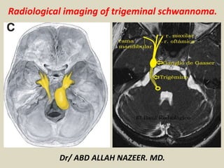
- 20. Trigeminal schwannoma. T1 and T2 weighted MR images show a bilobulated mass in right Meckel’s cave and cerebello-pontine cistern, with homogeneous intense post-contrast enhancement.
- 21. Giant Trigeminal Schwannoma Presenting with Obstructive hydrocephalus.
- 22. Representative axial T-1 weighted post-gadolinium images used for volumetric analysis showing the progressive postsurgical reduction of tumor volume: A. Preoperative; B. Three months postoperatively; C. One-year post-operatively.
- 23. (a-c) Preoperative MRI of brain showing right-sided huge trigeminal schwannoma (axial, coronal, and sagittal views sequentially). (d-f) Postoperative MRI of brain showing the complete removal of tumor (axial, coronal, and sagittal views sequentially)
- 24. Mixed schwannoma with meningioma of the trigeminal nerve.
- 25. Thank You.