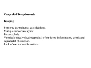
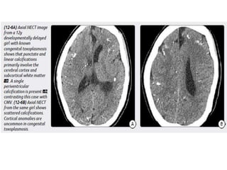
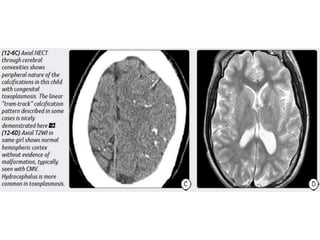
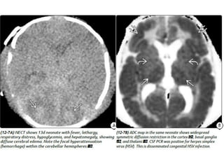
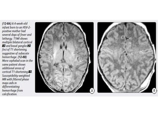
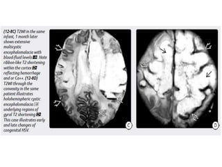
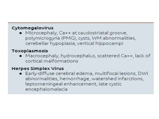
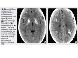
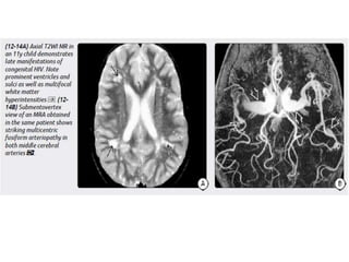
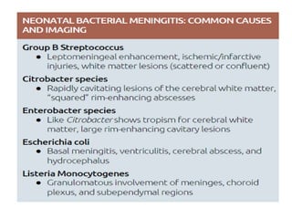
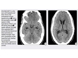
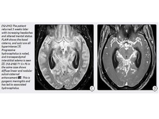
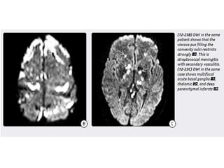
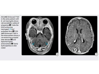
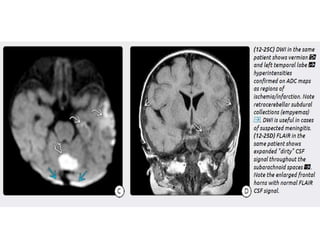
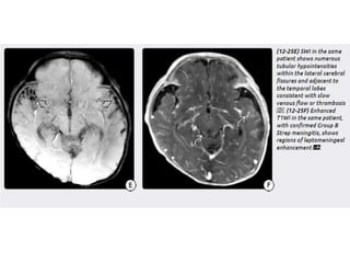
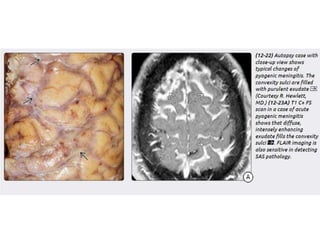
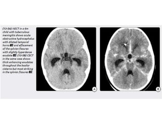
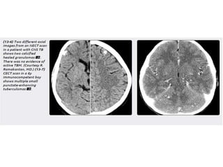
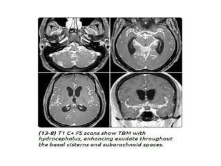
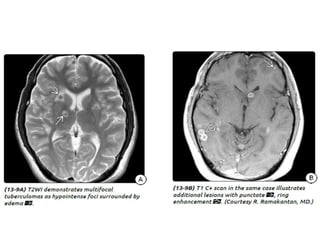
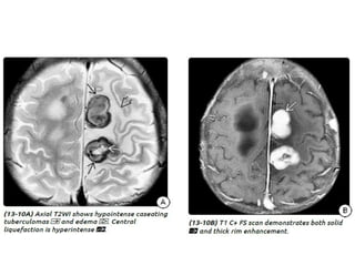
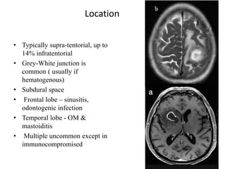
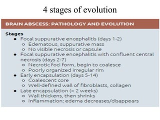
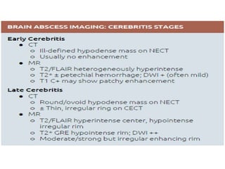
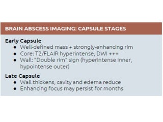
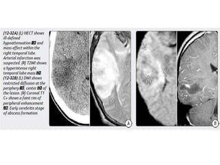
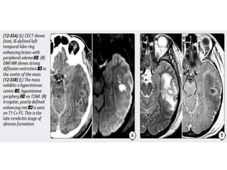
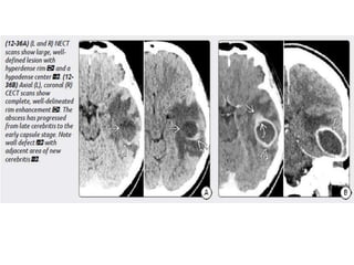
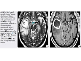
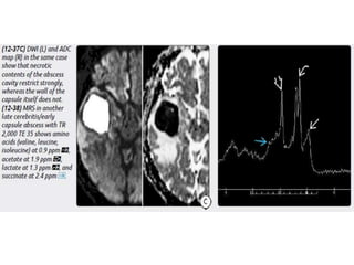
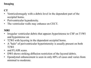
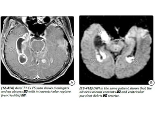
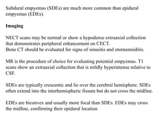
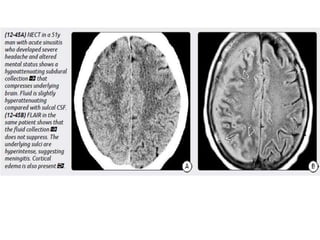
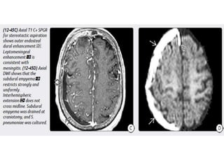
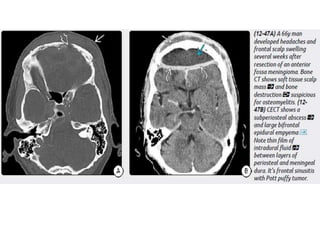
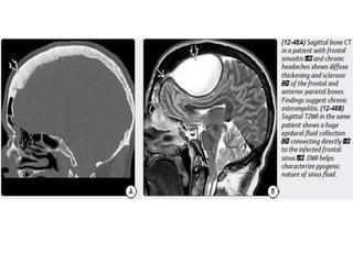
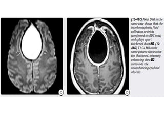
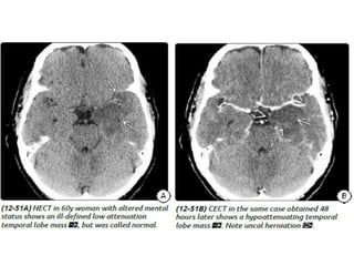
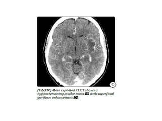
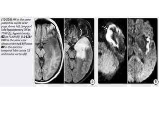
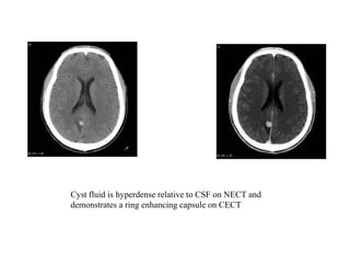
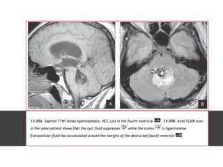
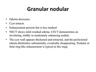
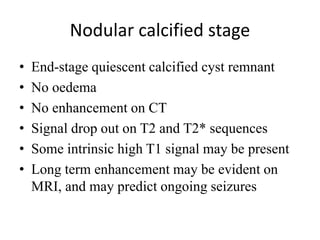
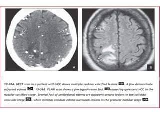
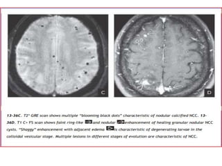
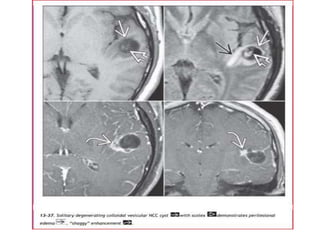
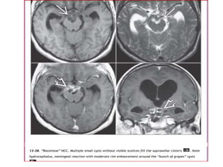
CNS Infections
Imaging
Dr. Yash Kumar Achantani
The concept that the brain was an "immune privileged" organ in which
the blood-brain barrier (BBB) was a relative fortress that restricted
pathogen entry and limited inflammation has recently undergone
significant revision.
A surprising large number of pathogens, including many neurotropic
viruses, can infect the CNS.
Routes of entry include
Transsynaptic spread (e.g., herpes viruses),
By "hiding" within blood-borne lymphocytes that access the brain (e.g.,
HIV and JC viruses),
By using the choroid plexus as a gateway into the CNS.
CNS infections can be classified in several ways. The most common
method is to divide them into
Congenital/neonatal
Acquired infections.
Congenital Infections
TORCH Infections
Congenital Cytomegalovirus
Congenital Toxoplasmosis
Herpes Simplex Virus: Congenital and Neonatal Infections
Congenital (Perinatal) HIV
Other Congenital Infections
Parenchymal calcifications are the hallmark of most congenital
infections.
Infections early in fetal development (e.g., during the first trimester)
usually result in miscarriage, severe brain destruction, and/or profound
malformations such as anencephaly, agyria, and lissencephaly.
When infections occur later in pregnancy, encephaloclastic
manifestations and myelination disturbance (e.g., demyelination,
dysmyelination, and hypomyelination) predominate.
With few exceptions (toxoplasmosis and syphilis), most
congenital/perinatal infections are viral and are usually secondary to
transplacental passage of the infectious agent.
TORCH Infections
TORCH infections—the acronym for Toxoplasmosis, Rubella,
Cytomegalovirus, and Herpes. If congenital Syphilis is included, the
grouping is called TORCH(S) or (S)TORCH.
Congenital Cytomegalovirus
Congenital Toxoplasmosis
Imaging
Scattered parenchymal calcifications.
Multiple subcortical cysts.
Porencephaly.
Ventriculomegaly (hydrocephalus) often due to inflammatory debris and
aqueductal obstruction.
Lack of cortical malformations.
Herpes Simplex Virus
Imaging
Unlike childhood or adult HSE, neonatal HSV CNS infection is much
more diffuse. Both gray and white matter are affected.
Consider neonatal HSV encephalitis when cranial imaging at 2-3 weeks of
neonatal life
shows unexplained diffuse cerebral edema, with leptomeningeal
enhancement, without or with cerebral parenchymal hemorrhage.
Congenital (Perinatal) HIV
Imaging
Atrophy, particularly in the frontal lobes.
Bilaterally symmetric basal ganglia calcifications.
Ectasia and fusiform enlargement of intracranial arteries.
Strokes with foci of restricted diffusion and subarachnoid hemorrhage
may occur as complications of the underlying vasculopathy.
Acquired Pyogenic
Infections
Meningitis
Meningitis is an acute or chronic inflammatory infiltrate of meninges
and CSF. Pachymeningitis involves the duraarachnoid; leptomeningitis
affects the pia and subarachnoid spaces.
Three bacteria account for the majority of cases
Haemophilus influenzae,
Streptococcus pneumoniae,
Neisseria meningitidis
Bacteria may arise at the CNS as a result of
Direct implantation,
Contagious infection from a local septic process (e.g. sinusitis) or an
infected foreign body (e.g. a shunting catheter),
Haematogenous spread
Imaging
General Features- The "gold standard" for the diagnosis of bacterial
meningitis is CSF analysis.
Remember: Imaging is neither sensitive nor specific for the detection of
meningitis!
Complications
• The complications of meningitis can be remembered using the
mnemonic HACTIVE :
• H: hydrocephalus
• A: abscess
• C: cerebritis / cranial nerve lesion
• T: thrombosis
• I: infarct
• V: ventriculitis / vasculopathy
• E: extra-axial collection: empyema and hygroma
Tuberculous meningitis
• Tuberculous meningitis is the most common presentation
of intracranial tuberculosis, and usually refers to infection of
the leptomeninges.
• Tuberculous meningitis, although seen in all age groups, has a
peak incidence in childhood (particularly 0-4 years of age) in
high prevalence areas.
Abscess
Abscess is initiated by focal intracranial infection as an area of cerebritis
and evolves into a collection of pus surrounded by a vascularized
capsule.
Size- 5 mm up to several centimetres.
Age – most common in 3rd and 4th decades.
Infants and neonates - its rare (may occur as complication of bacterial
meningitis)
Causative agents
• Adults : Streptococci, Staphylococci Gram-negative (Escherichia
coli, Klebsiella, Proteus, Pseudomonas, H. influenzae)
• Neonates and children : Citrobacter, Proteus, Pseudomonas, Serratia
and Staphyloccocus aureus
• AIDS patient – toxoplasmosis, Mycobacterium tuberculosis.
• Mostly the causative agents are bacteria but there can be fungal or
granulomatous or Parasitic agents .
Location
• Typically supra-tentorial, up to
14% infratentorial
• Grey-White junction is
common ( usually if
hematogenous)
• Subdural space
• Frontal lobe – sinusitis,
odontogenic infection
• Temporal lobe - OM &
mastoiditis
• Multiple uncommon except in
immunocompromised
4 stages of evolution
Ventriculitis
A collection of purulent material in the ventricle is more likely due to
intraventricular rupture of a brain abscess (IVRBA), a catastrophic
complication.
Ventriculitis also occurs as a complication of meningitis and
neurosurgical procedures such as external ventricular drainage.
Ventriculitis is also called ependymitis, pyocephalus, and (less
commonly) ventricular empyema.
Imaging
CT
• Ventriculomegaly with a debris level in the dependent part of the
occipital horns.
• Periventricular hypodensity.
• The ventricular walls may enhance on CECT.
MRI
• Irregular ventricular debris that appears hyperintense to CSF on T1WI
and hypointense on
• T2WI with layering in the dependent occipital horns.
• A "halo" of periventricular hyperintensity is usually present on both
T2WI
• and FLAIR scans.
• DWI shows striking diffusion restriction of the layered debris.
• Ependymal enhancement is seen in only 60% of cases and varies from
minimal to moderate.
Empyemas
Extraaxial infections of the CNS are rare but potentially life threatening
conditions.
Empyemas are pus collections that can occur in either the subdural or
epidural space.
Empyemas in infants and young children are most commonly
secondary to bacterial meningitis.
In older children and adults, over two-thirds of empyemas occur as
extension of infection from paranasal sinus disease.
Approximately 20% of empyemas in older children and adultsare
secondary to otomastoiditis.
Subdural empyemas (SDEs) are much more common than epidural
empyemas (EDEs).
Imaging
NECT scans may be normal or show a hypodense extraaxial collection
that demonstrates peripheral enhancement on CECT.
Bone CT should be evaluated for signs of sinusitis and otomastoiditis.
MR is the procedure of choice for evaluating potential empyemas. T1
scans show an extraaxial collection that is mildly hyperintense relative to
CSF.
SDEs are typically crescentic and lie over the cerebral hemisphere. SDEs
often extend into the interhemispheric fissure but do not cross the midline.
EDEs are biconvex and usually more focal than SDEs. EDEs may cross
the midline, confirming their epidural location
Acquired Viral Infections
Eight members of the herpes virus family are known to cause disease in
humans. These are
Herpes simplex virus 1 (HSV-1) and HSV-2,
Varicella-zoster virus (VZV),
Epstein-Barr virus (EBV),
Cytomegalovirus (CMV), and
Human herpes virus (HHV)-6, HHV-7, and HHV-8.
Each has its own disease spectrum, clinical setting, and imaging
findings.
Herpes Simplex Encephalitis
HHV-6 Encephalopathy
Parasitic infections of the Brain
Parasitic infections of the brain include
• Neurocysticercosis (Taenia solium)
• Echinococcosis
• Neurotoxoplasmosis (Toxoplasma gondii)
• Cerebral malaria (Plasmodium falciparum)
• Cerebral amoebiasis
- Primary amoebic meningoencephalitis (Naegleria fowleri)
- Granulomatous amoebic encephalitis (Acanthamoeba
spp, Balamuthia mandrillaris and Sappinia pedata)
• Cerebral sparganosis (Spirometra mansonoides)
Neurocysticercosis
Cysticercosis is the most common parasitic infection in the world, and
CNS lesions eventually develop in 60-90% of patients with
cysticercosis.
Clinical presentation includes :
• seizures: most common symptom and most common cause of
seizures in young adults in endemic areas
• headaches
• hydrocephalus
• altered mental status
• neurological deficits
LOCATION
• T. solium larvae are most common in
the CNS, eyes, muscles, and
subcutaneous tissue. The intracranial
subarachnoid spaces are the most
common CNS site, followed by the
brain parenchyma and ventricles
(fourth > third > lateral ventricles)
Stages
There are four main stages (also known as Escobar's pathological
stages):
• Vesicular: viable parasite with intact membrane and therefore no
host reaction.
• Colloidal vesicular: parasite dies within 4-5 years untreated, or
earlier with treatment and the cyst fluid becomes turbid. As the
membrane becomes leaky oedema surrounds the cyst. This is the
most symptomatic stage.
• Granular nodular: oedema decreases as the cyst retracts further;
enhancement persists.
• Nodular calcified: end-stage quiescent calcified cyst remnant; no
oedema.
Vesicular Stage
• Cyst with dot sign
• CSF density/intensity
• No enhancement is typical, although very faint enhancement of the
wall and enhancement of the scolex may be seen.
Colloidal vesicular
• Cyst fluid becomes turbid
– CT: hyperattenuating to CSF
– MRI T1: hyperintense to CSF
• Surrounding oedema
• Cyst and the wall become thickened and brightly enhances
• Scolex can often still be seen as an eccentric focus of enhancement
Cyst fluid is hyperdense relative to CSF on NECT and
demonstrates a ring enhancing capsule on CECT
Granular nodular
• Odema decreases
• Cyst retracts
• Enhancement persists but is less marked
• NECT shows mild residual edema. CECT demonstrates an
involuting, mildly to moderately enhancing nodule.
• The cyst wall appears thickened and retracted, and the perilesional
edema diminishes substantially, eventually disappearing. Nodular or
faint ring-like enhancement is typical at this stage
Nodular calcified stage
• End-stage quiescent calcified cyst remnant
• No oedema
• No enhancement on CT
• Signal drop out on T2 and T2* sequences
• Some intrinsic high T1 signal may be present
• Long term enhancement may be evident on
MRI, and may predict ongoing seizures
Echinococcosis
• Infection by Echinococcus is called echinococcosis.
• Two species of Echinococcus tapeworms, E. granulosis (EG) and E.
multilocularis/alveolaris (EM/EA), are responsible for most human
CNS infections.
• EG infestation is also called hydatid disease or hydatid cyst (HC).
Infection with EM/EA is also known as alveolar echinococcosis.
Clinical presentation
Symptoms and signs include:
• Focal neurological deficits
• Headaches
• Increased intracranial pressure
• Hydrocephalus
• Papilloedema and loss of vision
• Altered mental status
IMAGING The most common imaging appearance of HC is that of a
large, unilocular, thin-walled cyst without calcification, edema, or
enhancement on CT
Occasionally, a single large cyst will contain multiple “daughter cysts” MR shows
that cyst fluid is isointense with CSF on T1-and T2-weighted images. Sometimes
a detached germinal membrane and hydatid “sand” can be seen in the
dependent portion of the cyst.
Cerebral Malaria
• Cerebral malaria (CM) is caused by infection with the protozoan
parasite Plasmodium and is transmitted by infected Anopheles
mosquitoes. P. falciparum has the most severe morbidity and
mortality and causes 95% of all CM cases.
• Cerebral malaria is a rare intracranial complication of a malarial
infection.
• Cerebral malaria should be suspected when there are neurological
symptoms on a background of malarial infection.
• Clinical presentations include: headache, altered state of
consciousness, seizures, backache, vomiting, nausea, etc.
Imaging
Imaging findings on NECT vary from normal to striking.
• The most typical finding is focal infarcts in the cortex, basal
ganglia, and thalami.
• Gross hemorrhage can occur but is rare.
• Diffuse cerebral edema can occur in severe CM.
Imaging in CNS Infections

Imaging in CNS Infections

  • 1.
  • 2.
    The concept thatthe brain was an "immune privileged" organ in which the blood-brain barrier (BBB) was a relative fortress that restricted pathogen entry and limited inflammation has recently undergone significant revision. A surprising large number of pathogens, including many neurotropic viruses, can infect the CNS. Routes of entry include Transsynaptic spread (e.g., herpes viruses), By "hiding" within blood-borne lymphocytes that access the brain (e.g., HIV and JC viruses), By using the choroid plexus as a gateway into the CNS.
  • 3.
    CNS infections canbe classified in several ways. The most common method is to divide them into Congenital/neonatal Acquired infections. Congenital Infections TORCH Infections Congenital Cytomegalovirus Congenital Toxoplasmosis Herpes Simplex Virus: Congenital and Neonatal Infections Congenital (Perinatal) HIV Other Congenital Infections
  • 4.
    Parenchymal calcifications arethe hallmark of most congenital infections. Infections early in fetal development (e.g., during the first trimester) usually result in miscarriage, severe brain destruction, and/or profound malformations such as anencephaly, agyria, and lissencephaly. When infections occur later in pregnancy, encephaloclastic manifestations and myelination disturbance (e.g., demyelination, dysmyelination, and hypomyelination) predominate. With few exceptions (toxoplasmosis and syphilis), most congenital/perinatal infections are viral and are usually secondary to transplacental passage of the infectious agent.
  • 5.
    TORCH Infections TORCH infections—theacronym for Toxoplasmosis, Rubella, Cytomegalovirus, and Herpes. If congenital Syphilis is included, the grouping is called TORCH(S) or (S)TORCH. Congenital Cytomegalovirus
  • 8.
    Congenital Toxoplasmosis Imaging Scattered parenchymalcalcifications. Multiple subcortical cysts. Porencephaly. Ventriculomegaly (hydrocephalus) often due to inflammatory debris and aqueductal obstruction. Lack of cortical malformations.
  • 11.
    Herpes Simplex Virus Imaging Unlikechildhood or adult HSE, neonatal HSV CNS infection is much more diffuse. Both gray and white matter are affected. Consider neonatal HSV encephalitis when cranial imaging at 2-3 weeks of neonatal life shows unexplained diffuse cerebral edema, with leptomeningeal enhancement, without or with cerebral parenchymal hemorrhage.
  • 16.
    Congenital (Perinatal) HIV Imaging Atrophy,particularly in the frontal lobes. Bilaterally symmetric basal ganglia calcifications. Ectasia and fusiform enlargement of intracranial arteries. Strokes with foci of restricted diffusion and subarachnoid hemorrhage may occur as complications of the underlying vasculopathy.
  • 19.
    Acquired Pyogenic Infections Meningitis Meningitis isan acute or chronic inflammatory infiltrate of meninges and CSF. Pachymeningitis involves the duraarachnoid; leptomeningitis affects the pia and subarachnoid spaces. Three bacteria account for the majority of cases Haemophilus influenzae, Streptococcus pneumoniae, Neisseria meningitidis
  • 20.
    Bacteria may ariseat the CNS as a result of Direct implantation, Contagious infection from a local septic process (e.g. sinusitis) or an infected foreign body (e.g. a shunting catheter), Haematogenous spread Imaging General Features- The "gold standard" for the diagnosis of bacterial meningitis is CSF analysis. Remember: Imaging is neither sensitive nor specific for the detection of meningitis!
  • 29.
    Complications • The complicationsof meningitis can be remembered using the mnemonic HACTIVE : • H: hydrocephalus • A: abscess • C: cerebritis / cranial nerve lesion • T: thrombosis • I: infarct • V: ventriculitis / vasculopathy • E: extra-axial collection: empyema and hygroma
  • 30.
    Tuberculous meningitis • Tuberculousmeningitis is the most common presentation of intracranial tuberculosis, and usually refers to infection of the leptomeninges. • Tuberculous meningitis, although seen in all age groups, has a peak incidence in childhood (particularly 0-4 years of age) in high prevalence areas.
  • 38.
    Abscess Abscess is initiatedby focal intracranial infection as an area of cerebritis and evolves into a collection of pus surrounded by a vascularized capsule. Size- 5 mm up to several centimetres. Age – most common in 3rd and 4th decades. Infants and neonates - its rare (may occur as complication of bacterial meningitis)
  • 39.
    Causative agents • Adults: Streptococci, Staphylococci Gram-negative (Escherichia coli, Klebsiella, Proteus, Pseudomonas, H. influenzae) • Neonates and children : Citrobacter, Proteus, Pseudomonas, Serratia and Staphyloccocus aureus • AIDS patient – toxoplasmosis, Mycobacterium tuberculosis. • Mostly the causative agents are bacteria but there can be fungal or granulomatous or Parasitic agents .
  • 40.
    Location • Typically supra-tentorial,up to 14% infratentorial • Grey-White junction is common ( usually if hematogenous) • Subdural space • Frontal lobe – sinusitis, odontogenic infection • Temporal lobe - OM & mastoiditis • Multiple uncommon except in immunocompromised
  • 41.
    4 stages ofevolution
  • 49.
    Ventriculitis A collection ofpurulent material in the ventricle is more likely due to intraventricular rupture of a brain abscess (IVRBA), a catastrophic complication. Ventriculitis also occurs as a complication of meningitis and neurosurgical procedures such as external ventricular drainage. Ventriculitis is also called ependymitis, pyocephalus, and (less commonly) ventricular empyema.
  • 50.
    Imaging CT • Ventriculomegaly witha debris level in the dependent part of the occipital horns. • Periventricular hypodensity. • The ventricular walls may enhance on CECT. MRI • Irregular ventricular debris that appears hyperintense to CSF on T1WI and hypointense on • T2WI with layering in the dependent occipital horns. • A "halo" of periventricular hyperintensity is usually present on both T2WI • and FLAIR scans. • DWI shows striking diffusion restriction of the layered debris. • Ependymal enhancement is seen in only 60% of cases and varies from minimal to moderate.
  • 52.
    Empyemas Extraaxial infections ofthe CNS are rare but potentially life threatening conditions. Empyemas are pus collections that can occur in either the subdural or epidural space. Empyemas in infants and young children are most commonly secondary to bacterial meningitis. In older children and adults, over two-thirds of empyemas occur as extension of infection from paranasal sinus disease. Approximately 20% of empyemas in older children and adultsare secondary to otomastoiditis.
  • 53.
    Subdural empyemas (SDEs)are much more common than epidural empyemas (EDEs). Imaging NECT scans may be normal or show a hypodense extraaxial collection that demonstrates peripheral enhancement on CECT. Bone CT should be evaluated for signs of sinusitis and otomastoiditis. MR is the procedure of choice for evaluating potential empyemas. T1 scans show an extraaxial collection that is mildly hyperintense relative to CSF. SDEs are typically crescentic and lie over the cerebral hemisphere. SDEs often extend into the interhemispheric fissure but do not cross the midline. EDEs are biconvex and usually more focal than SDEs. EDEs may cross the midline, confirming their epidural location
  • 60.
    Acquired Viral Infections Eightmembers of the herpes virus family are known to cause disease in humans. These are Herpes simplex virus 1 (HSV-1) and HSV-2, Varicella-zoster virus (VZV), Epstein-Barr virus (EBV), Cytomegalovirus (CMV), and Human herpes virus (HHV)-6, HHV-7, and HHV-8. Each has its own disease spectrum, clinical setting, and imaging findings.
  • 61.
  • 66.
  • 69.
  • 70.
    Parasitic infections ofthe brain include • Neurocysticercosis (Taenia solium) • Echinococcosis • Neurotoxoplasmosis (Toxoplasma gondii) • Cerebral malaria (Plasmodium falciparum) • Cerebral amoebiasis - Primary amoebic meningoencephalitis (Naegleria fowleri) - Granulomatous amoebic encephalitis (Acanthamoeba spp, Balamuthia mandrillaris and Sappinia pedata) • Cerebral sparganosis (Spirometra mansonoides)
  • 71.
    Neurocysticercosis Cysticercosis is themost common parasitic infection in the world, and CNS lesions eventually develop in 60-90% of patients with cysticercosis. Clinical presentation includes : • seizures: most common symptom and most common cause of seizures in young adults in endemic areas • headaches • hydrocephalus • altered mental status • neurological deficits
  • 72.
    LOCATION • T. soliumlarvae are most common in the CNS, eyes, muscles, and subcutaneous tissue. The intracranial subarachnoid spaces are the most common CNS site, followed by the brain parenchyma and ventricles (fourth > third > lateral ventricles)
  • 73.
    Stages There are fourmain stages (also known as Escobar's pathological stages): • Vesicular: viable parasite with intact membrane and therefore no host reaction. • Colloidal vesicular: parasite dies within 4-5 years untreated, or earlier with treatment and the cyst fluid becomes turbid. As the membrane becomes leaky oedema surrounds the cyst. This is the most symptomatic stage. • Granular nodular: oedema decreases as the cyst retracts further; enhancement persists. • Nodular calcified: end-stage quiescent calcified cyst remnant; no oedema.
  • 74.
    Vesicular Stage • Cystwith dot sign • CSF density/intensity • No enhancement is typical, although very faint enhancement of the wall and enhancement of the scolex may be seen.
  • 76.
    Colloidal vesicular • Cystfluid becomes turbid – CT: hyperattenuating to CSF – MRI T1: hyperintense to CSF • Surrounding oedema • Cyst and the wall become thickened and brightly enhances • Scolex can often still be seen as an eccentric focus of enhancement
  • 77.
    Cyst fluid ishyperdense relative to CSF on NECT and demonstrates a ring enhancing capsule on CECT
  • 79.
    Granular nodular • Odemadecreases • Cyst retracts • Enhancement persists but is less marked • NECT shows mild residual edema. CECT demonstrates an involuting, mildly to moderately enhancing nodule. • The cyst wall appears thickened and retracted, and the perilesional edema diminishes substantially, eventually disappearing. Nodular or faint ring-like enhancement is typical at this stage
  • 80.
    Nodular calcified stage •End-stage quiescent calcified cyst remnant • No oedema • No enhancement on CT • Signal drop out on T2 and T2* sequences • Some intrinsic high T1 signal may be present • Long term enhancement may be evident on MRI, and may predict ongoing seizures
  • 85.
    Echinococcosis • Infection byEchinococcus is called echinococcosis. • Two species of Echinococcus tapeworms, E. granulosis (EG) and E. multilocularis/alveolaris (EM/EA), are responsible for most human CNS infections. • EG infestation is also called hydatid disease or hydatid cyst (HC). Infection with EM/EA is also known as alveolar echinococcosis.
  • 86.
    Clinical presentation Symptoms andsigns include: • Focal neurological deficits • Headaches • Increased intracranial pressure • Hydrocephalus • Papilloedema and loss of vision • Altered mental status
  • 87.
    IMAGING The mostcommon imaging appearance of HC is that of a large, unilocular, thin-walled cyst without calcification, edema, or enhancement on CT
  • 88.
    Occasionally, a singlelarge cyst will contain multiple “daughter cysts” MR shows that cyst fluid is isointense with CSF on T1-and T2-weighted images. Sometimes a detached germinal membrane and hydatid “sand” can be seen in the dependent portion of the cyst.
  • 89.
    Cerebral Malaria • Cerebralmalaria (CM) is caused by infection with the protozoan parasite Plasmodium and is transmitted by infected Anopheles mosquitoes. P. falciparum has the most severe morbidity and mortality and causes 95% of all CM cases. • Cerebral malaria is a rare intracranial complication of a malarial infection. • Cerebral malaria should be suspected when there are neurological symptoms on a background of malarial infection. • Clinical presentations include: headache, altered state of consciousness, seizures, backache, vomiting, nausea, etc.
  • 90.
    Imaging Imaging findings onNECT vary from normal to striking. • The most typical finding is focal infarcts in the cortex, basal ganglia, and thalami. • Gross hemorrhage can occur but is rare. • Diffuse cerebral edema can occur in severe CM.