SlideShare a Scribd company logo
Radiological Imaging of Anterior knee pain.
Dr/ ABD ALLAH NAZEER. MD.
Anterior knee pain that originates at the front of the
knee joint is a common complaint. This disorder has
a profound economic and social impact as it affects
approximately 15-33% of young and active people.
The role of physical examination in making
differential diagnosis is rather limited since different
conditions cause similar symptoms. Knee MRI is
considered the gold standard in evaluating the
damage to the anatomical structures of the knee,
such as ligament or tendon rupture, meniscus or
cartilage injury. It is often the case that MRI findings
determine the therapy, therefore radiologists have to
know what to look for in knee MRI images.
Anterior knee pain is common with a variety
of causes including:
Patella fracture.
Osteoarthritis.
Inflammatory and depositional arthritis
Bursitis around the knee.
Patellofemoral maltracking.
Excessive lateral pressure syndrome (ELPS).
Patellar cartilage defect.
Patellofemoral chondromalacia.
Patellar tendinitis.
Medial plica.
Hoffa's disease.
iliotibial band friction syndrome.
Patellar fracture
A direct blow or increased tension in a quadriceps and patellar tendon can cause patellar
fracture. In most cases, a fracture line is transverse and passes between the middle and
inferior third. Usually, the diagnosis is made on x-ray images. An MRI scan is useful in
excluding ligamentous injury or in cases of suspected stress fracture. On MRI images stress
fracture appears as low intensity line surrounded by bone marrow edema.
Inferior third of patella fracture without dislocation.
On MRI image the fracture line (arrows) extends across patella
with bone edema in the surrounding marrow.
Osteochondral sleeve fracture in a pediatric patient. (A) Lateral plain film of a
patellar avulsion fracture of the inferior pole of the patella demonstrating a
superiorly displaced patella and a small bone fragment (arrow). Sagittal T1-
weighted (B) and sagittal gradient recalled echo (C) MR images demonstrating an
osteochondral fracture of high signal intensity with a small bone fragment (arrow).
Patellar dislocation
It is one of the most common causes of anterior knee pain in young
people. A high-riding patella and femoral trochlear dysplasia are the
major predisposing factors. It is often the case that a dislocated
patella reduces spontaneously without patient's awareness of the
fact. The following MRI features of patellar dislocation are noted:
Medial retinaculum and medial capsule injury/tear
Lateral femoral condyle contusion and osteochondral damage
Medial patellar facet contusion with or without cartilaginous lesion
Medial retinaculum tears occur at the attachment site to the femur
or along the medial margin of the patella and subsequently may
cause avulsion injuries. Cartilaginous lesions can result in the
formation of loose bodies in the joint space.
An axial T2-weighted fast-spin-echo magnetic resonance imaging scan illustrates a
eighteen year-old female sustaining a primary traumatic lateral dislocation of the
patella while jumping. Complete avulsion of the medial patellofemoral ligament from
its femoral insertion can be seen (arrow) (Figure 2A). The bone contusions (stars)
(Figure 2B) of the lateral femoral condyle and the medial patellar facet are noted.
Axial (a) PD FS and sagittal (b) T2 shows medial retinaculum avulsion fracture
with patellar edge fragment (asterisk), medial patellar facet contusion
and cartilaginous lesion (black arrows), lateral femoral condyle contusion
(open arrow), loose cartilaginous body in anterolateral joint space.
Lateral patellar tilt and subluxation(red arrow)
and acute chondral defect(yellow arrow).
X-ray and MRI after luxation of the patella. There is a fragment and bone bruise
at the medial surface of the patella and in the corresponding surface of the
lateral condyle of the femur. The medial retinaculum of the patella is disrupted.
Coronal PD FS (a, b) shows lateral femoral condyle contusion and osteochondral damage
(white arrow), loose cartilaginous body (black arrows) in antero-lateral joint space.
Chondromalacia is defined as softening and
degeneration of the articular hyaline cartilage. Overuse
and trauma cause damage to the cartilage which over
time leads to fissuring and partial loss of cartilage
thickness. Subsequently, patients with high grade
osteoarthritis sustain cartilage destruction with underlying
bone changes. Cartilaginous lesions are graded according
to the Outer bridge classification which is based on a
correlation between MRI and arthroscopic findings.
Although arthroscopy is a gold standard for the evaluation
of the severity of cartilage defects, new MRI sequences
(T2 mapping, DW, dGEMRIC, T1rho,) allow early diagnosis,
contribute to preoperative planning and postoperative
follow-up.
Cartilage lesions on T2 images: a- focal area of hyperintensity with normal
contour; b- fragmentation of cartilage; c- partial thickness cartilage loss with focal
ulceration; d- full thickness cartilage loss with underlying bone reactive changes.
Chondromalacia patellae grades II–IV in various patients. (A) Axial fast spin echo
proton density fat saturated (FSE PD FS) MR image of chondromalacia patellae
grade II in a 46-year-old male. High signal is seen in the patellar cartilage in the
lateral patellar facet (arrows). (B) Axial FSE PD FS MR image of chondromalacia
patellae grade III in a 51-year-old female. There is full thickness focal signal intensity
change, contour irregularity and thinning of the cartilage (arrows). (C) Axial PD
Cartilage T2 mapping allows to evaluate the articular
cartilage of the knee joint. Red color shows cartilage lesion.
Patellar Abnormality.
-Bipartite patella.
-Multipartite patella.
Types of
bipartite
patella: type I-
inferior pole
(a,b); type II-
lateral margin
(c, d); type III-
superolateral
portion (e, f).
Osteochondritis dissecans of the patellofemoral joint is an
uncommon condition that may be the cause of anterior
knee pain or crepitus. The osteochondral lesions involve
the convex articular surfaces. The trochlear groove is the
rarest location for osteochondritis dissecans. MR is the
test of choice since it detects an osteochondral fragment
and evaluates its stability. A high signal line demarcating
the fragment from bone usually indicates an unstable
lesion. Cartilaginous lesions can be classified into three
main groups:
Subchondral- intact cartilage surface
Osteochondral fractures- disrupted articular surface with a
fragment of cortical bone
Chondral injury- underlying bone is intact
Bilateral osteochondritis dissecans of the knee.
Osteochondritis dissecans
Extensor mechanism of the knee pathology.
-Traumatic.
-Tendinosis.
-Patellar tendon tear.
-Quadriceps tendon tear.
.Intrinsic patellar tendon lesions. e.g. gout.
.Patellar enthesopathy.
.Osteochondrosis, e.g. Osgood-Schlatter disease.
Patellar tendinosis.
.Pain in the infrapatellar region.
.Commonly seen in athletes.
.MRI demonstrate thickening of the
patellar tendon with intermediate T1
and increased signal at T2 especially
with fat suppression sequences.
.Ultrasound show thickening, low
echogenicity and increase blood flow
at the Doppler study.
Chronic patellar tendinosis.
Patellar tendinopathy and Partial patellar tendon tear.
On sagittal (a) and axial (b) PD FS proximal end of patellar tendon is seen high intensity,
enlarged (white arrow) surrounding by inflammatory changes (black arrow).
Sagittal T2 (a) and PD SPIR (b) shows tibial tuberosity bony hypertrophy (black
arrow), fragmentation and inflammatory changes of overlying tissues.
Coronal (a) and sagittal (b) PD FS shows changes involving the inferior pole of the
patella which is characteristic for Sinding–Larsen–Johansson disease.
Complete Patellar tendon tear. Images show no continuity
between fibers and patella. The tendon is thickened.
Complete Patellar tendon tear. Image on the right shows
hemorrhagic bursitis ( low signal in bursa).
Quadriceps tendon tear.
Tear may be partial or complete.
Partial quadriceps tendon tear: T2W-images.LEFT: Abnormal attachment of tendon. Right :
Most of tendon is retracted (red arrow) deep part (vastus intermedius) is still intact.
Complete quadriceps tear. Sag T2W-images.
No continuity. Hematoma in between.
Sagittal PD FS (a) and T2 (b) shows quadriceps tendon full
thickness rupture as a gap filled with hyperintense fluid.
Intrinsic patellar tendon lesions : Gout
MRI show low T1 and mildly high T2 signal.
Fat pad syndromes
The infrapatellar (Hoffa's) fat pad is an intraarticular and yet
an extrasynovial structure which has an abundant vasculature
and innervation. Pathological changes are often associated
with other conditions, such as patellar tendinopathy, ligament
reconstruction or meniscal tear. Acute injury usually occurs in
the dorsal part of the fat pad and presents as edema or tears.
In chronic injury hemosiderin deposits or scarring are
observed. Lateral patellofemoral impingement involves of
edema of the superolateral part of the fat pat, a shallow
femoral trochlea, patellar malalignment and chondromalacia.
The inflammation of a quadriceps fat pad is rare and features
are similar to that of Hoffa’s fat pad.
Also, benign or malignant masses can be the cause of anterior
knee pain
Sagittal (a) and axial (b) IR shows high signal intensity at infrapatellar
fat pad (arrows). Sagittal image demonstrates patella alta.
Hoffa's Disease.
Sagittal T2 (a) and PD FS (b) shows heterointensive mass (arrows) at quadriceps fat pad
which was confirmed histologically as a metastasis of renocellular carcinoma
Sagittal MRI scans of the knee show an intermediate signal lesion in the apex of Hoffa's fat pad
between the inferior pole of the patella and the adjacent femoral condyle. The lesion's surface
retains the shape of the adjoining femoral condyle, indicating chronic wedging that reflects the
chronic nature of the lesion. The lesion was surgically excised. Histology showed adipose and
synovial tissue with inflammation and fibrosis, supporting a diagnosis of Hoffa's disease.
Pigmented villonodular synovitis is rare benign neoplastic synovial
proliferation with villous and nodular projections and hemosiderin deposition.
It mainly affects knee joint. On MRI images it appears as mass like lesion with
well- defined low signal intensity nodule. On both T1 and T2 images it is seen
low signal intensity with variable postcontrast enhancement.
A synovial nodular lesion is seen (arrows), with heterogenous signal characteristics.
Pigmented villonodular synovitis.
Localized Nodular Synovitis.
Intra-articular Chondroma.
A rare lesion occur around the knee typically at the infrapatellar fat pad.
May be calcified or erode the lower pole of patella.
Intra-articular Chondroma.
Infrapatellar Plica injury.
A thin fold of synovial tissue, extending from the inferior pole of the patella through
Hoffa's fat to the intercondylar notch anterior to the anterior cruciate ligament.
.High signal along the course of the plica indicates injury to the plica.
.Thickening of the plica even in the absence of edema or fluid suggestive of chronic injury.
Infrapatellar Plica injury.
Sagittal T2 (a) and axial PD FS (b) shows thickening of medial patellar place
(arrows) and insignificant Chondromalacia of femoral trochlear groove.
Iliotibial band syndrome, also known as Runner’s knee, is caused by an
excessive friction between the iliotibial band and the lateral femoral epicondyle. MRI
findings include the thickening of the iliotibial band and a deep bursa located over the
lateral epicondyle. Also, there is an inflammation of the surrounding soft tissues. MRI
is reserved to exclude other etiologies of pain such as lateral meniscal tear.
Coronal (a) and axial (b) PD FS shows thickened and edematous iliotibial band (white
arrow) and bursa (black arrow) between the band and lateral epicondyle of the femur.
Bursitis
There are four bursae anterior to the knee joint: a suprapatellar, a
subcutaneous prepatellar, a subcutaneous infrapatellar and a deep
infrapatellar bursa. Inflammation can be caused by acute or chronic trauma
and systemic diseases such as rheumatoid arthritis or metabolic disorders.
Axial PD FS (a) and sagittal GRE (b) shows hyperintense fluid in prepatellar region (black
arrows) and suprapatellar bursae (white arrows) - characteristic for prepatellar and suprapatellar bursitis
Prepatellar Bursitis.
Ligament injury
Cruciate ligaments stabilize the knee joint. Once they rupture, a shift in ligament
balance forces occurs and may lead to cartilage damage. An ACL rupture causes
rotational instability and secondary changes, such as medial compartment and
patellofemoral joint overload.
A PCL rupture causes posterior instability. When the tibia slips dorsally,
mechanical load to the patella increases and speeds up wear and tear. Following
an anterior cruciate ligament reconstruction, pain can be caused by postoperative
complications, such as:
Arthrofibrosis is a contracture of a infrapatellar fat pad and patellar tendon. A ill-
defined low signal area on T1 and T2 weighted images, similar to fibrous tissue.
Cyclops lesion is defined as a nodule located anteriorly to the distal end of an ACL
graft.
Two histologic types: “hard”- composed of bone or cartilage, more often acts as
obstacle and “soft”- only composed of fibrous tissue, rarely causes
impingement. A low signal intensity nodule on T1WI images, anteriorly to the
graft. It could show similar intensity to synovial fluid. The nodule is heterogeneous
on T2WI, usually hypointense and well differentiated from fluid. Differential
diagnosis should include focal pigmented villonodular synovitis, synovial
chondromatosis and loose bodies.
Coronal (frontal) IR FSE (a) and sagittal T2 FSE (b) shows unevenly thinning
of medial knee joint and femur trochlear groove cartilage (arrows)
caused by chronic rupture of anterior cruciate ligament.
Axial PDW SPIR (a) and sagittal PDW (b) shows fibrous nodule
(arrows) at distal part of anterior cruciate ligament transplant.
Thank You.

More Related Content

What's hot

MRI OF SHOULDER INJURY
MRI OF SHOULDER INJURYMRI OF SHOULDER INJURY
MRI OF SHOULDER INJURY
Krishna Kiran Karanth
 
MRI of the shoulder
MRI of the shoulderMRI of the shoulder
MRI of the shoulder
Thorsang Chayovan
 
Mri anatomy of ankle
Mri anatomy of ankleMri anatomy of ankle
Mri anatomy of ankle
Shrikant Nagare
 
Mri knee trauma
Mri knee traumaMri knee trauma
Mri knee trauma
Anish Choudhary
 
Mri anatomy of ankle radiology ppt pk
Mri anatomy of ankle radiology ppt pkMri anatomy of ankle radiology ppt pk
Mri anatomy of ankle radiology ppt pk
Dr pradeep Kumar
 
Presentation1.pptx, radiological imaging of skeletal dysplasia
Presentation1.pptx, radiological imaging of skeletal dysplasiaPresentation1.pptx, radiological imaging of skeletal dysplasia
Presentation1.pptx, radiological imaging of skeletal dysplasiaAbdellah Nazeer
 
Imaging of shoulder - Dr. Vishal Sankpal
Imaging of shoulder - Dr. Vishal SankpalImaging of shoulder - Dr. Vishal Sankpal
Imaging of shoulder - Dr. Vishal Sankpal
Vishal Sankpal
 
Radiology Spots PPT- 3 by Dr Chandni Wadhwani
 Radiology Spots PPT- 3 by Dr Chandni Wadhwani Radiology Spots PPT- 3 by Dr Chandni Wadhwani
Radiology Spots PPT- 3 by Dr Chandni Wadhwani
Chandni Wadhwani
 
Radiology Spots PPT- 2 by Dr Chandni Wadhwani
Radiology Spots PPT- 2 by Dr Chandni WadhwaniRadiology Spots PPT- 2 by Dr Chandni Wadhwani
Radiology Spots PPT- 2 by Dr Chandni Wadhwani
Chandni Wadhwani
 
Presentation1.pptx, radiological imaging of sacroiliac joint diseases.
Presentation1.pptx, radiological imaging of sacroiliac joint diseases.Presentation1.pptx, radiological imaging of sacroiliac joint diseases.
Presentation1.pptx, radiological imaging of sacroiliac joint diseases.Abdellah Nazeer
 
Presentation1.pptx, radiological imaging of spinal dysraphism.
Presentation1.pptx, radiological imaging of spinal dysraphism.Presentation1.pptx, radiological imaging of spinal dysraphism.
Presentation1.pptx, radiological imaging of spinal dysraphism.
Abdellah Nazeer
 
MRI Knee trauma
MRI Knee traumaMRI Knee trauma
MRI Knee trauma
Dr. Mohit Goel
 
Presentation1.pptx. shoulder joint.
Presentation1.pptx. shoulder joint.Presentation1.pptx. shoulder joint.
Presentation1.pptx. shoulder joint.
Abdellah Nazeer
 
Imaging in arthritis
Imaging in arthritisImaging in arthritis
Imaging in arthritis
Navni Garg
 
Radiology Spotters
Radiology Spotters Radiology Spotters
Radiology Spotters
Anish Choudhary
 
MRI SPINE ANATOMY
MRI SPINE ANATOMYMRI SPINE ANATOMY
MRI SPINE ANATOMY
NeurologyKota
 
Diagnostic Imaging of Bone Tumors
Diagnostic Imaging of Bone TumorsDiagnostic Imaging of Bone Tumors
Diagnostic Imaging of Bone Tumors
Mohamed M.A. Zaitoun
 
Presentation1, radiological imaging of popliteal fossa masses.
Presentation1, radiological imaging of popliteal fossa masses.Presentation1, radiological imaging of popliteal fossa masses.
Presentation1, radiological imaging of popliteal fossa masses.
Abdellah Nazeer
 

What's hot (20)

MRI OF SHOULDER INJURY
MRI OF SHOULDER INJURYMRI OF SHOULDER INJURY
MRI OF SHOULDER INJURY
 
MRI of the shoulder
MRI of the shoulderMRI of the shoulder
MRI of the shoulder
 
Mri anatomy of ankle
Mri anatomy of ankleMri anatomy of ankle
Mri anatomy of ankle
 
Knee mri
Knee mriKnee mri
Knee mri
 
Mri knee trauma
Mri knee traumaMri knee trauma
Mri knee trauma
 
Mri anatomy of ankle radiology ppt pk
Mri anatomy of ankle radiology ppt pkMri anatomy of ankle radiology ppt pk
Mri anatomy of ankle radiology ppt pk
 
Presentation1.pptx, radiological imaging of skeletal dysplasia
Presentation1.pptx, radiological imaging of skeletal dysplasiaPresentation1.pptx, radiological imaging of skeletal dysplasia
Presentation1.pptx, radiological imaging of skeletal dysplasia
 
Imaging of shoulder - Dr. Vishal Sankpal
Imaging of shoulder - Dr. Vishal SankpalImaging of shoulder - Dr. Vishal Sankpal
Imaging of shoulder - Dr. Vishal Sankpal
 
Radiology Spots PPT- 3 by Dr Chandni Wadhwani
 Radiology Spots PPT- 3 by Dr Chandni Wadhwani Radiology Spots PPT- 3 by Dr Chandni Wadhwani
Radiology Spots PPT- 3 by Dr Chandni Wadhwani
 
Radiology Spots PPT- 2 by Dr Chandni Wadhwani
Radiology Spots PPT- 2 by Dr Chandni WadhwaniRadiology Spots PPT- 2 by Dr Chandni Wadhwani
Radiology Spots PPT- 2 by Dr Chandni Wadhwani
 
Presentation1.pptx, radiological imaging of sacroiliac joint diseases.
Presentation1.pptx, radiological imaging of sacroiliac joint diseases.Presentation1.pptx, radiological imaging of sacroiliac joint diseases.
Presentation1.pptx, radiological imaging of sacroiliac joint diseases.
 
Presentation1.pptx, radiological imaging of spinal dysraphism.
Presentation1.pptx, radiological imaging of spinal dysraphism.Presentation1.pptx, radiological imaging of spinal dysraphism.
Presentation1.pptx, radiological imaging of spinal dysraphism.
 
MRI Knee trauma
MRI Knee traumaMRI Knee trauma
MRI Knee trauma
 
Presentation1.pptx. shoulder joint.
Presentation1.pptx. shoulder joint.Presentation1.pptx. shoulder joint.
Presentation1.pptx. shoulder joint.
 
Imaging in arthritis
Imaging in arthritisImaging in arthritis
Imaging in arthritis
 
Radiology Spotters
Radiology Spotters Radiology Spotters
Radiology Spotters
 
MRI SPINE ANATOMY
MRI SPINE ANATOMYMRI SPINE ANATOMY
MRI SPINE ANATOMY
 
Diagnostic Imaging of Bone Tumors
Diagnostic Imaging of Bone TumorsDiagnostic Imaging of Bone Tumors
Diagnostic Imaging of Bone Tumors
 
Presentation1, radiological imaging of popliteal fossa masses.
Presentation1, radiological imaging of popliteal fossa masses.Presentation1, radiological imaging of popliteal fossa masses.
Presentation1, radiological imaging of popliteal fossa masses.
 
Radiology question bank
Radiology question bankRadiology question bank
Radiology question bank
 

Similar to Presentation1, radiological imaging of anterior knee pain.

joints Radiological evaluation pdf.pdf
joints Radiological evaluation pdf.pdfjoints Radiological evaluation pdf.pdf
joints Radiological evaluation pdf.pdf
IhtPostPlanAll
 
23204928
2320492823204928
23204928
radgirl
 
Presentation1.pptx, ultrasound examination of the knee joint.
Presentation1.pptx, ultrasound examination of the knee joint.Presentation1.pptx, ultrasound examination of the knee joint.
Presentation1.pptx, ultrasound examination of the knee joint.Abdellah Nazeer
 
Skeletal Trauma And Healing Its Radiological Aspects
Skeletal Trauma And Healing Its Radiological  AspectsSkeletal Trauma And Healing Its Radiological  Aspects
Skeletal Trauma And Healing Its Radiological Aspectshari baskar
 
Dr. Pradeep Patil (M.D. Radiodiagnosis) Prof D Y Patil, Kolhapur
Dr. Pradeep Patil (M.D. Radiodiagnosis) Prof D Y Patil, KolhapurDr. Pradeep Patil (M.D. Radiodiagnosis) Prof D Y Patil, Kolhapur
Dr. Pradeep Patil (M.D. Radiodiagnosis) Prof D Y Patil, Kolhapur
dypradio
 
Ankle joint pathology imaging
Ankle joint pathology imagingAnkle joint pathology imaging
Ankle joint pathology imaging
macshrestha
 
Presentation1.pptx, radiological imaging of temporo mandibular joint diseases.
Presentation1.pptx, radiological imaging of temporo mandibular joint diseases.Presentation1.pptx, radiological imaging of temporo mandibular joint diseases.
Presentation1.pptx, radiological imaging of temporo mandibular joint diseases.Abdellah Nazeer
 
Mri of knee
Mri of kneeMri of knee
Mri of knee
DrHimanshu Bansal
 
osteoarthritis knee priyank
osteoarthritis knee priyankosteoarthritis knee priyank
osteoarthritis knee priyankDr Khushbu
 
Arthritis and arthroplasty- dr. Mahmoud Abdel Kareem
Arthritis and arthroplasty- dr. Mahmoud Abdel KareemArthritis and arthroplasty- dr. Mahmoud Abdel Kareem
Arthritis and arthroplasty- dr. Mahmoud Abdel Kareem
Ahmed-shedeed
 
Radiological evaluation of Arthritis
Radiological evaluation of  ArthritisRadiological evaluation of  Arthritis
Radiological evaluation of Arthritis
shopnilp
 
Msk Lecture2 1st Hospital
Msk Lecture2 1st HospitalMsk Lecture2 1st Hospital
Msk Lecture2 1st HospitalSumit Prajapati
 
Presentation1, radiological imaging of diabetic foor and charcot joint.
Presentation1, radiological imaging of diabetic foor and charcot joint.Presentation1, radiological imaging of diabetic foor and charcot joint.
Presentation1, radiological imaging of diabetic foor and charcot joint.
Abdellah Nazeer
 
Presentation1.pptx mri of elbow joint
Presentation1.pptx mri of elbow jointPresentation1.pptx mri of elbow joint
Presentation1.pptx mri of elbow jointAbdellah Nazeer
 
Fractures of MSK
Fractures of MSKFractures of MSK
Fractures of MSK
shama101p
 
Muculoskeletal Pediatic Imaging..pptx
Muculoskeletal Pediatic Imaging..pptxMuculoskeletal Pediatic Imaging..pptx
Muculoskeletal Pediatic Imaging..pptx
Abdellah Nazeer
 
Role of sonography in knee joint diseases
Role of sonography in knee joint diseasesRole of sonography in knee joint diseases
Role of sonography in knee joint diseases
REKHAKHARE
 
MRI Knee Interpretation - Some Insights.pptx
MRI Knee Interpretation - Some Insights.pptxMRI Knee Interpretation - Some Insights.pptx
MRI Knee Interpretation - Some Insights.pptx
ssuser80b0e8
 
bone tumors-1.pptx
bone tumors-1.pptxbone tumors-1.pptx
bone tumors-1.pptx
kattabharath
 
Special types of trauma
Special types of traumaSpecial types of trauma
Special types of trauma
airwave12
 

Similar to Presentation1, radiological imaging of anterior knee pain. (20)

joints Radiological evaluation pdf.pdf
joints Radiological evaluation pdf.pdfjoints Radiological evaluation pdf.pdf
joints Radiological evaluation pdf.pdf
 
23204928
2320492823204928
23204928
 
Presentation1.pptx, ultrasound examination of the knee joint.
Presentation1.pptx, ultrasound examination of the knee joint.Presentation1.pptx, ultrasound examination of the knee joint.
Presentation1.pptx, ultrasound examination of the knee joint.
 
Skeletal Trauma And Healing Its Radiological Aspects
Skeletal Trauma And Healing Its Radiological  AspectsSkeletal Trauma And Healing Its Radiological  Aspects
Skeletal Trauma And Healing Its Radiological Aspects
 
Dr. Pradeep Patil (M.D. Radiodiagnosis) Prof D Y Patil, Kolhapur
Dr. Pradeep Patil (M.D. Radiodiagnosis) Prof D Y Patil, KolhapurDr. Pradeep Patil (M.D. Radiodiagnosis) Prof D Y Patil, Kolhapur
Dr. Pradeep Patil (M.D. Radiodiagnosis) Prof D Y Patil, Kolhapur
 
Ankle joint pathology imaging
Ankle joint pathology imagingAnkle joint pathology imaging
Ankle joint pathology imaging
 
Presentation1.pptx, radiological imaging of temporo mandibular joint diseases.
Presentation1.pptx, radiological imaging of temporo mandibular joint diseases.Presentation1.pptx, radiological imaging of temporo mandibular joint diseases.
Presentation1.pptx, radiological imaging of temporo mandibular joint diseases.
 
Mri of knee
Mri of kneeMri of knee
Mri of knee
 
osteoarthritis knee priyank
osteoarthritis knee priyankosteoarthritis knee priyank
osteoarthritis knee priyank
 
Arthritis and arthroplasty- dr. Mahmoud Abdel Kareem
Arthritis and arthroplasty- dr. Mahmoud Abdel KareemArthritis and arthroplasty- dr. Mahmoud Abdel Kareem
Arthritis and arthroplasty- dr. Mahmoud Abdel Kareem
 
Radiological evaluation of Arthritis
Radiological evaluation of  ArthritisRadiological evaluation of  Arthritis
Radiological evaluation of Arthritis
 
Msk Lecture2 1st Hospital
Msk Lecture2 1st HospitalMsk Lecture2 1st Hospital
Msk Lecture2 1st Hospital
 
Presentation1, radiological imaging of diabetic foor and charcot joint.
Presentation1, radiological imaging of diabetic foor and charcot joint.Presentation1, radiological imaging of diabetic foor and charcot joint.
Presentation1, radiological imaging of diabetic foor and charcot joint.
 
Presentation1.pptx mri of elbow joint
Presentation1.pptx mri of elbow jointPresentation1.pptx mri of elbow joint
Presentation1.pptx mri of elbow joint
 
Fractures of MSK
Fractures of MSKFractures of MSK
Fractures of MSK
 
Muculoskeletal Pediatic Imaging..pptx
Muculoskeletal Pediatic Imaging..pptxMuculoskeletal Pediatic Imaging..pptx
Muculoskeletal Pediatic Imaging..pptx
 
Role of sonography in knee joint diseases
Role of sonography in knee joint diseasesRole of sonography in knee joint diseases
Role of sonography in knee joint diseases
 
MRI Knee Interpretation - Some Insights.pptx
MRI Knee Interpretation - Some Insights.pptxMRI Knee Interpretation - Some Insights.pptx
MRI Knee Interpretation - Some Insights.pptx
 
bone tumors-1.pptx
bone tumors-1.pptxbone tumors-1.pptx
bone tumors-1.pptx
 
Special types of trauma
Special types of traumaSpecial types of trauma
Special types of trauma
 

More from Abdellah Nazeer

Presentation1, Ultrasound of the bowel loops and the lymph nodes..pptx
Presentation1, Ultrasound of the bowel loops and the lymph nodes..pptxPresentation1, Ultrasound of the bowel loops and the lymph nodes..pptx
Presentation1, Ultrasound of the bowel loops and the lymph nodes..pptx
Abdellah Nazeer
 
Presentation1 Short cases MD..pptx
Presentation1 Short cases MD..pptxPresentation1 Short cases MD..pptx
Presentation1 Short cases MD..pptx
Abdellah Nazeer
 
Presentation1, MD MCQ Cases..pptx
Presentation1, MD MCQ Cases..pptxPresentation1, MD MCQ Cases..pptx
Presentation1, MD MCQ Cases..pptx
Abdellah Nazeer
 
Presentation1, Short Cases Quiz..pptx
Presentation1, Short Cases Quiz..pptxPresentation1, Short Cases Quiz..pptx
Presentation1, Short Cases Quiz..pptx
Abdellah Nazeer
 
Presentation1, radiological imaging of lateral hindfoot impingement.
Presentation1, radiological imaging of lateral hindfoot impingement.Presentation1, radiological imaging of lateral hindfoot impingement.
Presentation1, radiological imaging of lateral hindfoot impingement.
Abdellah Nazeer
 
Presentation2, radiological anatomy of the liver and spleen.
Presentation2, radiological anatomy of the liver and spleen.Presentation2, radiological anatomy of the liver and spleen.
Presentation2, radiological anatomy of the liver and spleen.
Abdellah Nazeer
 
Presentation1, artifacts and pitfalls of the wrist and elbow joints.
Presentation1, artifacts and pitfalls of the wrist and elbow joints.Presentation1, artifacts and pitfalls of the wrist and elbow joints.
Presentation1, artifacts and pitfalls of the wrist and elbow joints.
Abdellah Nazeer
 
Presentation1, artifact and pitfalls of the knee, hip and ankle joints.
Presentation1, artifact and pitfalls of the knee, hip and ankle joints.Presentation1, artifact and pitfalls of the knee, hip and ankle joints.
Presentation1, artifact and pitfalls of the knee, hip and ankle joints.
Abdellah Nazeer
 
Presentation1, radiological imaging of artifact and pitfalls in shoulder join...
Presentation1, radiological imaging of artifact and pitfalls in shoulder join...Presentation1, radiological imaging of artifact and pitfalls in shoulder join...
Presentation1, radiological imaging of artifact and pitfalls in shoulder join...
Abdellah Nazeer
 
Presentation1, radiological imaging of internal abdominal hernia.
Presentation1, radiological imaging of internal abdominal hernia.Presentation1, radiological imaging of internal abdominal hernia.
Presentation1, radiological imaging of internal abdominal hernia.
Abdellah Nazeer
 
Presentation11, radiological imaging of ovarian torsion.
Presentation11, radiological imaging of ovarian torsion.Presentation11, radiological imaging of ovarian torsion.
Presentation11, radiological imaging of ovarian torsion.
Abdellah Nazeer
 
Presentation1, musculoskeletal anatomy.
Presentation1, musculoskeletal anatomy.Presentation1, musculoskeletal anatomy.
Presentation1, musculoskeletal anatomy.
Abdellah Nazeer
 
Presentation1, new mri techniques in the diagnosis and monitoring of multiple...
Presentation1, new mri techniques in the diagnosis and monitoring of multiple...Presentation1, new mri techniques in the diagnosis and monitoring of multiple...
Presentation1, new mri techniques in the diagnosis and monitoring of multiple...
Abdellah Nazeer
 
Presentation1, radiological application of diffusion weighted mri in neck mas...
Presentation1, radiological application of diffusion weighted mri in neck mas...Presentation1, radiological application of diffusion weighted mri in neck mas...
Presentation1, radiological application of diffusion weighted mri in neck mas...
Abdellah Nazeer
 
Presentation1, radiological application of diffusion weighted images in breas...
Presentation1, radiological application of diffusion weighted images in breas...Presentation1, radiological application of diffusion weighted images in breas...
Presentation1, radiological application of diffusion weighted images in breas...
Abdellah Nazeer
 
Presentation1, radiological application of diffusion weighted images in abdom...
Presentation1, radiological application of diffusion weighted images in abdom...Presentation1, radiological application of diffusion weighted images in abdom...
Presentation1, radiological application of diffusion weighted images in abdom...
Abdellah Nazeer
 
Presentation1, radiological application of diffusion weighted imges in neuror...
Presentation1, radiological application of diffusion weighted imges in neuror...Presentation1, radiological application of diffusion weighted imges in neuror...
Presentation1, radiological application of diffusion weighted imges in neuror...
Abdellah Nazeer
 
Presentation1, mr physics.
Presentation1, mr physics.Presentation1, mr physics.
Presentation1, mr physics.
Abdellah Nazeer
 
Presentation1. ct physics.
Presentation1. ct physics.Presentation1. ct physics.
Presentation1. ct physics.
Abdellah Nazeer
 
Presentation1, ultrasound physics.
Presentation1, ultrasound physics.Presentation1, ultrasound physics.
Presentation1, ultrasound physics.
Abdellah Nazeer
 

More from Abdellah Nazeer (20)

Presentation1, Ultrasound of the bowel loops and the lymph nodes..pptx
Presentation1, Ultrasound of the bowel loops and the lymph nodes..pptxPresentation1, Ultrasound of the bowel loops and the lymph nodes..pptx
Presentation1, Ultrasound of the bowel loops and the lymph nodes..pptx
 
Presentation1 Short cases MD..pptx
Presentation1 Short cases MD..pptxPresentation1 Short cases MD..pptx
Presentation1 Short cases MD..pptx
 
Presentation1, MD MCQ Cases..pptx
Presentation1, MD MCQ Cases..pptxPresentation1, MD MCQ Cases..pptx
Presentation1, MD MCQ Cases..pptx
 
Presentation1, Short Cases Quiz..pptx
Presentation1, Short Cases Quiz..pptxPresentation1, Short Cases Quiz..pptx
Presentation1, Short Cases Quiz..pptx
 
Presentation1, radiological imaging of lateral hindfoot impingement.
Presentation1, radiological imaging of lateral hindfoot impingement.Presentation1, radiological imaging of lateral hindfoot impingement.
Presentation1, radiological imaging of lateral hindfoot impingement.
 
Presentation2, radiological anatomy of the liver and spleen.
Presentation2, radiological anatomy of the liver and spleen.Presentation2, radiological anatomy of the liver and spleen.
Presentation2, radiological anatomy of the liver and spleen.
 
Presentation1, artifacts and pitfalls of the wrist and elbow joints.
Presentation1, artifacts and pitfalls of the wrist and elbow joints.Presentation1, artifacts and pitfalls of the wrist and elbow joints.
Presentation1, artifacts and pitfalls of the wrist and elbow joints.
 
Presentation1, artifact and pitfalls of the knee, hip and ankle joints.
Presentation1, artifact and pitfalls of the knee, hip and ankle joints.Presentation1, artifact and pitfalls of the knee, hip and ankle joints.
Presentation1, artifact and pitfalls of the knee, hip and ankle joints.
 
Presentation1, radiological imaging of artifact and pitfalls in shoulder join...
Presentation1, radiological imaging of artifact and pitfalls in shoulder join...Presentation1, radiological imaging of artifact and pitfalls in shoulder join...
Presentation1, radiological imaging of artifact and pitfalls in shoulder join...
 
Presentation1, radiological imaging of internal abdominal hernia.
Presentation1, radiological imaging of internal abdominal hernia.Presentation1, radiological imaging of internal abdominal hernia.
Presentation1, radiological imaging of internal abdominal hernia.
 
Presentation11, radiological imaging of ovarian torsion.
Presentation11, radiological imaging of ovarian torsion.Presentation11, radiological imaging of ovarian torsion.
Presentation11, radiological imaging of ovarian torsion.
 
Presentation1, musculoskeletal anatomy.
Presentation1, musculoskeletal anatomy.Presentation1, musculoskeletal anatomy.
Presentation1, musculoskeletal anatomy.
 
Presentation1, new mri techniques in the diagnosis and monitoring of multiple...
Presentation1, new mri techniques in the diagnosis and monitoring of multiple...Presentation1, new mri techniques in the diagnosis and monitoring of multiple...
Presentation1, new mri techniques in the diagnosis and monitoring of multiple...
 
Presentation1, radiological application of diffusion weighted mri in neck mas...
Presentation1, radiological application of diffusion weighted mri in neck mas...Presentation1, radiological application of diffusion weighted mri in neck mas...
Presentation1, radiological application of diffusion weighted mri in neck mas...
 
Presentation1, radiological application of diffusion weighted images in breas...
Presentation1, radiological application of diffusion weighted images in breas...Presentation1, radiological application of diffusion weighted images in breas...
Presentation1, radiological application of diffusion weighted images in breas...
 
Presentation1, radiological application of diffusion weighted images in abdom...
Presentation1, radiological application of diffusion weighted images in abdom...Presentation1, radiological application of diffusion weighted images in abdom...
Presentation1, radiological application of diffusion weighted images in abdom...
 
Presentation1, radiological application of diffusion weighted imges in neuror...
Presentation1, radiological application of diffusion weighted imges in neuror...Presentation1, radiological application of diffusion weighted imges in neuror...
Presentation1, radiological application of diffusion weighted imges in neuror...
 
Presentation1, mr physics.
Presentation1, mr physics.Presentation1, mr physics.
Presentation1, mr physics.
 
Presentation1. ct physics.
Presentation1. ct physics.Presentation1. ct physics.
Presentation1. ct physics.
 
Presentation1, ultrasound physics.
Presentation1, ultrasound physics.Presentation1, ultrasound physics.
Presentation1, ultrasound physics.
 

Recently uploaded

Physiology of Special Chemical Sensation of Taste
Physiology of Special Chemical Sensation of TastePhysiology of Special Chemical Sensation of Taste
Physiology of Special Chemical Sensation of Taste
MedicoseAcademics
 
basicmodesofventilation2022-220313203758.pdf
basicmodesofventilation2022-220313203758.pdfbasicmodesofventilation2022-220313203758.pdf
basicmodesofventilation2022-220313203758.pdf
aljamhori teaching hospital
 
New Drug Discovery and Development .....
New Drug Discovery and Development .....New Drug Discovery and Development .....
New Drug Discovery and Development .....
NEHA GUPTA
 
BRACHYTHERAPY OVERVIEW AND APPLICATORS
BRACHYTHERAPY OVERVIEW  AND  APPLICATORSBRACHYTHERAPY OVERVIEW  AND  APPLICATORS
BRACHYTHERAPY OVERVIEW AND APPLICATORS
Krishan Murari
 
NVBDCP.pptx Nation vector borne disease control program
NVBDCP.pptx Nation vector borne disease control programNVBDCP.pptx Nation vector borne disease control program
NVBDCP.pptx Nation vector borne disease control program
Sapna Thakur
 
Flu Vaccine Alert in Bangalore Karnataka
Flu Vaccine Alert in Bangalore KarnatakaFlu Vaccine Alert in Bangalore Karnataka
Flu Vaccine Alert in Bangalore Karnataka
addon Scans
 
Novas diretrizes da OMS para os cuidados perinatais de mais qualidade
Novas diretrizes da OMS para os cuidados perinatais de mais qualidadeNovas diretrizes da OMS para os cuidados perinatais de mais qualidade
Novas diretrizes da OMS para os cuidados perinatais de mais qualidade
Prof. Marcus Renato de Carvalho
 
KDIGO 2024 guidelines for diabetologists
KDIGO 2024 guidelines for diabetologistsKDIGO 2024 guidelines for diabetologists
KDIGO 2024 guidelines for diabetologists
د.محمود نجيب
 
Report Back from SGO 2024: What’s the Latest in Cervical Cancer?
Report Back from SGO 2024: What’s the Latest in Cervical Cancer?Report Back from SGO 2024: What’s the Latest in Cervical Cancer?
Report Back from SGO 2024: What’s the Latest in Cervical Cancer?
bkling
 
Prix Galien International 2024 Forum Program
Prix Galien International 2024 Forum ProgramPrix Galien International 2024 Forum Program
Prix Galien International 2024 Forum Program
Levi Shapiro
 
263778731218 Abortion Clinic /Pills In Harare ,
263778731218 Abortion Clinic /Pills In Harare ,263778731218 Abortion Clinic /Pills In Harare ,
263778731218 Abortion Clinic /Pills In Harare ,
sisternakatoto
 
TEST BANK for Operations Management, 14th Edition by William J. Stevenson, Ve...
TEST BANK for Operations Management, 14th Edition by William J. Stevenson, Ve...TEST BANK for Operations Management, 14th Edition by William J. Stevenson, Ve...
TEST BANK for Operations Management, 14th Edition by William J. Stevenson, Ve...
kevinkariuki227
 
Evaluation of antidepressant activity of clitoris ternatea in animals
Evaluation of antidepressant activity of clitoris ternatea in animalsEvaluation of antidepressant activity of clitoris ternatea in animals
Evaluation of antidepressant activity of clitoris ternatea in animals
Shweta
 
Maxilla, Mandible & Hyoid Bone & Clinical Correlations by Dr. RIG.pptx
Maxilla, Mandible & Hyoid Bone & Clinical Correlations by Dr. RIG.pptxMaxilla, Mandible & Hyoid Bone & Clinical Correlations by Dr. RIG.pptx
Maxilla, Mandible & Hyoid Bone & Clinical Correlations by Dr. RIG.pptx
Dr. Rabia Inam Gandapore
 
How to Give Better Lectures: Some Tips for Doctors
How to Give Better Lectures: Some Tips for DoctorsHow to Give Better Lectures: Some Tips for Doctors
How to Give Better Lectures: Some Tips for Doctors
LanceCatedral
 
Lung Cancer: Artificial Intelligence, Synergetics, Complex System Analysis, S...
Lung Cancer: Artificial Intelligence, Synergetics, Complex System Analysis, S...Lung Cancer: Artificial Intelligence, Synergetics, Complex System Analysis, S...
Lung Cancer: Artificial Intelligence, Synergetics, Complex System Analysis, S...
Oleg Kshivets
 
Pulmonary Thromboembolism - etilogy, types, medical- Surgical and nursing man...
Pulmonary Thromboembolism - etilogy, types, medical- Surgical and nursing man...Pulmonary Thromboembolism - etilogy, types, medical- Surgical and nursing man...
Pulmonary Thromboembolism - etilogy, types, medical- Surgical and nursing man...
VarunMahajani
 
Alcohol_Dr. Jeenal Mistry MD Pharmacology.pdf
Alcohol_Dr. Jeenal Mistry MD Pharmacology.pdfAlcohol_Dr. Jeenal Mistry MD Pharmacology.pdf
Alcohol_Dr. Jeenal Mistry MD Pharmacology.pdf
Dr Jeenal Mistry
 
Tom Selleck Health: A Comprehensive Look at the Iconic Actor’s Wellness Journey
Tom Selleck Health: A Comprehensive Look at the Iconic Actor’s Wellness JourneyTom Selleck Health: A Comprehensive Look at the Iconic Actor’s Wellness Journey
Tom Selleck Health: A Comprehensive Look at the Iconic Actor’s Wellness Journey
greendigital
 
New Directions in Targeted Therapeutic Approaches for Older Adults With Mantl...
New Directions in Targeted Therapeutic Approaches for Older Adults With Mantl...New Directions in Targeted Therapeutic Approaches for Older Adults With Mantl...
New Directions in Targeted Therapeutic Approaches for Older Adults With Mantl...
i3 Health
 

Recently uploaded (20)

Physiology of Special Chemical Sensation of Taste
Physiology of Special Chemical Sensation of TastePhysiology of Special Chemical Sensation of Taste
Physiology of Special Chemical Sensation of Taste
 
basicmodesofventilation2022-220313203758.pdf
basicmodesofventilation2022-220313203758.pdfbasicmodesofventilation2022-220313203758.pdf
basicmodesofventilation2022-220313203758.pdf
 
New Drug Discovery and Development .....
New Drug Discovery and Development .....New Drug Discovery and Development .....
New Drug Discovery and Development .....
 
BRACHYTHERAPY OVERVIEW AND APPLICATORS
BRACHYTHERAPY OVERVIEW  AND  APPLICATORSBRACHYTHERAPY OVERVIEW  AND  APPLICATORS
BRACHYTHERAPY OVERVIEW AND APPLICATORS
 
NVBDCP.pptx Nation vector borne disease control program
NVBDCP.pptx Nation vector borne disease control programNVBDCP.pptx Nation vector borne disease control program
NVBDCP.pptx Nation vector borne disease control program
 
Flu Vaccine Alert in Bangalore Karnataka
Flu Vaccine Alert in Bangalore KarnatakaFlu Vaccine Alert in Bangalore Karnataka
Flu Vaccine Alert in Bangalore Karnataka
 
Novas diretrizes da OMS para os cuidados perinatais de mais qualidade
Novas diretrizes da OMS para os cuidados perinatais de mais qualidadeNovas diretrizes da OMS para os cuidados perinatais de mais qualidade
Novas diretrizes da OMS para os cuidados perinatais de mais qualidade
 
KDIGO 2024 guidelines for diabetologists
KDIGO 2024 guidelines for diabetologistsKDIGO 2024 guidelines for diabetologists
KDIGO 2024 guidelines for diabetologists
 
Report Back from SGO 2024: What’s the Latest in Cervical Cancer?
Report Back from SGO 2024: What’s the Latest in Cervical Cancer?Report Back from SGO 2024: What’s the Latest in Cervical Cancer?
Report Back from SGO 2024: What’s the Latest in Cervical Cancer?
 
Prix Galien International 2024 Forum Program
Prix Galien International 2024 Forum ProgramPrix Galien International 2024 Forum Program
Prix Galien International 2024 Forum Program
 
263778731218 Abortion Clinic /Pills In Harare ,
263778731218 Abortion Clinic /Pills In Harare ,263778731218 Abortion Clinic /Pills In Harare ,
263778731218 Abortion Clinic /Pills In Harare ,
 
TEST BANK for Operations Management, 14th Edition by William J. Stevenson, Ve...
TEST BANK for Operations Management, 14th Edition by William J. Stevenson, Ve...TEST BANK for Operations Management, 14th Edition by William J. Stevenson, Ve...
TEST BANK for Operations Management, 14th Edition by William J. Stevenson, Ve...
 
Evaluation of antidepressant activity of clitoris ternatea in animals
Evaluation of antidepressant activity of clitoris ternatea in animalsEvaluation of antidepressant activity of clitoris ternatea in animals
Evaluation of antidepressant activity of clitoris ternatea in animals
 
Maxilla, Mandible & Hyoid Bone & Clinical Correlations by Dr. RIG.pptx
Maxilla, Mandible & Hyoid Bone & Clinical Correlations by Dr. RIG.pptxMaxilla, Mandible & Hyoid Bone & Clinical Correlations by Dr. RIG.pptx
Maxilla, Mandible & Hyoid Bone & Clinical Correlations by Dr. RIG.pptx
 
How to Give Better Lectures: Some Tips for Doctors
How to Give Better Lectures: Some Tips for DoctorsHow to Give Better Lectures: Some Tips for Doctors
How to Give Better Lectures: Some Tips for Doctors
 
Lung Cancer: Artificial Intelligence, Synergetics, Complex System Analysis, S...
Lung Cancer: Artificial Intelligence, Synergetics, Complex System Analysis, S...Lung Cancer: Artificial Intelligence, Synergetics, Complex System Analysis, S...
Lung Cancer: Artificial Intelligence, Synergetics, Complex System Analysis, S...
 
Pulmonary Thromboembolism - etilogy, types, medical- Surgical and nursing man...
Pulmonary Thromboembolism - etilogy, types, medical- Surgical and nursing man...Pulmonary Thromboembolism - etilogy, types, medical- Surgical and nursing man...
Pulmonary Thromboembolism - etilogy, types, medical- Surgical and nursing man...
 
Alcohol_Dr. Jeenal Mistry MD Pharmacology.pdf
Alcohol_Dr. Jeenal Mistry MD Pharmacology.pdfAlcohol_Dr. Jeenal Mistry MD Pharmacology.pdf
Alcohol_Dr. Jeenal Mistry MD Pharmacology.pdf
 
Tom Selleck Health: A Comprehensive Look at the Iconic Actor’s Wellness Journey
Tom Selleck Health: A Comprehensive Look at the Iconic Actor’s Wellness JourneyTom Selleck Health: A Comprehensive Look at the Iconic Actor’s Wellness Journey
Tom Selleck Health: A Comprehensive Look at the Iconic Actor’s Wellness Journey
 
New Directions in Targeted Therapeutic Approaches for Older Adults With Mantl...
New Directions in Targeted Therapeutic Approaches for Older Adults With Mantl...New Directions in Targeted Therapeutic Approaches for Older Adults With Mantl...
New Directions in Targeted Therapeutic Approaches for Older Adults With Mantl...
 

Presentation1, radiological imaging of anterior knee pain.

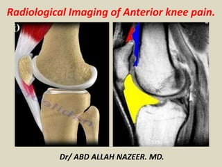
  • 1. Radiological Imaging of Anterior knee pain. Dr/ ABD ALLAH NAZEER. MD.
  • 2. Anterior knee pain that originates at the front of the knee joint is a common complaint. This disorder has a profound economic and social impact as it affects approximately 15-33% of young and active people. The role of physical examination in making differential diagnosis is rather limited since different conditions cause similar symptoms. Knee MRI is considered the gold standard in evaluating the damage to the anatomical structures of the knee, such as ligament or tendon rupture, meniscus or cartilage injury. It is often the case that MRI findings determine the therapy, therefore radiologists have to know what to look for in knee MRI images.
  • 3. Anterior knee pain is common with a variety of causes including: Patella fracture. Osteoarthritis. Inflammatory and depositional arthritis Bursitis around the knee. Patellofemoral maltracking. Excessive lateral pressure syndrome (ELPS). Patellar cartilage defect. Patellofemoral chondromalacia. Patellar tendinitis. Medial plica. Hoffa's disease. iliotibial band friction syndrome.
  • 4. Patellar fracture A direct blow or increased tension in a quadriceps and patellar tendon can cause patellar fracture. In most cases, a fracture line is transverse and passes between the middle and inferior third. Usually, the diagnosis is made on x-ray images. An MRI scan is useful in excluding ligamentous injury or in cases of suspected stress fracture. On MRI images stress fracture appears as low intensity line surrounded by bone marrow edema. Inferior third of patella fracture without dislocation.
  • 5. On MRI image the fracture line (arrows) extends across patella with bone edema in the surrounding marrow.
  • 6. Osteochondral sleeve fracture in a pediatric patient. (A) Lateral plain film of a patellar avulsion fracture of the inferior pole of the patella demonstrating a superiorly displaced patella and a small bone fragment (arrow). Sagittal T1- weighted (B) and sagittal gradient recalled echo (C) MR images demonstrating an osteochondral fracture of high signal intensity with a small bone fragment (arrow).
  • 7. Patellar dislocation It is one of the most common causes of anterior knee pain in young people. A high-riding patella and femoral trochlear dysplasia are the major predisposing factors. It is often the case that a dislocated patella reduces spontaneously without patient's awareness of the fact. The following MRI features of patellar dislocation are noted: Medial retinaculum and medial capsule injury/tear Lateral femoral condyle contusion and osteochondral damage Medial patellar facet contusion with or without cartilaginous lesion Medial retinaculum tears occur at the attachment site to the femur or along the medial margin of the patella and subsequently may cause avulsion injuries. Cartilaginous lesions can result in the formation of loose bodies in the joint space.
  • 8. An axial T2-weighted fast-spin-echo magnetic resonance imaging scan illustrates a eighteen year-old female sustaining a primary traumatic lateral dislocation of the patella while jumping. Complete avulsion of the medial patellofemoral ligament from its femoral insertion can be seen (arrow) (Figure 2A). The bone contusions (stars) (Figure 2B) of the lateral femoral condyle and the medial patellar facet are noted.
  • 9. Axial (a) PD FS and sagittal (b) T2 shows medial retinaculum avulsion fracture with patellar edge fragment (asterisk), medial patellar facet contusion and cartilaginous lesion (black arrows), lateral femoral condyle contusion (open arrow), loose cartilaginous body in anterolateral joint space.
  • 10. Lateral patellar tilt and subluxation(red arrow) and acute chondral defect(yellow arrow).
  • 11. X-ray and MRI after luxation of the patella. There is a fragment and bone bruise at the medial surface of the patella and in the corresponding surface of the lateral condyle of the femur. The medial retinaculum of the patella is disrupted.
  • 12. Coronal PD FS (a, b) shows lateral femoral condyle contusion and osteochondral damage (white arrow), loose cartilaginous body (black arrows) in antero-lateral joint space.
  • 13. Chondromalacia is defined as softening and degeneration of the articular hyaline cartilage. Overuse and trauma cause damage to the cartilage which over time leads to fissuring and partial loss of cartilage thickness. Subsequently, patients with high grade osteoarthritis sustain cartilage destruction with underlying bone changes. Cartilaginous lesions are graded according to the Outer bridge classification which is based on a correlation between MRI and arthroscopic findings. Although arthroscopy is a gold standard for the evaluation of the severity of cartilage defects, new MRI sequences (T2 mapping, DW, dGEMRIC, T1rho,) allow early diagnosis, contribute to preoperative planning and postoperative follow-up.
  • 14. Cartilage lesions on T2 images: a- focal area of hyperintensity with normal contour; b- fragmentation of cartilage; c- partial thickness cartilage loss with focal ulceration; d- full thickness cartilage loss with underlying bone reactive changes.
  • 15. Chondromalacia patellae grades II–IV in various patients. (A) Axial fast spin echo proton density fat saturated (FSE PD FS) MR image of chondromalacia patellae grade II in a 46-year-old male. High signal is seen in the patellar cartilage in the lateral patellar facet (arrows). (B) Axial FSE PD FS MR image of chondromalacia patellae grade III in a 51-year-old female. There is full thickness focal signal intensity change, contour irregularity and thinning of the cartilage (arrows). (C) Axial PD
  • 16. Cartilage T2 mapping allows to evaluate the articular cartilage of the knee joint. Red color shows cartilage lesion.
  • 18. Types of bipartite patella: type I- inferior pole (a,b); type II- lateral margin (c, d); type III- superolateral portion (e, f).
  • 19.
  • 20. Osteochondritis dissecans of the patellofemoral joint is an uncommon condition that may be the cause of anterior knee pain or crepitus. The osteochondral lesions involve the convex articular surfaces. The trochlear groove is the rarest location for osteochondritis dissecans. MR is the test of choice since it detects an osteochondral fragment and evaluates its stability. A high signal line demarcating the fragment from bone usually indicates an unstable lesion. Cartilaginous lesions can be classified into three main groups: Subchondral- intact cartilage surface Osteochondral fractures- disrupted articular surface with a fragment of cortical bone Chondral injury- underlying bone is intact
  • 23. Extensor mechanism of the knee pathology. -Traumatic. -Tendinosis. -Patellar tendon tear. -Quadriceps tendon tear. .Intrinsic patellar tendon lesions. e.g. gout. .Patellar enthesopathy. .Osteochondrosis, e.g. Osgood-Schlatter disease.
  • 24. Patellar tendinosis. .Pain in the infrapatellar region. .Commonly seen in athletes. .MRI demonstrate thickening of the patellar tendon with intermediate T1 and increased signal at T2 especially with fat suppression sequences. .Ultrasound show thickening, low echogenicity and increase blood flow at the Doppler study.
  • 26. Patellar tendinopathy and Partial patellar tendon tear.
  • 27. On sagittal (a) and axial (b) PD FS proximal end of patellar tendon is seen high intensity, enlarged (white arrow) surrounding by inflammatory changes (black arrow).
  • 28. Sagittal T2 (a) and PD SPIR (b) shows tibial tuberosity bony hypertrophy (black arrow), fragmentation and inflammatory changes of overlying tissues.
  • 29. Coronal (a) and sagittal (b) PD FS shows changes involving the inferior pole of the patella which is characteristic for Sinding–Larsen–Johansson disease.
  • 30. Complete Patellar tendon tear. Images show no continuity between fibers and patella. The tendon is thickened. Complete Patellar tendon tear. Image on the right shows hemorrhagic bursitis ( low signal in bursa).
  • 31. Quadriceps tendon tear. Tear may be partial or complete. Partial quadriceps tendon tear: T2W-images.LEFT: Abnormal attachment of tendon. Right : Most of tendon is retracted (red arrow) deep part (vastus intermedius) is still intact.
  • 32. Complete quadriceps tear. Sag T2W-images. No continuity. Hematoma in between.
  • 33. Sagittal PD FS (a) and T2 (b) shows quadriceps tendon full thickness rupture as a gap filled with hyperintense fluid.
  • 34. Intrinsic patellar tendon lesions : Gout MRI show low T1 and mildly high T2 signal.
  • 35.
  • 36.
  • 37. Fat pad syndromes The infrapatellar (Hoffa's) fat pad is an intraarticular and yet an extrasynovial structure which has an abundant vasculature and innervation. Pathological changes are often associated with other conditions, such as patellar tendinopathy, ligament reconstruction or meniscal tear. Acute injury usually occurs in the dorsal part of the fat pad and presents as edema or tears. In chronic injury hemosiderin deposits or scarring are observed. Lateral patellofemoral impingement involves of edema of the superolateral part of the fat pat, a shallow femoral trochlea, patellar malalignment and chondromalacia. The inflammation of a quadriceps fat pad is rare and features are similar to that of Hoffa’s fat pad. Also, benign or malignant masses can be the cause of anterior knee pain
  • 38. Sagittal (a) and axial (b) IR shows high signal intensity at infrapatellar fat pad (arrows). Sagittal image demonstrates patella alta.
  • 40. Sagittal T2 (a) and PD FS (b) shows heterointensive mass (arrows) at quadriceps fat pad which was confirmed histologically as a metastasis of renocellular carcinoma
  • 41. Sagittal MRI scans of the knee show an intermediate signal lesion in the apex of Hoffa's fat pad between the inferior pole of the patella and the adjacent femoral condyle. The lesion's surface retains the shape of the adjoining femoral condyle, indicating chronic wedging that reflects the chronic nature of the lesion. The lesion was surgically excised. Histology showed adipose and synovial tissue with inflammation and fibrosis, supporting a diagnosis of Hoffa's disease.
  • 42. Pigmented villonodular synovitis is rare benign neoplastic synovial proliferation with villous and nodular projections and hemosiderin deposition. It mainly affects knee joint. On MRI images it appears as mass like lesion with well- defined low signal intensity nodule. On both T1 and T2 images it is seen low signal intensity with variable postcontrast enhancement. A synovial nodular lesion is seen (arrows), with heterogenous signal characteristics.
  • 45. Intra-articular Chondroma. A rare lesion occur around the knee typically at the infrapatellar fat pad. May be calcified or erode the lower pole of patella. Intra-articular Chondroma.
  • 46. Infrapatellar Plica injury. A thin fold of synovial tissue, extending from the inferior pole of the patella through Hoffa's fat to the intercondylar notch anterior to the anterior cruciate ligament. .High signal along the course of the plica indicates injury to the plica. .Thickening of the plica even in the absence of edema or fluid suggestive of chronic injury. Infrapatellar Plica injury.
  • 47. Sagittal T2 (a) and axial PD FS (b) shows thickening of medial patellar place (arrows) and insignificant Chondromalacia of femoral trochlear groove.
  • 48. Iliotibial band syndrome, also known as Runner’s knee, is caused by an excessive friction between the iliotibial band and the lateral femoral epicondyle. MRI findings include the thickening of the iliotibial band and a deep bursa located over the lateral epicondyle. Also, there is an inflammation of the surrounding soft tissues. MRI is reserved to exclude other etiologies of pain such as lateral meniscal tear. Coronal (a) and axial (b) PD FS shows thickened and edematous iliotibial band (white arrow) and bursa (black arrow) between the band and lateral epicondyle of the femur.
  • 49. Bursitis There are four bursae anterior to the knee joint: a suprapatellar, a subcutaneous prepatellar, a subcutaneous infrapatellar and a deep infrapatellar bursa. Inflammation can be caused by acute or chronic trauma and systemic diseases such as rheumatoid arthritis or metabolic disorders. Axial PD FS (a) and sagittal GRE (b) shows hyperintense fluid in prepatellar region (black arrows) and suprapatellar bursae (white arrows) - characteristic for prepatellar and suprapatellar bursitis
  • 52. Ligament injury Cruciate ligaments stabilize the knee joint. Once they rupture, a shift in ligament balance forces occurs and may lead to cartilage damage. An ACL rupture causes rotational instability and secondary changes, such as medial compartment and patellofemoral joint overload. A PCL rupture causes posterior instability. When the tibia slips dorsally, mechanical load to the patella increases and speeds up wear and tear. Following an anterior cruciate ligament reconstruction, pain can be caused by postoperative complications, such as: Arthrofibrosis is a contracture of a infrapatellar fat pad and patellar tendon. A ill- defined low signal area on T1 and T2 weighted images, similar to fibrous tissue. Cyclops lesion is defined as a nodule located anteriorly to the distal end of an ACL graft. Two histologic types: “hard”- composed of bone or cartilage, more often acts as obstacle and “soft”- only composed of fibrous tissue, rarely causes impingement. A low signal intensity nodule on T1WI images, anteriorly to the graft. It could show similar intensity to synovial fluid. The nodule is heterogeneous on T2WI, usually hypointense and well differentiated from fluid. Differential diagnosis should include focal pigmented villonodular synovitis, synovial chondromatosis and loose bodies.
  • 53. Coronal (frontal) IR FSE (a) and sagittal T2 FSE (b) shows unevenly thinning of medial knee joint and femur trochlear groove cartilage (arrows) caused by chronic rupture of anterior cruciate ligament.
  • 54. Axial PDW SPIR (a) and sagittal PDW (b) shows fibrous nodule (arrows) at distal part of anterior cruciate ligament transplant.