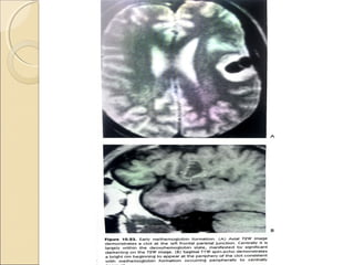
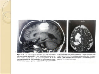
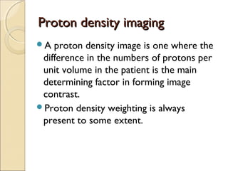
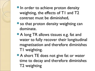
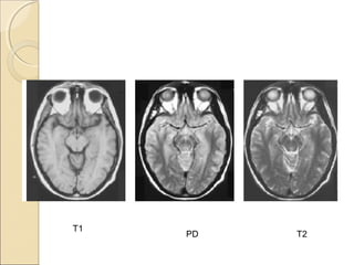
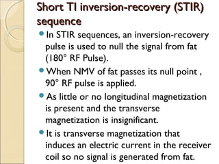
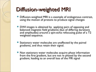
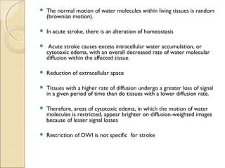
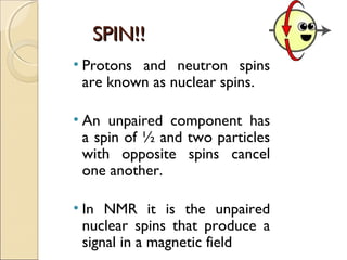
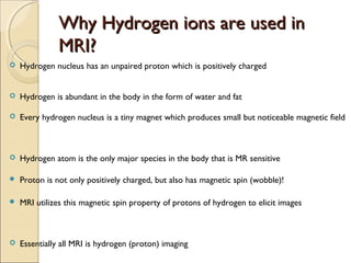
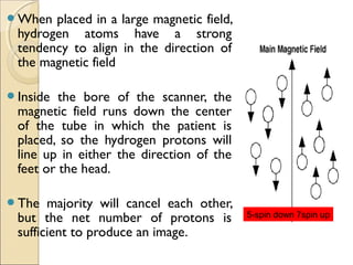
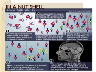
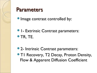
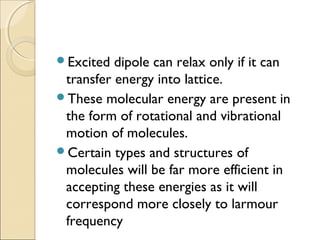
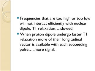
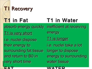
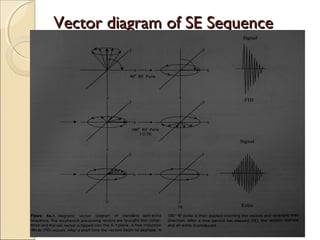
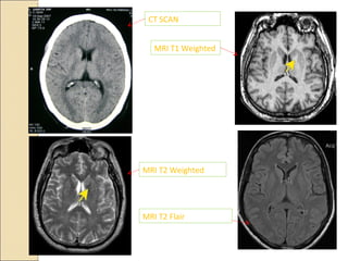
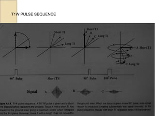
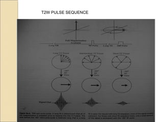
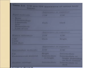
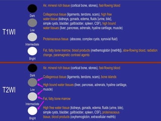
MRI uses magnetic fields and radio waves to produce detailed images of the brain and detect abnormalities. It is based on nuclear magnetic resonance, where hydrogen protons in the body are aligned by a strong magnetic field. When hit with radio waves of a specific frequency, the protons absorb energy and spin, and emit radio signals as they relax back to baseline. These signals are used to construct images, with different tissues appearing different intensities based on their relaxation times T1 and T2. MRI provides valuable information to assess many neurological conditions without using ionizing radiation.
Magnetic resonance imaging (MRI) is an imaging technique used primarily in medical settings to produce high quality images of the soft tissues of the human body.
1. Brain Facts
2. MRI Imaging Techniques
3. How the Various Imaging Helps Us in Treatment
Objectives
Planning
4. Locate and Identify Pertinent Cerebral Anatomy
on CT and MRI Images
5. What is a Functional MRI and What it Tells Us
This is a presentation describing in brief regarding the physics behind MRI and it's application from dental point of view. It contains few videos as well.
Training Material inherited form Philips Basics of Ultrasonography. Covers the fundamentals of Ultrasound Waveform, Piezoelectric Effect, Phased Echo Concept, Goal of Ultrasound, Ultrasound Image Construction process, Types of Resolution, Probe Internals, The Doppler Effect, Spectrum Waveform and concept, Color Doppler, Components of Ultrasound.
Here I discussed about the concept,types, types of images obtained, the advantages and disadvantages of MRI shortly...anyone who wants to know about MRI just go through it. I just prepared it in very simple language for the convenience of the readers all over the world. Thank you.
MRI uses a strong magnetic field and radio waves to create detailed images of the organs and tissues within the body.
Developed by the Lauterbur in 1972 at Stony brook in New York.
MRI does not involve radiation
MRI contrasting agent is less likely to produce an allergic reaction that may occur when iodine-based substances are used for x-rays and CT scans
MRI gives extremely clear, detailed images of soft-tissue structures that other imaging techniques cannot achieve
The MRI machine cannot just simply “see the hydrogen nuclei which lie “hidden” in the water molecules distributed in the patient.
It needs to do ‘something’ to the hydrogen nuclei to detect their presence.
NMR - Nuclear magnetic resonance (NMR).pptxmuskaangandhi1
Nuclear magnetic resonance (NMR) spectroscopy is the study of molecules by recording the interaction of radiofrequency (Rf) electromagnetic radiations with the nuclei of molecules placed in a strong magnetic field.
It is concerned with absorption of certain amount of energy
by spinning nuclei in a magnetic field when irradiated with
radiofrequency radiation (RFR) of equivalent energy.
NMR gives the information about the number and configuration of
magnetically active atoms, like positions of different types
of Hydrogen over the C- skeleton of an organic molecule.
Absorption of RFR occurs when the nucleus undergoes
transition from one alignment in the applied magnetic field
to the opposite alignment, i.e. from parallel (ground state)
orientation to anti-parallel (excited state) orientation.
When the frequency of the oscillating electric field of the
incoming RFR just matches the frequency of the electric field
generated by the precising nucleus, then the 2 fields can
couple, and the energy can be transferred from the
incoming radiation to the nucleus, thus causing a spin change
(clock-wise to anti-clock-wise).
This condition is called "resonance", and the nucleus is said to
have resonance with the incoming electromagnetic wave
(RFR).
In NMR technique, the frequency of the RFR is kept constant
(60MHz) and the strength of magnetic field is varied.
At certain value of the applied field strength, depending
upon the nature of proton or nucleus, the energy required to
flip the proton matches the energy of radiation.
As a result, absorption takes place and a signal is observed
in the spectrum. Such a signal or peak is called an NMR
Spectrum.
NMR spectrum is graphical plot of relative intensity
(Y axis) and the δ value (x axis).
The nucleus of a hydrogen atom (proton) behaves as a spinning bar magnet because it possesses both electric and magnetic spin.
Like any other spinning charged body, the nucleus of hydrogen atom also generates a magnetic field.
Nuclear magnetic resonance Involves the interaction between an oscillating magnetic field of electromagnetic radiation and the magnetic energy of the hydrogen nucleus or some other type of nuclei when these are placed in an external static magnetic field.
The sample absorbs electromagnetic radiations in radio wave region at different frequencies since absorption depends upon the type of protons or certain nuclei contained in the sample)
Consider a spinning top. It also performs a slower waltz like motion,
in which the spinning axis of the top moves slowly around
the vertical.
This is processional motion & the top is said to be processing around the vertical axis of earth's gravitational field.
The precession arises from the interaction of spin with earth's gravity acting vertically downwards.
It is called Gyroscopic motion.
Proton will be showing processional motion due to interaction of Spin &
Gravitational force of Earth
ARTIFICIAL INTELLIGENCE IN HEALTHCARE.pdfAnujkumaranit
Artificial intelligence (AI) refers to the simulation of human intelligence processes by machines, especially computer systems. It encompasses tasks such as learning, reasoning, problem-solving, perception, and language understanding. AI technologies are revolutionizing various fields, from healthcare to finance, by enabling machines to perform tasks that typically require human intelligence.
MANAGEMENT OF ATRIOVENTRICULAR CONDUCTION BLOCK.pdfJim Jacob Roy
Cardiac conduction defects can occur due to various causes.
Atrioventricular conduction blocks ( AV blocks ) are classified into 3 types.
This document describes the acute management of AV block.
Factory Supply Best Quality Pmk Oil CAS 28578–16–7 PMK Powder in Stockrebeccabio
Factory Supply Best Quality Pmk Oil CAS 28578–16–7 PMK Powder in Stock
Telegram: bmksupplier
signal: +85264872720
threema: TUD4A6YC
You can contact me on Telegram or Threema
Communicate promptly and reply
Free of customs clearance, Double Clearance 100% pass delivery to USA, Canada, Spain, Germany, Netherland, Poland, Italy, Sweden, UK, Czech Republic, Australia, Mexico, Russia, Ukraine, Kazakhstan.Door to door service
Hot Selling Organic intermediates
Ethanol (CH3CH2OH), or beverage alcohol, is a two-carbon alcohol
that is rapidly distributed in the body and brain. Ethanol alters many
neurochemical systems and has rewarding and addictive properties. It
is the oldest recreational drug and likely contributes to more morbidity,
mortality, and public health costs than all illicit drugs combined. The
5th edition of the Diagnostic and Statistical Manual of Mental Disorders
(DSM-5) integrates alcohol abuse and alcohol dependence into a single
disorder called alcohol use disorder (AUD), with mild, moderate,
and severe subclassifications (American Psychiatric Association, 2013).
In the DSM-5, all types of substance abuse and dependence have been
combined into a single substance use disorder (SUD) on a continuum
from mild to severe. A diagnosis of AUD requires that at least two of
the 11 DSM-5 behaviors be present within a 12-month period (mild
AUD: 2–3 criteria; moderate AUD: 4–5 criteria; severe AUD: 6–11 criteria).
The four main behavioral effects of AUD are impaired control over
drinking, negative social consequences, risky use, and altered physiological
effects (tolerance, withdrawal). This chapter presents an overview
of the prevalence and harmful consequences of AUD in the U.S.,
the systemic nature of the disease, neurocircuitry and stages of AUD,
comorbidities, fetal alcohol spectrum disorders, genetic risk factors, and
pharmacotherapies for AUD.
Explore natural remedies for syphilis treatment in Singapore. Discover alternative therapies, herbal remedies, and lifestyle changes that may complement conventional treatments. Learn about holistic approaches to managing syphilis symptoms and supporting overall health.
Tom Selleck Health: A Comprehensive Look at the Iconic Actor’s Wellness Journeygreendigital
Tom Selleck, an enduring figure in Hollywood. has captivated audiences for decades with his rugged charm, iconic moustache. and memorable roles in television and film. From his breakout role as Thomas Magnum in Magnum P.I. to his current portrayal of Frank Reagan in Blue Bloods. Selleck's career has spanned over 50 years. But beyond his professional achievements. fans have often been curious about Tom Selleck Health. especially as he has aged in the public eye.
Follow us on: Pinterest
Introduction
Many have been interested in Tom Selleck health. not only because of his enduring presence on screen but also because of the challenges. and lifestyle choices he has faced and made over the years. This article delves into the various aspects of Tom Selleck health. exploring his fitness regimen, diet, mental health. and the challenges he has encountered as he ages. We'll look at how he maintains his well-being. the health issues he has faced, and his approach to ageing .
Early Life and Career
Childhood and Athletic Beginnings
Tom Selleck was born on January 29, 1945, in Detroit, Michigan, and grew up in Sherman Oaks, California. From an early age, he was involved in sports, particularly basketball. which played a significant role in his physical development. His athletic pursuits continued into college. where he attended the University of Southern California (USC) on a basketball scholarship. This early involvement in sports laid a strong foundation for his physical health and disciplined lifestyle.
Transition to Acting
Selleck's transition from an athlete to an actor came with its physical demands. His first significant role in "Magnum P.I." required him to perform various stunts and maintain a fit appearance. This role, which he played from 1980 to 1988. necessitated a rigorous fitness routine to meet the show's demands. setting the stage for his long-term commitment to health and wellness.
Fitness Regimen
Workout Routine
Tom Selleck health and fitness regimen has evolved. adapting to his changing roles and age. During his "Magnum, P.I." days. Selleck's workouts were intense and focused on building and maintaining muscle mass. His routine included weightlifting, cardiovascular exercises. and specific training for the stunts he performed on the show.
Selleck adjusted his fitness routine as he aged to suit his body's needs. Today, his workouts focus on maintaining flexibility, strength, and cardiovascular health. He incorporates low-impact exercises such as swimming, walking, and light weightlifting. This balanced approach helps him stay fit without putting undue strain on his joints and muscles.
Importance of Flexibility and Mobility
In recent years, Selleck has emphasized the importance of flexibility and mobility in his fitness regimen. Understanding the natural decline in muscle mass and joint flexibility with age. he includes stretching and yoga in his routine. These practices help prevent injuries, improve posture, and maintain mobilit
Couples presenting to the infertility clinic- Do they really have infertility...Sujoy Dasgupta
Dr Sujoy Dasgupta presented the study on "Couples presenting to the infertility clinic- Do they really have infertility? – The unexplored stories of non-consummation" in the 13th Congress of the Asia Pacific Initiative on Reproduction (ASPIRE 2024) at Manila on 24 May, 2024.
Title: Sense of Taste
Presenter: Dr. Faiza, Assistant Professor of Physiology
Qualifications:
MBBS (Best Graduate, AIMC Lahore)
FCPS Physiology
ICMT, CHPE, DHPE (STMU)
MPH (GC University, Faisalabad)
MBA (Virtual University of Pakistan)
Learning Objectives:
Describe the structure and function of taste buds.
Describe the relationship between the taste threshold and taste index of common substances.
Explain the chemical basis and signal transduction of taste perception for each type of primary taste sensation.
Recognize different abnormalities of taste perception and their causes.
Key Topics:
Significance of Taste Sensation:
Differentiation between pleasant and harmful food
Influence on behavior
Selection of food based on metabolic needs
Receptors of Taste:
Taste buds on the tongue
Influence of sense of smell, texture of food, and pain stimulation (e.g., by pepper)
Primary and Secondary Taste Sensations:
Primary taste sensations: Sweet, Sour, Salty, Bitter, Umami
Chemical basis and signal transduction mechanisms for each taste
Taste Threshold and Index:
Taste threshold values for Sweet (sucrose), Salty (NaCl), Sour (HCl), and Bitter (Quinine)
Taste index relationship: Inversely proportional to taste threshold
Taste Blindness:
Inability to taste certain substances, particularly thiourea compounds
Example: Phenylthiocarbamide
Structure and Function of Taste Buds:
Composition: Epithelial cells, Sustentacular/Supporting cells, Taste cells, Basal cells
Features: Taste pores, Taste hairs/microvilli, and Taste nerve fibers
Location of Taste Buds:
Found in papillae of the tongue (Fungiform, Circumvallate, Foliate)
Also present on the palate, tonsillar pillars, epiglottis, and proximal esophagus
Mechanism of Taste Stimulation:
Interaction of taste substances with receptors on microvilli
Signal transduction pathways for Umami, Sweet, Bitter, Sour, and Salty tastes
Taste Sensitivity and Adaptation:
Decrease in sensitivity with age
Rapid adaptation of taste sensation
Role of Saliva in Taste:
Dissolution of tastants to reach receptors
Washing away the stimulus
Taste Preferences and Aversions:
Mechanisms behind taste preference and aversion
Influence of receptors and neural pathways
Impact of Sensory Nerve Damage:
Degeneration of taste buds if the sensory nerve fiber is cut
Abnormalities of Taste Detection:
Conditions: Ageusia, Hypogeusia, Dysgeusia (parageusia)
Causes: Nerve damage, neurological disorders, infections, poor oral hygiene, adverse drug effects, deficiencies, aging, tobacco use, altered neurotransmitter levels
Neurotransmitters and Taste Threshold:
Effects of serotonin (5-HT) and norepinephrine (NE) on taste sensitivity
Supertasters:
25% of the population with heightened sensitivity to taste, especially bitterness
Increased number of fungiform papillae
Report Back from SGO 2024: What’s the Latest in Cervical Cancer?bkling
Are you curious about what’s new in cervical cancer research or unsure what the findings mean? Join Dr. Emily Ko, a gynecologic oncologist at Penn Medicine, to learn about the latest updates from the Society of Gynecologic Oncology (SGO) 2024 Annual Meeting on Women’s Cancer. Dr. Ko will discuss what the research presented at the conference means for you and answer your questions about the new developments.
Report Back from SGO 2024: What’s the Latest in Cervical Cancer?
Ppt mri brain
1. BASICS OF MRI BRAINBASICS OF MRI BRAIN
PRESENTER:DR. ARSHAD YAHYA
MODERATOR: DR. K S ANAND
2. HISTORYHISTORY
►Dr Isidor Rabi (Nobel in 1944!) He succeeded in
detecting and measuring single states of rotation of
atoms and molecules, and in determining the
magnetic moments of the nuclei .
►CJ Gorter, coined the term ‘Nuclear Magnetic
Resonance’ in 1942.
►Bloch and Purcell were awarded the Nobel Prize
for Physics in 1952
►Found that when certain nuclei were placed in
magnetic field they absorbed energy in
electromagnetic spectrum and re emit energy when
regained their original position.
3. ““We are so close to the man behindWe are so close to the man behind
MRI”MRI”
►Prof Peter Mansfield was awarded
Nobel in 2003 for his discoveries in MRI
(with Prof Paul C Lauterbur of USA)
►Peter Mansfield is from Nottingham
University, UK
►Described the use of magnetic field
gradients to acquire spatial information in
NMR experiments
4. MRI principleMRI principle
MRI is based on the principle of nuclear magnetic
resonance (NMR)
Two basic principles of NMR
1. Atoms with an odd number of protons or neutrons
have spin
2. A moving electric charge, be it positive or negative,
produces a magnetic field
5. We all are made up of elementsWe all are made up of elements
►92 elements occur naturally on earth.
►Human body is built of only 26 elements.
►Oxygen, hydrogen, carbon, nitrogen
elements constitute 96 % of human body
mass.
►Oxygen is 65 % of body mass; carbon is
18.5 %, hydrogen 9.5 %, nitrogen 3.2 %.
6. Nucleus needs to have 2 properties:
◦ Spin
◦ charge
Nuclei are made of protons(3 quark) and neutrons
◦ Both have spin ½
◦ Protons have charge
Pairs of spins tend to cancel, so only atoms with an
odd number of protons or neutrons have spin
◦ Good MR nuclei are 1
H, 13
C, 19
F, 23
Na, 31
P
7. SPIN!!SPIN!!
• Protons and neutron spins
are known as nuclear spins.
• An unpaired component has
a spin of ½ and two particles
with opposite spins cancel
one another.
• In NMR it is the unpaired
nuclear spins that produce a
signal in a magnetic field
8. Why Hydrogen ions are used inWhy Hydrogen ions are used in
MRI?MRI?
Hydrogen nucleus has an unpaired proton which is positively charged
Hydrogen is abundant in the body in the form of water and fat
Every hydrogen nucleus is a tiny magnet which produces small but noticeable magnetic field
Hydrogen atom is the only major species in the body that is MR sensitive
Proton is not only positively charged, but also has magnetic spin (wobble)!
MRI utilizes this magnetic spin property of protons of hydrogen to elicit images
Essentially all MRI is hydrogen (proton) imaging
9. But why we can’t act like magnets?But why we can’t act like magnets?
The protons (i.e.
Hydrogen ions) in body
are spinning in a
haphazard fashion, and
cancel all the magnetism.
That is our natural state!
10. When placed in a large magnetic field,
hydrogen atoms have a strong
tendency to align in the direction of
the magnetic field
Inside the bore of the scanner, the
magnetic field runs down the center
of the tube in which the patient is
placed, so the hydrogen protons will
line up in either the direction of the
feet or the head.
The majority will cancel each other,
but the net number of protons is
sufficient to produce an image.
5-spin down 7spin up
11. Net magnetizationNet magnetization
Half of the protons align along the magnetic field and rest are aligned opposite
.
At room temperature, the
population ratio of anti-
parallel versus parallel
protons is roughly 100,000
to 100,006 per Tesla of B0
These extra protons produce net magnetization vector (M)
Net magnetization depends on B0 and temperature
12. PrecessionPrecession
The static field causes the spinning proton to
‘wobble’ in a regular manner from it’s original
axis to axis of magnetic field called
‘PRECESSION’
14. Manipulating the net magnetizationManipulating the net magnetization
Magnetization can be manipulated by changing the magnetic field
environment (static, gradient, and RF fields)
Static field vary in space but not over time during image
acquisation.by magnet itself or perturbation by materials with
different magnetic permeability.
RF waves are used to manipulate the magnetization of H nuclei
Externally applied RF waves perturb magnetization into different
axis (transverse axis). Only transverse magnetization produces
signal.
When perturbed nuclei return to their original state they emit RF
signals which can be detected with the help of receiving coils
16. Measuring the MR Signal:
◦ the moving proton vector
induces a signal in the RF
antenna
◦ The signal is picked up by a coil
and sent to the computer
system.
the received signal is
sinusoidal in nature
◦ The computer receives
mathematical data, which is
converted through the use of a
Fourier transform into an image.
MeasuringtheMRSignal
z
y x
RFsignalfrom
precessingprotons
RFantenna
17. Now, we re-transmit the energy forNow, we re-transmit the energy for
image processingimage processing
►The emitted energy is too small (despite
2500 times the magnetic field with
resonance RF pulse) to convert them into
images.
►Hence, repeated “ON-OFF” of RF pulses
are required.
►The emitted energy is stored (K-space),
analysed and converted into images.
19. Energy Absorption:
◦ The MRI machine applies
radio frequency (RF) pulse
that is specific to hydrogen.
◦ The RF pulses are applied
through a coil that is
specific to the part of the
body being scanned.
20. T1 and T2 relaxationT1 and T2 relaxation
When RF pulse is stopped higher energy gained by proton is
retransmitted and hydrogen nuclei relax by two mechanisms
T1 or spin lattice relaxation- by which original magnetization
(Mz) begins to recover.
T2 relaxation or spin spin relaxation - by which magnetization
in X-Y plane decays towards zero in an exponential fashion. It
is due to incoherence of H nuclei.
T2 values of CNS tissues are shorter than T1 values
21. T1 relaxationT1 relaxation
After protons are
Excited with RF pulse
They move out of
Alignment with B0
But once the RF Pulse
is stopped they Realign
after some Time And
this is called t1 relaxation
T1 is defined as the time it takes for the hydrogen nucleus to recover
63% of its longitudinal magnetization
22. T2 relaxation time is the time for 63% of the protons to become dephased
owing to interactions among nearby protons.
23. T2* RelaxationT2* Relaxation
Due to combined loss of phase coherence from both
static and time varying magnetic field in homogenity.
Static magnet field in homogenity are constant in time
signal loss can be recovered by use of 2nd
180*
pulse..rephasing the nuclei
Time varying magnetic field-water molecule move
rapidly …acquire different area of different magnetic
field…precess at different rate..goes out of phase
Not static so can’t be reversed in to phase of
coherence
24. TR and TETR and TE
TR (repetition time) : the time between two excitations is called repetition
time
TE (echo time) : time interval in which signals are measured after RF
excitation
By varying the TR and TE one can obtain T1WI and T2WI
In general a short TR (<1000ms) and short TE (<45 ms) scan is T1WI
Long TR (>2000ms) and long TE (>45ms) scan is T2WI
Long TR (>2000ms) and short TE (<45ms) scan is proton density image
26. T1 RecoveryT1 Recovery
Caused by EXCHANGE OF ENERGY
FROM
NUCLEI TO THEIR SURROUNDING
ENVIRONMENT OR LATTICE
"Spin Lattice Energy Transfer"
and realign in B0
this occur in exponential process
at different rates in different tissue
NB: Molecules are constantly in motion;
Rotational and Transitional
27. Excited dipole can relax only if it can
transfer energy into lattice.
These molecular energy are present in
the form of rotational and vibrational
motion of molecules.
Certain types and structures of
molecules will be far more efficient in
accepting these energies as it will
correspond more closely to larmour
frequency
28. Frequencies that are too high or too low
will not interact efficiently with nuclear
dipole, T1 relaxation….slowed.
When proton dipole undergo faster T1
relaxation more of their longitudinal
vector is available with each succeeding
pulse…..more signal.
29. T1 RecoveryT1 Recovery
T1 in FatT1 in Fat T1 in WaterT1 in Water
absorb energy quicklyabsorb energy quickly
T1 is very shortT1 is very short
i.e. nuclei disposei.e. nuclei dispose
their energy totheir energy to
surrounding fat tissuesurrounding fat tissue
and return to B0 inand return to B0 in
very short timevery short time
inefficient at receivinginefficient at receiving
energyenergy
T1 is longerT1 is longer
i.e. nuclei take a loti.e. nuclei take a lot
longer to disposelonger to dispose
energy to surroundingenergy to surrounding
water tissuewater tissue
30. Myelin has slowing effect on motion of
adjacent H2O….relatively bright on T1W
Very large, solid structure such as bone
or protein(ligament) have proton
…..immobile
Low signal, because rotational and
vibrational frequencies have been slowed
to point, they are no longer optimal for
relaxation.
31. Proton on cholesterol and lipid
membrane have poor mobility …long T1
as opposed to adipose fat having
molecule in oil, mobile relax more quickly
Paramagnetic material also decreases T1
relaxation as can non paramagnetic
calcium salts
32.
33. T2 DecayT2 Decay
Those tissues having constituent which
moves fast loses coherence very late
….long T2 (water)
Water tumbles rapidly in space..any
magnetic field distortion are rapidly
averaged out.
Adjacent water molecule have similar
magnetic field …nuclear dipole dephase
very slowly.
34. Fat being larger molecule than water
moves slowly in space…loses coherence
early than free water.
Less coherent movement give low signal
on T2.
Paramagnetic substances(iron)distort
magnetic field…..loss of coherence
35. Different tissues have different
relaxation times. These relaxation time
differences is used to generate image
contrast.
41. Shorter the TE less decay of signal
Best signal to noise ratio with short TE
T1 and proton-W ….highest anatomical
detail
T1 field strength dependent
Also varies in different instrument and
investigator
Not useful for absolute comparison with
disease process.
48. Confusing…evolution of hematomaConfusing…evolution of hematoma
Hyper acute(mts…hrs):oxyHb…O2
binds to 6th
location on iron in heme of Hb
O2…strong ligand, significantly split energy
level of iron….in low spin state…not
paramagnetic
O2Hb blood will have long T1…dark on T1W
image
Any T1 shortening occurs due to water binding
transiently to hydration layer of
protein..shortening T1…iso intense
Due to free flowing without paramagnetic
49.
50.
51. Acute phase deoxyHb(hrs….days):
Fe in deoxyHb is in ferrous state….no
O2 present
High spin state, paramagnetic because of
presence of 4 unpaired electron….should
cause shortening of T2 with brightness on
T1W.
When O2 falls of Hb , iron molecule
retract slightly inside the porphyrin
ring…allosteric shift of HB
52.
53. Iron is pulled out of plane of porphyrin,
water is excluded from sensing the
paramagnetic effects of iron.
For significant paramagnetic effect water
must transiently bind to iron…
On T1W deoxy Hb …isointense with
water.
Immobilization of fibrin,Hb, paramagnetic
iron molecule with in semisolid
gelatinaous clot reduces the mobility of
these substances….disturbing local
magnetic field
Rapid dephasing and loss of signal inT2W
54.
55. MetHb-subacute phase(3d…18
month)
metHb is formed with oxidation of iron
from ferrous to ferric form
With loss of additional electron, smaller
ferric ion moves back into porphyrin
ring…
Available to exchange with relax bound
water molecules
Paramagnetic effect …relaxation of T1
becomes short…bright onT1W
56.
57. In early stage red cell membrane is still
intact and paramagnetic substance remain
in RBC …immobilized in space
Due to early loss of coherence T2
short…dark in T2W.
In later part as clot dissolve local
variation in magnetic field averaged out
by tumbling of water and iron
Dephasing lessened…image bright on
T2W because of increased water content
58.
59.
60. Chronic phase hemosiderin
formation
Blood breakdown occurs..iron falls off the heme
molecule….hemosiderin formed with microglia
Hemosiderin insoluble and rigidly held in
space…little effect on T1 relaxation….minimal
darkening on T1W
With al SE imaging,T1W image have
contribution of T2 W because of echo
time..minimal darkening onT1W.
On T2W due to interferance of local magnetic
field by large immoble fibrin…dephasing occurs
early …dark on T2W
61. InfarctInfarct
Acute : T1W –Isointense hypo
intense
T2W-Hyper intense
Sub acute: T1W-Low
signal,increasedsignal in peripheral
region..hemorrhage(metHb)
T2W- High signal
Chronic:T1W-low signal
T2W-High siignal
62. Dark on T1Dark on T1
Edema, tumor, infection, inflammation,
hemorrhage(hyperacute, chronic)
Low proton density, calcification
Flow void
63. Bright on T1Bright on T1
Fat, subacute hemorrhage, melanin,
protein rich fluid.
Slowly flowing blood
Paramagnetic
substances(gadolinium,copper,manganese)
64. Bright on T2Bright on T2
Edema, tumor, Infection, inflammation,
subdural collection
Met hemoglobin in late sub acute
hemorrhage
65. Dark on T2Dark on T2
Low proton density,calcification,fibrous
tissue
Paramagnetic substances(deoxy
hemoglobin,methemoglobin(intracellular),
ferritin,hemosiderin,melanin.
Protein rich fluid
Flow void
66. Which scan best defines theWhich scan best defines the
abnormalityabnormality
T1 W Images:
Subacute Hemorrhage
Fat-containing structures
Anatomical Details
T2 W Images:
Edema
Demyelination
Infarction
Chronic Hemorrhage
67. Proton density imagingProton density imaging
A proton density image is one where the
difference in the numbers of protons per
unit volume in the patient is the main
determining factor in forming image
contrast.
Proton density weighting is always
present to some extent.
69. In order to achieve proton density
weighting, the effects of T1 and T2
contrast must be diminished,
so that proton density weighting can
dominate.
A long TR allows tissues e.g. fat and
water to fully recover their longitudinal
magnetisation and therefore diminishes
T1 weighting.
A short TE does not give fat or water
time to decay and therefore diminishes
T2 weighting
72. Short TI inversion-recovery (STIR)Short TI inversion-recovery (STIR)
sequencesequence
In STIR sequences, an inversion-recovery
pulse is used to null the signal from fat
(180° RF Pulse).
When NMV of fat passes its null point ,
90° RF pulse is applied.
As little or no longitudinal magnetization
is present and the transverse
magnetization is insignificant.
It is transverse magnetization that
induces an electric current in the receiver
coil so no signal is generated from fat.
74. STIR allows only short time between 180
deg. Pulse and second 90 deg. Pulse.
If TE is kept prolonged, effect of T1 and
T2 on lesion detection can be additive.
T1 and T2 of most pathologic lesion are
prolonged.
Long TE selection…substances having
both LONG T1 and T2 will be bright.
STIR also suppress substances with short
T1….hemorrhage, gd-enhancement.
75.
76. Fluid-attenuated inversion recoveryFluid-attenuated inversion recovery
(FLAIR)(FLAIR)
First described in 1992 and has become
one of the corner stones of brain MR
imaging protocols
An IR sequence with a long TR and TE
and an inversion time (TI) that is tailored
to null the signal from CSF
77.
78. Particularly helpful in evaluating
periventricular white matter lesion.
Water bound to complex molecule with
in plaque has relatively shorter T1 than
free water with in ventricle.
Long inversion time effectively suppress
free water…csf….nulled.
Lesion that contain complex, partially
bound water (less mobile)…shorter T1
than free water….not nulled.
79. Long TE …used…result in T2W
Sequence…..additional effect for contrast
effect of tissue with prolonged T2 and
short T1(White matter lesion)
Effective in high lightening lesion…
demyelination, stroke, Ischemic gliosis
and tumor
Sensitive but less specific
80. Normal partially myelinated white matter
tract….highlighted
Protein rich pituitary stalk….normally
bright on FLAIR.
More sensitive for detection of acute
infarct…differentiate it from cystic
encephalomalacia
Useful in SAH …removes CSF signal
86. GREGRE
In a GRE sequence, an RF pulse is applied that
partly flips the NMV into the transverse plane
(variable flip angle).
Gradients, as opposed to RF pulses, are used to
dephase (negative gradient) and rephase (positive
gradients) transverse magnetization.
Because gradients do not refocus field
inhomogeneities, GRE sequences with long TEs
are T2* weighted (because of magnetic
susceptibility) rather than T2 weighted like SE
sequences
87.
88. GRE Sequences contd:
This feature of GRE sequences is exploited- in
detection of hemorrhage, as the iron in Hb becomes
magnetized locally (produces its own local magnetic
field) and thus dephases the spinning nuclei.
The technique is particularly helpful for diagnosing
hemorrhagic contusions such as those in the brain .
SE sequences, on the other hand- relatively immune
from magnetic susceptibility artifacts, and also less
sensitive in depicting hemorrhage and calcification.
92. Diffusion-weighted MRIDiffusion-weighted MRI
Diffusion-weighted MRI is a example of endogenous contrast,
using the motion of protons to produce signal changes
DWI images is obtained by applying pairs of opposing and
balanced magnetic field gradients (but of differing durations
and amplitudes) around a spin-echo refocusing pulse of a T2
weighted sequence.
Stationary water molecules are unaffected by the paired
gradients, and thus retain their signal.
Non stationary water molecules acquire phase information
from the first gradient, but are not re phased by the second
gradient, leading to an overall loss of the MR signal
93. The normal motion of water molecules within living tissues is random
(brownian motion).
In acute stroke, there is an alteration of homeostasis
Acute stroke causes excess intracellular water accumulation, or
cytotoxic edema, with an overall decreased rate of water molecular
diffusion within the affected tissue.
Reduction of extracellular space
Tissues with a higher rate of diffusion undergo a greater loss of signal
in a given period of time than do tissues with a lower diffusion rate.
Therefore, areas of cytotoxic edema, in which the motion of water
molecules is restricted, appear brighter on diffusion-weighted images
because of lesser signal losses
Restriction of DWI is not specific for stroke
94. The primary application of DW MR
imaging has been in brain imaging, mainly
because of its exquisite sensitivity to early
detection of ischemic stroke
95. The increased sensitivity of diffusion-
weighted MRI in detecting acute ischemia
is thought to be the result of the water
shift intracellularly restricting motion of
water protons (cytotoxic edema)
whereas the conventional T2 weighted
images show signal alteration mostly as a
result of vasogenic edema
96. Core of infarct = irreversible damage
Surrounding ischemic area may be
salvaged
DWI: open a window of opportunity
during which Tt is beneficial
Regions of high mobility “rapid diffusion”
dark
Regions of low mobility “slow diffusion”
bright
97.
98.
99. T2 shine throughT2 shine through
T2 shine-through refers to high signal
on DWI images that is not due to
restricted diffusion,
high T2 signal which 'shines through' to
the DWI image.
T2 shine through occurs because of long
T2 decay time in some normal tissue.
This is most often seen with subacute
infarctions due to vasogenic edema but
can be seen in other pathologic
abnormalities i.e epidermoid cyst.
100. Apparent Diffusion CoefficientApparent Diffusion Coefficient
It is a measure of diffusion
Calculated by acquiring two or more images
with a different gradient duration and
amplitude .
To differentiate T2 shine through effects or
artifacts from real ischemic lesions.
The lower ADC measurements seen with
early ischemia,
An ADC map shows parametric images
containing the apparent diffusion coefficients
of diffusion weighted images. Also called
diffusion map
101. The ADC may be useful for estimating the lesion age and
distinguishing acute from subacute DWI lesions.
Acute ischemic lesions can be divided into hyperacute lesions
(low ADC and DWI-positive) and subacute lesions
(normalized ADC).
Chronic lesions can be differentiated from acute lesions by
normalization of ADC and DWI.
a tumour would exhibit more restricted apparent diffusion
compared with a cyst because intact cellular membranes in a
tumour would hinder the free movement of water molecules
102. Nonischemic causes for decreased ADCNonischemic causes for decreased ADC
Abscess
Lymphoma and other tumors
Multiple sclerosis
Seizures
Metabolic (Canavans )
104. Evaluation of acute stroke on DWIEvaluation of acute stroke on DWI
The DWI and ADC maps show changes
in ischemic brain within minutes to few
hours
The signal intensity of acute stroke on
DW images increase during the first
week after symptom onset and decrease
thereafter, but signal remains hyper
intense for a long period (up to 72 days in
the study by Lausberg et al)
The ADC values decline rapidly after the
onset of ischemia and subsequently
increase from dark to bright 7-10 days
later .
105. This property may be used to
differentiate the lesion older than 10 days
from more acute ones .
Chronic infarcts are characterized by
elevated diffusion and appear hypo, iso or
hyper intense on DW images and
hyperintense on ADC maps
106.
107. DW MR imaging characteristics of Various Disease Entities
MR Signal Intensity
Disease DW Image ADC Image ADC Cause
Acute Stroke High Low Restricted Cytotoxic edema
Chronic Strokes Variable High Elevated Gliosis
Hypertensive
encephalopathy
Variable High Elevated Vasogenic edema
Arachnoid cyst Low High Elevated Free water
Epidermoid mass High Low Restricted Cellular tumor
Herpes encephalitis High Low Restricted Cytotoxic edema
CJD High Low Restricted Cytotoxic edema
MS acute lesions Variable High Elevated Vasogenic edema
Chronic lesions Variable High Elevated Gliosis