Downloaded 3,730 times

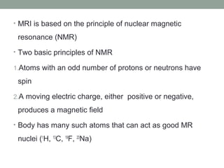


MRI BRAIN BASICS AND RADIOLOGICAL ANATOMY 1. MRI uses strong magnetic fields and radio waves to produce detailed images of the brain and detect abnormalities. It has largely replaced CT for evaluating many conditions due to its superior soft tissue contrast. 2. Different MRI sequences such as T1-weighted, T2-weighted, FLAIR and DWI highlight various tissues and pathologies based on their relaxation properties. T1 highlights anatomy while T2 highlights abnormalities like tumors and inflammation. 3. Key anatomical structures are clearly visualized on MRI slices through different levels of the brain. Axial slices progress from the brainstem to the cortex, while sagittal slices show deep midline structures


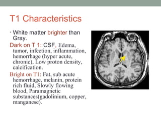
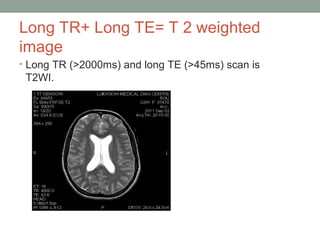
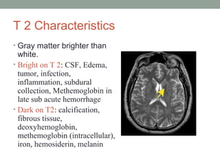
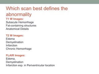
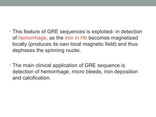
![CASE_PRESENTATION_ON_subdural_hematoma(SDH)[1 FINAL PPT]-1.pptx](https://cdn.slidesharecdn.com/ss_thumbnails/casepresentationonsubduralhematomasdh1finalppt-1-260129172522-d405d375-thumbnail.jpg?width=640&height=640&fit=bounds)


