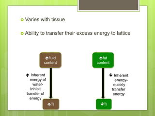
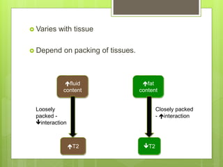
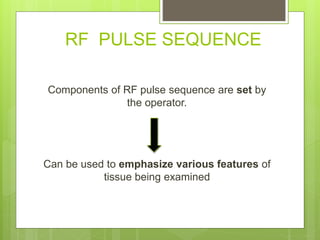
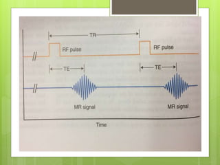
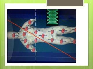
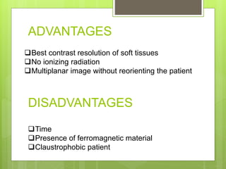
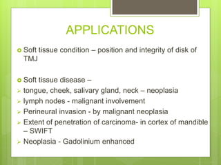
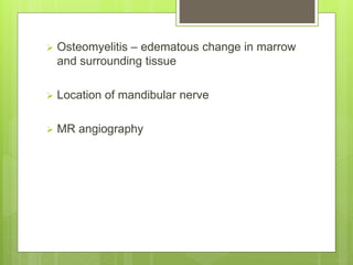
This is a presentation describing in brief regarding the physics behind MRI and it's application from dental point of view. It contains few videos as well.
4. PROTONS
Proton – spin – angular momentum
Spin – associated with charge – B generated in
nuclei – act as magnet– magnet dipole has
magnetic moment.
9. PRECESSION
Magnetic moment of H+ in a Bext – do not align
exactly with the direction of Bext
– TILT from a
position exactly parallel to Bext
10.
11. Bext – axis of spinning of H+ to wobble(precess) around
the lines of B ext.
This frequency of precession
• PRECESSION FREQUENCY/
• RESONANCE FREQUENCY/
• LARMOR FREQUENCY
Depends on
• Atom
• Strength of Bext- 0.1-4 Tesla
12. RESONANCE
Energy to energy –
provided by radiofrequency(RF) spectrum of
EM spectrum.
When RF = larmor frequency resonance.
13. COIL PATIENT RESONANCE
Spin up to spin
down
BH+ longitudinal
ses
RF
Causes the
H+ to
precess in
phase
BH+
transverse
14.
15.
16. Longer RF pulse –BH+ longitudinal
BH+ transverse
90 degree RF pulse/flip angle of 90 degree
Significant intensity and duration –
BH+ longitudinal sed to 0
22. BH+ longitudinal
returned to
original
Dephase
RELAXATION
RF turned
off
BH+
Transverse-
DECAY
T2 -
magnetic
moment of H+
interact with
each other
SPIN SPIN
RELAXATION
TIME
T1 – energy
absorbed by
lattice
SPIN –
LATTICE
REALAXATIO
N TIME
T1 and T2 RELAXATION
23. T1 RELAXATION TIME
It is an exponential process and the time
required for 63% of the magnetization to
return to equilibrium (the time constant) by
this transfer of energy.
24. Varies with tissue
Ability to transfer their excess energy to lattice
fluid
content
TI TI
fat
content
Inherent
energy of
water-
Inhibit
transfer of
energy
Inherent
energy-
quickly
transfer
energy
25. T2 RELAXATION TIME
Time constant that describes the exponential
rate of loss of transverse magnetization.
26. Varies with tissue
Depend on packing of tissues.
fluid
content
T2 T2
fat
content
Closely packed
- interaction
Loosely
packed -
interaction
28. RF PULSE SEQUENCE
Components of RF pulse sequence are set by
the operator.
Can be used to emphasize various features of
tissue being examined
29.
30. TR and TE
TR(REPETITION TIME) : duration between
repeat RF pulses.
TE (ECHO TIME) : time after application of
the RF pulse when the MR signal is read.
37. OTHER METHODS
STIR(short tau inversion recovery) – dec signal
from fat(FAT SATURATION) – better visualization
of adjacent structures.
FLAIR(fluid attenuation inversion recovery) - dec
signal from fluid – better visualization of
pathology.
CONTRAST AGENT – gadolinium.
It dec T1 of enhancing tissue – they appear
bright.
38. SCANNER GRADIENT
3 gradient coils
1. X-axis : left to right
2. Y-axis : anterior to posterior
3. Z-axis : head to toe
Intensity of Bext can be modified when gradient applied
Z gradient turned on
precessional frequency of H+ varies linearly along Bext
RF pulse applied – nuclei with RF = PF – resonate
Hence, desired slice selected.
Z gradient coil turned off – X(phase encoding) and Y(frequency
encoding) gradients are turned on – location of signal within xand y
is determined.
39.
40. ADVANTAGES
Time
Presence of ferromagnetic material
Claustrophobic patient
Best contrast resolution of soft tissues
No ionizing radiation
Multiplanar image without reorienting the patient
DISADVANTAGES
41. APPLICATIONS
Soft tissue condition – position and integrity of disk of
TMJ
Soft tissue disease –
tongue, cheek, salivary gland, neck – neoplasia
lymph nodes - malignant involvement
Perineural invasion - by malignant neoplasia
Extent of penetration of carcinoma- in cortex of mandible
– SWIFT
Neoplasia - Gadolinium enhanced
42. Osteomyelitis – edematous change in marrow
and surrounding tissue
Location of mandibular nerve
MR angiography
55. 1. RF pulse causes
a. B transverse
b. B longitudinal
c. Both
d. None
2. T1 weighted image is used for studying
a. Anatomy
b. pathology
3. T1 weighted image uses
a. Short TR, short TE
b. Long TR, long TE
c. Short TR, long TE
d. Long TR, short TE
56. 4. In T2 weighted image, tissue with long T2 (eg- TMJ fluid) appears
a. Bright
b. Dark
c. Both
d. None
5. Which diagnostic method would you prefer for diagnosing collection of effusion in
TMJ
a. T1
b. T2
c. Both
d. None
6. Which diagnostic method would you prefer for diagnosing disk displacement in
TMJ?
a. T1
b. T2
c. Both
d. none
57. 7. This is a case of lymphoepithelial cyst in right parotid
space, which is T2?
a. A
b. B
58. 8. MRI uses
a. Ionizing radiation
b. Non-ionizing radiation
c. Both
d. None
9. MRI is used for-
a. Neoplasia of soft tissue
b. Mallignant involvement of lymph nodes
c. Both
d. none
10. Contrast agent used is ________?