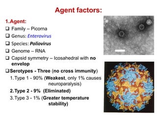
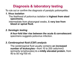
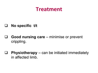
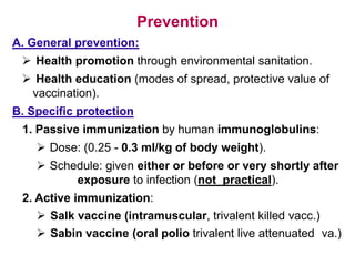
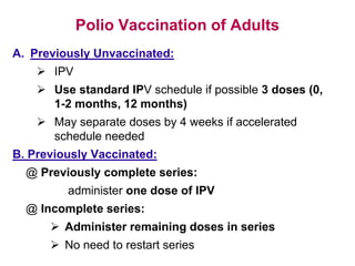
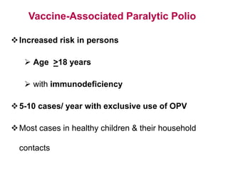
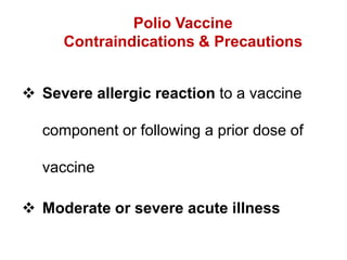
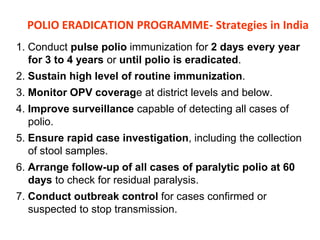
Rukhsar Khatoon
Elimination Vs Eradication
Poliomyelitis
Epidemiology & control measures
Introduction
๏ฑ Highly infectious disease caused by three serotypes
of poliovirus.
๏ฑ Primarily an infection of GIT, & about 1% get CNS
infection.
๏ฑ Spectrum of clinical manifestations:
Inapparent infection - non-specific febrile illness -
aseptic meningitis - paralytic disease - death.
๏ฑ Infection most often recognized by acute onset of
flaccid paralysis.
Problem
๏ฑ Pre-vaccination era โ€“ worldwide.
๏ฑ Since introduction of vaccine in 1954 โ€“ eliminated in
developed countries.
๏ฑ 1988 โ€“ world health assembly resolved to eradicate
polio globally.
๏ฑ In 1988 it was endemic 125 countries, by 2008 in 4
countries (India Pakistan, Afghanistan & Nigeria)
๏ฑ Since February 2012 โ€“ India polio free
Global Status 1988
Epidemiological determinants
EPIDEMIOLOGICAL TRIAD
ENVIRONMENT
(Rainy season, Poor sanitation)
AGENT HOST
(Poliovirus- 3 Serotypes) (Children, male)
Agent factors:
1.Agent:
๏ฑ Family โ€“ Picorna
๏ฑ Genus: Enterovirus
๏ฑ Species: Poliovirus
๏ฑ Genome โ€“ RNA
๏ฑ Capsid symmetry โ€“ Icosahedral with no
envelop
๏ฑSerotypes - Three (no cross immunity)
1.Type 1 - 90% (Weakest, only 1% causes
neuroparalysis)
2.Type 2 - 9% (Eliminated)
3.Type 3 - 1% (Greater temperature
stability)
Agent factors:
2. Reservoir of infection:
๏ƒ˜ Man only (clinical or sub-clinical (mostly))
๏ƒ˜ For every clinical cases โ€“ 1000 sub-clinical cases in
children & 75 in adults.
๏ƒ˜ No chronic carriers.
3. Infectious material:
๏ƒ˜ Faeces & oropharyngeal secretion of infected
persons.
4. Period of communicability:
๏ƒ˜ 7 to 10 days before & after onset of symptoms.
๏ƒ˜ In faeces virus may be excreted as long as 3 to 4
months.
Host factors
1. Age:
๏ƒ˜ All age groups
๏ƒ˜ Children more susceptible (more vulnerable in 6 months
to 3 years)
2. Sex:
๏ƒ˜ M/F ratio, 3 : 1
3. Risk factors:
๏ƒ˜ Fatigue, trauma, IM injection, Operation during
epidemics, DPT vaccine.
4. Immunity:
๏ƒ˜ Maternal antibody protect up to first 6 months.
๏ƒ˜ No cross immunity with other sero-types.
๏ƒ˜ Type 2 PV โ€“ most effective antigen.
Environmental factors
โ€ข Common in rainy season (June to
September)
โ€ข Over-crowiding & poor sanitation
favors
Sewage Drinking water
Sewage
๏ถ MODE OF TRANSMISSION:
A. Faeco-oral route (mainly in developing con.):
๏ƒ˜ Directly โ€“ by contaminated fingers,
๏ƒ˜ Indirectly โ€“ by contaminated water, milk, food,
flies, fomites.
B. Droplet infection:
In acute phase of dis.(virus in throat)
๏ถ INCUBATION PERIOD: 7 to 14 days( 3 - 35 days)
Pathology
Polio virus
Infect intestinal
epithelium
Replication of virus
in epithelial cells
Infect Peyr's patches &
secondary multiplication
Viremia
Antibody production
CNS infection
IgA secretory antibodies
Sequelae of polio infection
Polio infection
In apparent infection
(91 to 96%)
Clinical poliomyelitis
Abortive polio
(minor illness)
(4 to 8%)
Involvement of CNS
(major illness)
Paralytic polio (<1%) Non-paralytic
polio (2 to 10%)
Spinal polio
Bulbar polio
Bulbospinal polio
Clinical poliomyelitis
1. Inapparent (subclinical) infection
No clinical manifestations, but infection is associated
with acquired immunity, & carrier state.
2. Abortive polio (minor illness):
๏ƒ˜ Mild systemic manifestations for 1or 2 days only,
then clears up, giving immunity.
๏ƒ˜ Manifestations:
๏ฑ Moderate fever
๏ฑ Pharyngitis & sore throat
๏ฑ Vomiting, abdominal pain, & diarrhea.
Clinical poliomyelitis (cont.)
3. Involvement of the CNS (major illness):
A. Nonparalytic polio:
๏ฑ Manifested by:
๏ƒ˜ Fever, headache, nausea, vomiting, & abdominal pain.
๏ƒ˜ Pain & stiffness in the neck back & limbs may also
occur.
๏ฑ The case either recovers or passes to the paralytic
stage.
Clinical poliomyelitis (cont.)
B. Paralytic poliomyelitis:
๏ฑ Paralysis usually appears around 7-10 days from
onset of disease.
๏ฑ Presented with:
๏ƒ˜ Fever, anorexia, nausea, vomiting, headache, sore
throat, constipation, abdominal pain.
๏ƒ˜ May be sign of meningeal irritation.
๏ƒ˜ Tripod sign (difficulties in sitting & sits by supporting
hands at back & by partially flexing the hip & knees.
Clinical poliomyelitis (cont.)
๏ฑ Different paralytic manifestations according to
the part of the CNS involved, with destruction
of the motor nerve cells, but not the sensory
nerve cells (i. e. no sensory loss).
๏ฑ Paralysis reach maximum in less than 4 days
in majority of cases (4 โ€“ 7 days).
๏ฑ Forms: 1. Spinal, 2. Bulbar, 3. Bulbospinal.
1. Spinal polio
๏ƒ˜ Different spinal nerves are involved, due to injury
of the anterior horn cells of the spinal cord,
causing tenderness, weakness, & flaccid paralysis
of the corresponding striated muscles
๏ƒ˜ The lower limbs are the most commonly affected.
๏ƒ˜ Paralysis:
o Characterized as descending i.e. start at hip &
then moving down to the distal parts of the
extremity.
o Asymmetrical patchy,
o Muscle strength varies in different muscle groups of
different limbs.
o Proximal muscle groups are more involved than
distal one.
2. Bulbar polio
๏ƒ˜ Nuclei of the cranial nerves are involved, causing
weakness of the supplied muscles, & maybe
encephalitis.
๏ƒ˜ Bulbar manifestations include dysphagia, nasal voice,
fluid regurgitation from the nose, difficult chewing,
facial weakness & diplopia
๏ƒ˜ Paralysis of the muscles of respiration is the most serious
life-threatening manifestation.
3. Bulbospinal polio
๏ƒ˜ Combination of both spinal & bulbar forms
Among children who are paralyzed by
polio:
๏ƒ˜ 30% make a full recovery
๏ƒ˜ 30% are left with mild paralysis
๏ƒ˜ 30% have medium to severe
paralysis
๏ƒ˜ 10% die
Complications & case fatality
๏ถ Respiratory complications: pneumonia, pulmonary
edema
๏ถ Cardiovascular complications: myocarditis, cor-
pulmonale.
๏ถ Late complications: soft tissue & bone deformities,
osteoporosis, chronic distension of the colon.
๏ถ Case fatality: varies from 1% to 10% according to the
form of disease (higher in bulbar), complications &
age (fatality increases with age).
Cold Chain Vs Reverse Cold chain
Diagnosis & laboratory testing
To rule out or confirm the diagnosis of paralytic poliomyelitis.
1. Virus isolation
The likelihood of poliovirus isolation is highest from stool
specimens,
intermediate from pharyngeal swabs, & very low from
blood or spinal fluid.
2. Serologic testing
A four-fold titer rise between the acute & convalescent
specimens suggests poliovirus infection.
3. Cerebrospinal fluid (CSF) analysis
The cerebrospinal fluid usually contains an increased
number of leukocytesโ€”from 10 to 200 cells/mm3
(primarily lymphocytes) & a mildly elevated protein, from
40 to 50 mg/100 ml.
Treatment
๏ฑ No specific t/t
๏ฑ Good nursing care โ€“ minimise or prevent
crippling.
๏ฑ Physiotherapy โ€“ can be initiated immediately
in affected limb.
Prevention
A. General prevention:
๏ƒ˜ Health promotion through environmental sanitation.
๏ƒ˜ Health education (modes of spread, protective value of
vaccination).
B. Specific protection
1. Passive immunization by human immunoglobulins:
๏ƒ˜ Dose: (0.25 - 0.3 ml/kg of body weight).
๏ƒ˜ Schedule: given either or before or very shortly after
exposure to infection (not practical).
2. Active immunization:
๏ƒ˜ Salk vaccine (intramuscular, trivalent killed vacc.)
๏ƒ˜ Sabin vaccine (oral polio trivalent live attenuated va.)
Inactivated Polio Vaccine
๏ƒ˜ Contains 3 serotypes & inactivated with formaldehyde
๏ƒ˜ Contains 2-phenoxyethanol, neomycin, streptomycin,
polymyxin B
A. Classical IPV:
๏ƒ˜ Contain 20, 2 & 4D antigen unit of type 1, 2 & 3
serotype respectively.
๏ƒ˜ Dose: Primary 4 doses at interval of 1 - 2 months &
Booster โ€“ every 5 years
B. Improved IPV:
๏ƒ˜ Contain 40, 8 & 32D antigen unit of type 1, 2 & 3
serotype respectively
๏ƒ˜ 100% effective after 2nd dose.
๏ƒ˜ Can be combined with DPT.
Oral Polio Vaccine by Sabin in 1957
๏ถ Contains 3 serotypes of vaccine virus
๏ถ Live attenuated virus grown on monkey kidney (Vero) cells
๏ถ Contain:
1. Over 3LakhTCDI 50 of type 1PV
2. Over 1LakhTCDI 50 of type 2PV
3. Over 3LakhTCDI 50 of type 3PV
๏ถ Contains neomycin & streptomycin
๏ถ Vaccine virus shed in stool for up to 6 weeks following
vaccination
๏ถ Dose: 2 drops/dose
1. Primary 4 doses (at 0, 6, 10 & 14 weeks)
2. Booster โ€“ at age of 16 to 24 months.
VACCINE VIAL MONITOR
Salk versus Sabin vaccine
IPV (Salk)
1. Killed formolised virus
2. Given SC or IM
3. Induces circulating antibodies, but
not local (intestinal immunity)
4. Prevents paralysis but does not
prevent re-infection.
5. Not useful in controlling epidemics
6. More difficult to manufacture & is
relatively costly
7. Does not require stringent
conditions during storage &
transportation. Has a longer shelf
life.
OPV (Sabin
1. Live attenuated virus
2. Given orally
3. Immunity is both humoral and
intestinal. induces antibody quickly
4. Prevents paralysis and prevents re-
infection
5. Can be effectively used in
controlling epidemics.
6. Easy to manufacture and is cheaper
7. Requires to be stored &
transported at subzero
temperatures, & is damaged easily.
Polio Vaccination of Adults
A. Previously Unvaccinated:
๏ƒ˜ IPV
๏ƒ˜ Use standard IPV schedule if possible 3 doses (0,
1-2 months, 12 months)
๏ƒ˜ May separate doses by 4 weeks if accelerated
schedule needed
B. Previously Vaccinated:
@ Previously complete series:
administer one dose of IPV
@ Incomplete series:
๏ƒ˜ Administer remaining doses in series
๏ƒ˜ No need to restart series
Polio Vaccine Adverse Reactions
โ€ข Rare local reactions (IPV)
โ€ข No serious reactions to IPV have been
documented
โ€ข Paralytic poliomyelitis (OPV)
Vaccine-Associated Paralytic Polio
๏ถIncreased risk in persons
๏ƒ˜ Age >18 years
๏ƒ˜ with immunodeficiency
๏ถ5-10 cases/ year with exclusive use of OPV
๏ถMost cases in healthy children & their household
contacts
Polio Vaccine
Contraindications & Precautions
๏ถ Severe allergic reaction to a vaccine
component or following a prior dose of
vaccine
๏ถ Moderate or severe acute illness
POLIO ERADICATION PROGRAMME- Strategies in India
1. Conduct pulse polio immunization for 2 days every year
for 3 to 4 years or until polio is eradicated.
2. Sustain high level of routine immunization.
3. Monitor OPV coverage at district levels and below.
4. Improve surveillance capable of detecting all cases of
polio.
5. Ensure rapid case investigation, including the collection
of stool samples.
6. Arrange follow-up of all cases of paralytic polio at 60
days to check for residual paralysis.
7. Conduct outbreak control for cases confirmed or
suspected to stop transmission.
GOAL:
Immunize every child against polio until polio
transmission has stopped, so that the world can be
certified polio-free.
Objective:
Replacement of wild PV with Vaccine virus in the
community
Strategy:
Intensified pulse polio immunization (From 1996-97
to all children under the age of 5 years were
covered).
AFP Surveillance
๏ถ Acute flaccid paralysis is defined as:
Any case of AFP in a child aged <15 years, or any
case of paralytic illness in a person of any age when
polio is suspected.
๏ƒ˜ Acute: rapid progression of paralysis from onset to
maximum paralysis
๏ƒ˜ Flaccid: loss of muscle tone, โ€œfloppyโ€ โ€“ as opposed
to spastic or rigid
๏ƒ˜ Paralysis: weakness, loss of voluntary movement
๏ถ Any case meeting this definition undergoes a thorough
investigation to determine if the paralysis is caused by
polio.
COMPONENTS OF AFP SURVEILANCE
1. The AFP surveillance network & case notification
2. Case & laboratory investigation
3. Outbreak response & active case search in the
community
4. 60-day follow-up, cross-notification & tracking of
cases
5. Data management & case classification
6. Virologic case classification scheme
7. Surveillance performance indicators
Sample collectionA
Collection of 2 adequate stool samples from all
cases.
1. Collected within 14 days of paralysis onset & at
least 24 hours apart;
2. Adequate volume (8-10g)
3. Arrives at a WHO-accredited laboratory in good
condition (ie, no desiccation, no leakage), with
adequate documentation & evidence of cold-chain
maintenance.
Thank You

poliomyelitis 12 04-2016

  • 1.
  • 4.
  • 5.
  • 6.
    Introduction ๏ฑ Highly infectiousdisease caused by three serotypes of poliovirus. ๏ฑ Primarily an infection of GIT, & about 1% get CNS infection. ๏ฑ Spectrum of clinical manifestations: Inapparent infection - non-specific febrile illness - aseptic meningitis - paralytic disease - death. ๏ฑ Infection most often recognized by acute onset of flaccid paralysis.
  • 7.
    Problem ๏ฑ Pre-vaccination eraโ€“ worldwide. ๏ฑ Since introduction of vaccine in 1954 โ€“ eliminated in developed countries. ๏ฑ 1988 โ€“ world health assembly resolved to eradicate polio globally. ๏ฑ In 1988 it was endemic 125 countries, by 2008 in 4 countries (India Pakistan, Afghanistan & Nigeria) ๏ฑ Since February 2012 โ€“ India polio free
  • 8.
  • 10.
  • 11.
    EPIDEMIOLOGICAL TRIAD ENVIRONMENT (Rainy season,Poor sanitation) AGENT HOST (Poliovirus- 3 Serotypes) (Children, male)
  • 12.
    Agent factors: 1.Agent: ๏ฑ Familyโ€“ Picorna ๏ฑ Genus: Enterovirus ๏ฑ Species: Poliovirus ๏ฑ Genome โ€“ RNA ๏ฑ Capsid symmetry โ€“ Icosahedral with no envelop ๏ฑSerotypes - Three (no cross immunity) 1.Type 1 - 90% (Weakest, only 1% causes neuroparalysis) 2.Type 2 - 9% (Eliminated) 3.Type 3 - 1% (Greater temperature stability)
  • 13.
    Agent factors: 2. Reservoirof infection: ๏ƒ˜ Man only (clinical or sub-clinical (mostly)) ๏ƒ˜ For every clinical cases โ€“ 1000 sub-clinical cases in children & 75 in adults. ๏ƒ˜ No chronic carriers. 3. Infectious material: ๏ƒ˜ Faeces & oropharyngeal secretion of infected persons. 4. Period of communicability: ๏ƒ˜ 7 to 10 days before & after onset of symptoms. ๏ƒ˜ In faeces virus may be excreted as long as 3 to 4 months.
  • 14.
    Host factors 1. Age: ๏ƒ˜All age groups ๏ƒ˜ Children more susceptible (more vulnerable in 6 months to 3 years) 2. Sex: ๏ƒ˜ M/F ratio, 3 : 1 3. Risk factors: ๏ƒ˜ Fatigue, trauma, IM injection, Operation during epidemics, DPT vaccine. 4. Immunity: ๏ƒ˜ Maternal antibody protect up to first 6 months. ๏ƒ˜ No cross immunity with other sero-types. ๏ƒ˜ Type 2 PV โ€“ most effective antigen.
  • 15.
    Environmental factors โ€ข Commonin rainy season (June to September) โ€ข Over-crowiding & poor sanitation favors Sewage Drinking water Sewage
  • 16.
    ๏ถ MODE OFTRANSMISSION: A. Faeco-oral route (mainly in developing con.): ๏ƒ˜ Directly โ€“ by contaminated fingers, ๏ƒ˜ Indirectly โ€“ by contaminated water, milk, food, flies, fomites. B. Droplet infection: In acute phase of dis.(virus in throat) ๏ถ INCUBATION PERIOD: 7 to 14 days( 3 - 35 days)
  • 17.
    Pathology Polio virus Infect intestinal epithelium Replicationof virus in epithelial cells Infect Peyr's patches & secondary multiplication Viremia Antibody production CNS infection IgA secretory antibodies
  • 18.
    Sequelae of polioinfection Polio infection In apparent infection (91 to 96%) Clinical poliomyelitis Abortive polio (minor illness) (4 to 8%) Involvement of CNS (major illness) Paralytic polio (<1%) Non-paralytic polio (2 to 10%) Spinal polio Bulbar polio Bulbospinal polio
  • 19.
    Clinical poliomyelitis 1. Inapparent(subclinical) infection No clinical manifestations, but infection is associated with acquired immunity, & carrier state. 2. Abortive polio (minor illness): ๏ƒ˜ Mild systemic manifestations for 1or 2 days only, then clears up, giving immunity. ๏ƒ˜ Manifestations: ๏ฑ Moderate fever ๏ฑ Pharyngitis & sore throat ๏ฑ Vomiting, abdominal pain, & diarrhea.
  • 20.
    Clinical poliomyelitis (cont.) 3.Involvement of the CNS (major illness): A. Nonparalytic polio: ๏ฑ Manifested by: ๏ƒ˜ Fever, headache, nausea, vomiting, & abdominal pain. ๏ƒ˜ Pain & stiffness in the neck back & limbs may also occur. ๏ฑ The case either recovers or passes to the paralytic stage.
  • 21.
    Clinical poliomyelitis (cont.) B.Paralytic poliomyelitis: ๏ฑ Paralysis usually appears around 7-10 days from onset of disease. ๏ฑ Presented with: ๏ƒ˜ Fever, anorexia, nausea, vomiting, headache, sore throat, constipation, abdominal pain. ๏ƒ˜ May be sign of meningeal irritation. ๏ƒ˜ Tripod sign (difficulties in sitting & sits by supporting hands at back & by partially flexing the hip & knees.
  • 23.
    Clinical poliomyelitis (cont.) ๏ฑDifferent paralytic manifestations according to the part of the CNS involved, with destruction of the motor nerve cells, but not the sensory nerve cells (i. e. no sensory loss). ๏ฑ Paralysis reach maximum in less than 4 days in majority of cases (4 โ€“ 7 days). ๏ฑ Forms: 1. Spinal, 2. Bulbar, 3. Bulbospinal.
  • 24.
    1. Spinal polio ๏ƒ˜Different spinal nerves are involved, due to injury of the anterior horn cells of the spinal cord, causing tenderness, weakness, & flaccid paralysis of the corresponding striated muscles ๏ƒ˜ The lower limbs are the most commonly affected. ๏ƒ˜ Paralysis: o Characterized as descending i.e. start at hip & then moving down to the distal parts of the extremity. o Asymmetrical patchy, o Muscle strength varies in different muscle groups of different limbs. o Proximal muscle groups are more involved than distal one.
  • 25.
    2. Bulbar polio ๏ƒ˜Nuclei of the cranial nerves are involved, causing weakness of the supplied muscles, & maybe encephalitis. ๏ƒ˜ Bulbar manifestations include dysphagia, nasal voice, fluid regurgitation from the nose, difficult chewing, facial weakness & diplopia ๏ƒ˜ Paralysis of the muscles of respiration is the most serious life-threatening manifestation. 3. Bulbospinal polio ๏ƒ˜ Combination of both spinal & bulbar forms
  • 26.
    Among children whoare paralyzed by polio: ๏ƒ˜ 30% make a full recovery ๏ƒ˜ 30% are left with mild paralysis ๏ƒ˜ 30% have medium to severe paralysis ๏ƒ˜ 10% die
  • 27.
    Complications & casefatality ๏ถ Respiratory complications: pneumonia, pulmonary edema ๏ถ Cardiovascular complications: myocarditis, cor- pulmonale. ๏ถ Late complications: soft tissue & bone deformities, osteoporosis, chronic distension of the colon. ๏ถ Case fatality: varies from 1% to 10% according to the form of disease (higher in bulbar), complications & age (fatality increases with age).
  • 28.
    Cold Chain VsReverse Cold chain
  • 29.
    Diagnosis & laboratorytesting To rule out or confirm the diagnosis of paralytic poliomyelitis. 1. Virus isolation The likelihood of poliovirus isolation is highest from stool specimens, intermediate from pharyngeal swabs, & very low from blood or spinal fluid. 2. Serologic testing A four-fold titer rise between the acute & convalescent specimens suggests poliovirus infection. 3. Cerebrospinal fluid (CSF) analysis The cerebrospinal fluid usually contains an increased number of leukocytesโ€”from 10 to 200 cells/mm3 (primarily lymphocytes) & a mildly elevated protein, from 40 to 50 mg/100 ml.
  • 30.
    Treatment ๏ฑ No specifict/t ๏ฑ Good nursing care โ€“ minimise or prevent crippling. ๏ฑ Physiotherapy โ€“ can be initiated immediately in affected limb.
  • 31.
    Prevention A. General prevention: ๏ƒ˜Health promotion through environmental sanitation. ๏ƒ˜ Health education (modes of spread, protective value of vaccination). B. Specific protection 1. Passive immunization by human immunoglobulins: ๏ƒ˜ Dose: (0.25 - 0.3 ml/kg of body weight). ๏ƒ˜ Schedule: given either or before or very shortly after exposure to infection (not practical). 2. Active immunization: ๏ƒ˜ Salk vaccine (intramuscular, trivalent killed vacc.) ๏ƒ˜ Sabin vaccine (oral polio trivalent live attenuated va.)
  • 32.
    Inactivated Polio Vaccine ๏ƒ˜Contains 3 serotypes & inactivated with formaldehyde ๏ƒ˜ Contains 2-phenoxyethanol, neomycin, streptomycin, polymyxin B A. Classical IPV: ๏ƒ˜ Contain 20, 2 & 4D antigen unit of type 1, 2 & 3 serotype respectively. ๏ƒ˜ Dose: Primary 4 doses at interval of 1 - 2 months & Booster โ€“ every 5 years B. Improved IPV: ๏ƒ˜ Contain 40, 8 & 32D antigen unit of type 1, 2 & 3 serotype respectively ๏ƒ˜ 100% effective after 2nd dose. ๏ƒ˜ Can be combined with DPT.
  • 33.
    Oral Polio Vaccineby Sabin in 1957 ๏ถ Contains 3 serotypes of vaccine virus ๏ถ Live attenuated virus grown on monkey kidney (Vero) cells ๏ถ Contain: 1. Over 3LakhTCDI 50 of type 1PV 2. Over 1LakhTCDI 50 of type 2PV 3. Over 3LakhTCDI 50 of type 3PV ๏ถ Contains neomycin & streptomycin ๏ถ Vaccine virus shed in stool for up to 6 weeks following vaccination ๏ถ Dose: 2 drops/dose 1. Primary 4 doses (at 0, 6, 10 & 14 weeks) 2. Booster โ€“ at age of 16 to 24 months.
  • 34.
  • 35.
    Salk versus Sabinvaccine IPV (Salk) 1. Killed formolised virus 2. Given SC or IM 3. Induces circulating antibodies, but not local (intestinal immunity) 4. Prevents paralysis but does not prevent re-infection. 5. Not useful in controlling epidemics 6. More difficult to manufacture & is relatively costly 7. Does not require stringent conditions during storage & transportation. Has a longer shelf life. OPV (Sabin 1. Live attenuated virus 2. Given orally 3. Immunity is both humoral and intestinal. induces antibody quickly 4. Prevents paralysis and prevents re- infection 5. Can be effectively used in controlling epidemics. 6. Easy to manufacture and is cheaper 7. Requires to be stored & transported at subzero temperatures, & is damaged easily.
  • 36.
    Polio Vaccination ofAdults A. Previously Unvaccinated: ๏ƒ˜ IPV ๏ƒ˜ Use standard IPV schedule if possible 3 doses (0, 1-2 months, 12 months) ๏ƒ˜ May separate doses by 4 weeks if accelerated schedule needed B. Previously Vaccinated: @ Previously complete series: administer one dose of IPV @ Incomplete series: ๏ƒ˜ Administer remaining doses in series ๏ƒ˜ No need to restart series
  • 37.
    Polio Vaccine AdverseReactions โ€ข Rare local reactions (IPV) โ€ข No serious reactions to IPV have been documented โ€ข Paralytic poliomyelitis (OPV)
  • 38.
    Vaccine-Associated Paralytic Polio ๏ถIncreasedrisk in persons ๏ƒ˜ Age >18 years ๏ƒ˜ with immunodeficiency ๏ถ5-10 cases/ year with exclusive use of OPV ๏ถMost cases in healthy children & their household contacts
  • 39.
    Polio Vaccine Contraindications &Precautions ๏ถ Severe allergic reaction to a vaccine component or following a prior dose of vaccine ๏ถ Moderate or severe acute illness
  • 40.
    POLIO ERADICATION PROGRAMME-Strategies in India 1. Conduct pulse polio immunization for 2 days every year for 3 to 4 years or until polio is eradicated. 2. Sustain high level of routine immunization. 3. Monitor OPV coverage at district levels and below. 4. Improve surveillance capable of detecting all cases of polio. 5. Ensure rapid case investigation, including the collection of stool samples. 6. Arrange follow-up of all cases of paralytic polio at 60 days to check for residual paralysis. 7. Conduct outbreak control for cases confirmed or suspected to stop transmission.
  • 41.
    GOAL: Immunize every childagainst polio until polio transmission has stopped, so that the world can be certified polio-free. Objective: Replacement of wild PV with Vaccine virus in the community Strategy: Intensified pulse polio immunization (From 1996-97 to all children under the age of 5 years were covered).
  • 43.
    AFP Surveillance ๏ถ Acuteflaccid paralysis is defined as: Any case of AFP in a child aged <15 years, or any case of paralytic illness in a person of any age when polio is suspected. ๏ƒ˜ Acute: rapid progression of paralysis from onset to maximum paralysis ๏ƒ˜ Flaccid: loss of muscle tone, โ€œfloppyโ€ โ€“ as opposed to spastic or rigid ๏ƒ˜ Paralysis: weakness, loss of voluntary movement ๏ถ Any case meeting this definition undergoes a thorough investigation to determine if the paralysis is caused by polio.
  • 44.
    COMPONENTS OF AFPSURVEILANCE 1. The AFP surveillance network & case notification 2. Case & laboratory investigation 3. Outbreak response & active case search in the community 4. 60-day follow-up, cross-notification & tracking of cases 5. Data management & case classification 6. Virologic case classification scheme 7. Surveillance performance indicators
  • 45.
    Sample collectionA Collection of2 adequate stool samples from all cases. 1. Collected within 14 days of paralysis onset & at least 24 hours apart; 2. Adequate volume (8-10g) 3. Arrives at a WHO-accredited laboratory in good condition (ie, no desiccation, no leakage), with adequate documentation & evidence of cold-chain maintenance.
  • 48.