Physiology of Parathyroid glands
Outline :
- Location of Parathyroid glands.
- Who discovered the glands.
- Some info. about it.
- Parathyroid hormone.
- Histology of the gland.
- PTH biosynthesis.
- The calcium-sensing receptors (CaSR)
- Why Calcium is so Important?
- Calcitonin
- vitamin D
-Metabolic bone diseases (Hypercalcaemia and hypocalcaemia)
Parathyroid hormone by Dr. Amruta Nitin Kumbhar, Asst. Professor Dept. of Phy...Physiology Dept
FUNCTIONAL ANATOMY OF PARATHYROID GLANDS
Histological structure
STRUCTURE, SYNTHESIS AND SECRETION OF PTH
REGULATION OF PTH SECRETION
MECHANISM OF ACTION AND ACTIONS OF PTH
Applied physiology
Parathyroid hormone (The Guyton and Hall physiology)Maryam Fida
Parathyroid hormone
Calcium salts in bone provide structural integrity of the skeleton
Calcium ions in extracellular and cellular fluids is essential to normal function of a host of biochemical processes
Neuoromuscular excitability
Blood coagulation
Hormonal secretion
Enzymatic regulation
The important role that calcium plays in so many processes dictates that its concentration, both extracellulary and intracellulary, be maintained within a very narrow range.
Normal level of calcium is about 9.4 mg/dl.
0.1 % extracellular fluid
1 % stored in cells (mitochondria and ER)
99% stored in bones in hydroxyapatite crystals. Very little Ca2+ can be released from the bone– though it is the major reservoir of Ca2+ in the body.
Calcium in Plasma is present in three forms:
1. Ionized and diffusible calcium 50%
2. Protein-bound calcium 41% non diffusible form
90% bound to albumin
Remainder bound to globulins
3. Calcium complexed to serum constituents 9%
Citrate and phosphate
a brief on thyroid gland covering following titles:
Introduction
Anatomy and physiology of thyroid gland
Synthesis of thyroid hormones
Regulation
Mechanism of action
Biological function
LOCATION: WALL OF GUT
NEURONS: 100 MILLIONS
GIT MOVEMENTS AND SECRETIONS
COMPOSED: TWO PLEXUSES
OUTER PLEXUS (MYENTERIC AND AUERBACH'S PLEXUS)
INNER PLEXUS (MEISSNER'S PLEXUS AND SUBMUCOSAL PLEXUS)
MYENTERIC PLEXUS
GI MOVEMENTS
SUBMUCOSAL PLEXUS
SECRETION AND LOCAL BLOOD FLOW
Short and brief presentation of anatomy, physiology , disorder and management of parathyroid glands.
management of MEN syndrome, hyper and hypoparathyroidism.
disorder of calcium metabolism like tetany,.
surgical steps of parathyroidectomy with indication and complications
This presentation is about Parathyroid Disorders which are hypo and hyperparathyroidism and their relationship to teeth and oral cavity including oral and dental manifestation of these disorders , and correct management patients seeking dental care with these disorders.
Parathyroid hormone (PTH), parathormone or parathyrin, is secreted by the chief cells of the parathyroid glands as a polypeptide containing 84 amino acids. It acts to increase the concentration of calcium (Ca2+) in the blood, whereas calcitonin (a hormone produced by the parafollicular cells (C cells) of the thyroid gland) acts to decrease calcium concentration. PTH acts to increase the concentration of calcium in the blood by acting upon the parathyroid hormone 1 receptor (high levels in bone and kidney) and the parathyroid hormone 2 receptor (high levels in the central nervous system, pancreas, testis, and placenta).PTH half-life is approximately 4 minutes. It has a molecular mass of 9.4 kDa.
Hyperparathyroidism exists in three different forms: primary, secondary and tertiary. Primary hyperparathyroidism (pHPT) is the most frequent pathological condition of the parathyroid glands and one of the most frequent endocrine disorders overall. The most probable location of parathyroid gland is posterior to the thyroid gland. The parathyroid glands produce parathyroid hormone (PTH), which is important for maintaining calcium, phosphate and vitamin D homeostasis, and ultimately bone health.
Primary hyperparathyroidism is characterized by increased production and secretion of parathyroid hormone. This condition causes nephrocalcinosis, urolithiasis, osteoporosis, gastrointestinal disturbances, neuromuscular manifestation and neuropsychiatric disorders. Parathyroidectomy is the only curative treatment for pHPT. pHPT is typically caused by a solitary parathyroid adenoma (80%-90%), hyperplasia (10%) and less frequently parathyroid carcinoma (5%).
Secondary hyperparathyroidism develops as a consequent to a chronic hypocalcemic condition that can be caused by renal failure, gastroinstinal malabsorption, dietary rickets and ingestion of drugs. Secondary hyperparathyroidism is a frequent and serious complication in haemodialysis patients. Tertiary hyperparathyroidism is a condition where parathyroid hyperplasia, secondary to chronic hypocalcemia, becomes autonomous with development of hypercalcemia. Tertiary hyperparathyroidism is used to designate hyperparathyroidism that persists or develops after renal transplantation.
Localization of hyperfunctioning parathyroid tissue (adenomas or hyperplasia) in primary hyperparathyroidism is useful before surgery to help the surgeon localize the lesion, thus shortening the time of the procedure. Parathyroid glands can be imaged with multiple modalities, including scintigraphy, high-resolution ultrasonograhy, thin-section CT and MRI. Parathyroid scintigraphy may also be indicated for localization of hyperfunctioning parathyroid tissue in patients with persistent or
recurrent disease. For this situation scintigraphy is superior to any other radiological modalities, including MRI, CT scan, ultrasonography combined with needle aspiration and also some invasive techniques like arteriography, selective venography and mediastinoscopy.
The prevalence of well-documented, permanent adrenal insufficiency is 5 in 10,000 in the general population. Hypothalamic-pituitary origin of disease is most frequent, with a prevalence of 3 in 10,000, whereas primary adrenal insufficiency has a prevalence of 2 in 10,000. Approximately one-half of the latter cases are acquired, mostly caused by autoimmune destruction of the adrenal glands; the other one-half are genetic, most commonly caused by distinct enzymatic blocks in adrenal steroidogenesis affecting glucocorticoid synthesis (i.e. congenital adrenal hyperplasia.)
Adrenal insufficiency arising from suppression of the HPA axis as a consequence of exogenous glucocorticoid treatment is much more common, occurring in 0.5–2% of the population in developed countries.
Hyperparahyroidsm is an endocrinal disorder majorly affecting the parathyroid glands which secrete parathyroid hormone and calcitonin.
A condition characterised by excessive secretion of calcium in blood and Bone resorption and inanbility to metabolise calcium in blood.
The Indian Dental Academy is the Leader in continuing dental education , training dentists in all aspects of dentistry and
offering a wide range of dental certified courses in different formats.
Nutraceutical market, scope and growth: Herbal drug technologyLokesh Patil
As consumer awareness of health and wellness rises, the nutraceutical market—which includes goods like functional meals, drinks, and dietary supplements that provide health advantages beyond basic nutrition—is growing significantly. As healthcare expenses rise, the population ages, and people want natural and preventative health solutions more and more, this industry is increasing quickly. Further driving market expansion are product formulation innovations and the use of cutting-edge technology for customized nutrition. With its worldwide reach, the nutraceutical industry is expected to keep growing and provide significant chances for research and investment in a number of categories, including vitamins, minerals, probiotics, and herbal supplements.
Seminar of U.V. Spectroscopy by SAMIR PANDASAMIR PANDA
Spectroscopy is a branch of science dealing the study of interaction of electromagnetic radiation with matter.
Ultraviolet-visible spectroscopy refers to absorption spectroscopy or reflect spectroscopy in the UV-VIS spectral region.
Ultraviolet-visible spectroscopy is an analytical method that can measure the amount of light received by the analyte.
(May 29th, 2024) Advancements in Intravital Microscopy- Insights for Preclini...Scintica Instrumentation
Intravital microscopy (IVM) is a powerful tool utilized to study cellular behavior over time and space in vivo. Much of our understanding of cell biology has been accomplished using various in vitro and ex vivo methods; however, these studies do not necessarily reflect the natural dynamics of biological processes. Unlike traditional cell culture or fixed tissue imaging, IVM allows for the ultra-fast high-resolution imaging of cellular processes over time and space and were studied in its natural environment. Real-time visualization of biological processes in the context of an intact organism helps maintain physiological relevance and provide insights into the progression of disease, response to treatments or developmental processes.
In this webinar we give an overview of advanced applications of the IVM system in preclinical research. IVIM technology is a provider of all-in-one intravital microscopy systems and solutions optimized for in vivo imaging of live animal models at sub-micron resolution. The system’s unique features and user-friendly software enables researchers to probe fast dynamic biological processes such as immune cell tracking, cell-cell interaction as well as vascularization and tumor metastasis with exceptional detail. This webinar will also give an overview of IVM being utilized in drug development, offering a view into the intricate interaction between drugs/nanoparticles and tissues in vivo and allows for the evaluation of therapeutic intervention in a variety of tissues and organs. This interdisciplinary collaboration continues to drive the advancements of novel therapeutic strategies.
The increased availability of biomedical data, particularly in the public domain, offers the opportunity to better understand human health and to develop effective therapeutics for a wide range of unmet medical needs. However, data scientists remain stymied by the fact that data remain hard to find and to productively reuse because data and their metadata i) are wholly inaccessible, ii) are in non-standard or incompatible representations, iii) do not conform to community standards, and iv) have unclear or highly restricted terms and conditions that preclude legitimate reuse. These limitations require a rethink on data can be made machine and AI-ready - the key motivation behind the FAIR Guiding Principles. Concurrently, while recent efforts have explored the use of deep learning to fuse disparate data into predictive models for a wide range of biomedical applications, these models often fail even when the correct answer is already known, and fail to explain individual predictions in terms that data scientists can appreciate. These limitations suggest that new methods to produce practical artificial intelligence are still needed.
In this talk, I will discuss our work in (1) building an integrative knowledge infrastructure to prepare FAIR and "AI-ready" data and services along with (2) neurosymbolic AI methods to improve the quality of predictions and to generate plausible explanations. Attention is given to standards, platforms, and methods to wrangle knowledge into simple, but effective semantic and latent representations, and to make these available into standards-compliant and discoverable interfaces that can be used in model building, validation, and explanation. Our work, and those of others in the field, creates a baseline for building trustworthy and easy to deploy AI models in biomedicine.
Bio
Dr. Michel Dumontier is the Distinguished Professor of Data Science at Maastricht University, founder and executive director of the Institute of Data Science, and co-founder of the FAIR (Findable, Accessible, Interoperable and Reusable) data principles. His research explores socio-technological approaches for responsible discovery science, which includes collaborative multi-modal knowledge graphs, privacy-preserving distributed data mining, and AI methods for drug discovery and personalized medicine. His work is supported through the Dutch National Research Agenda, the Netherlands Organisation for Scientific Research, Horizon Europe, the European Open Science Cloud, the US National Institutes of Health, and a Marie-Curie Innovative Training Network. He is the editor-in-chief for the journal Data Science and is internationally recognized for his contributions in bioinformatics, biomedical informatics, and semantic technologies including ontologies and linked data.
Richard's aventures in two entangled wonderlandsRichard Gill
Since the loophole-free Bell experiments of 2020 and the Nobel prizes in physics of 2022, critics of Bell's work have retreated to the fortress of super-determinism. Now, super-determinism is a derogatory word - it just means "determinism". Palmer, Hance and Hossenfelder argue that quantum mechanics and determinism are not incompatible, using a sophisticated mathematical construction based on a subtle thinning of allowed states and measurements in quantum mechanics, such that what is left appears to make Bell's argument fail, without altering the empirical predictions of quantum mechanics. I think however that it is a smoke screen, and the slogan "lost in math" comes to my mind. I will discuss some other recent disproofs of Bell's theorem using the language of causality based on causal graphs. Causal thinking is also central to law and justice. I will mention surprising connections to my work on serial killer nurse cases, in particular the Dutch case of Lucia de Berk and the current UK case of Lucy Letby.
Introduction:
RNA interference (RNAi) or Post-Transcriptional Gene Silencing (PTGS) is an important biological process for modulating eukaryotic gene expression.
It is highly conserved process of posttranscriptional gene silencing by which double stranded RNA (dsRNA) causes sequence-specific degradation of mRNA sequences.
dsRNA-induced gene silencing (RNAi) is reported in a wide range of eukaryotes ranging from worms, insects, mammals and plants.
This process mediates resistance to both endogenous parasitic and exogenous pathogenic nucleic acids, and regulates the expression of protein-coding genes.
What are small ncRNAs?
micro RNA (miRNA)
short interfering RNA (siRNA)
Properties of small non-coding RNA:
Involved in silencing mRNA transcripts.
Called “small” because they are usually only about 21-24 nucleotides long.
Synthesized by first cutting up longer precursor sequences (like the 61nt one that Lee discovered).
Silence an mRNA by base pairing with some sequence on the mRNA.
Discovery of siRNA?
The first small RNA:
In 1993 Rosalind Lee (Victor Ambros lab) was studying a non- coding gene in C. elegans, lin-4, that was involved in silencing of another gene, lin-14, at the appropriate time in the
development of the worm C. elegans.
Two small transcripts of lin-4 (22nt and 61nt) were found to be complementary to a sequence in the 3' UTR of lin-14.
Because lin-4 encoded no protein, she deduced that it must be these transcripts that are causing the silencing by RNA-RNA interactions.
Types of RNAi ( non coding RNA)
MiRNA
Length (23-25 nt)
Trans acting
Binds with target MRNA in mismatch
Translation inhibition
Si RNA
Length 21 nt.
Cis acting
Bind with target Mrna in perfect complementary sequence
Piwi-RNA
Length ; 25 to 36 nt.
Expressed in Germ Cells
Regulates trnasposomes activity
MECHANISM OF RNAI:
First the double-stranded RNA teams up with a protein complex named Dicer, which cuts the long RNA into short pieces.
Then another protein complex called RISC (RNA-induced silencing complex) discards one of the two RNA strands.
The RISC-docked, single-stranded RNA then pairs with the homologous mRNA and destroys it.
THE RISC COMPLEX:
RISC is large(>500kD) RNA multi- protein Binding complex which triggers MRNA degradation in response to MRNA
Unwinding of double stranded Si RNA by ATP independent Helicase
Active component of RISC is Ago proteins( ENDONUCLEASE) which cleave target MRNA.
DICER: endonuclease (RNase Family III)
Argonaute: Central Component of the RNA-Induced Silencing Complex (RISC)
One strand of the dsRNA produced by Dicer is retained in the RISC complex in association with Argonaute
ARGONAUTE PROTEIN :
1.PAZ(PIWI/Argonaute/ Zwille)- Recognition of target MRNA
2.PIWI (p-element induced wimpy Testis)- breaks Phosphodiester bond of mRNA.)RNAse H activity.
MiRNA:
The Double-stranded RNAs are naturally produced in eukaryotic cells during development, and they have a key role in regulating gene expression .
A brief information about the SCOP protein database used in bioinformatics.
The Structural Classification of Proteins (SCOP) database is a comprehensive and authoritative resource for the structural and evolutionary relationships of proteins. It provides a detailed and curated classification of protein structures, grouping them into families, superfamilies, and folds based on their structural and sequence similarities.
This pdf is about the Schizophrenia.
For more details visit on YouTube; @SELF-EXPLANATORY;
https://www.youtube.com/channel/UCAiarMZDNhe1A3Rnpr_WkzA/videos
Thanks...!
Slide 1: Title Slide
Extrachromosomal Inheritance
Slide 2: Introduction to Extrachromosomal Inheritance
Definition: Extrachromosomal inheritance refers to the transmission of genetic material that is not found within the nucleus.
Key Components: Involves genes located in mitochondria, chloroplasts, and plasmids.
Slide 3: Mitochondrial Inheritance
Mitochondria: Organelles responsible for energy production.
Mitochondrial DNA (mtDNA): Circular DNA molecule found in mitochondria.
Inheritance Pattern: Maternally inherited, meaning it is passed from mothers to all their offspring.
Diseases: Examples include Leber’s hereditary optic neuropathy (LHON) and mitochondrial myopathy.
Slide 4: Chloroplast Inheritance
Chloroplasts: Organelles responsible for photosynthesis in plants.
Chloroplast DNA (cpDNA): Circular DNA molecule found in chloroplasts.
Inheritance Pattern: Often maternally inherited in most plants, but can vary in some species.
Examples: Variegation in plants, where leaf color patterns are determined by chloroplast DNA.
Slide 5: Plasmid Inheritance
Plasmids: Small, circular DNA molecules found in bacteria and some eukaryotes.
Features: Can carry antibiotic resistance genes and can be transferred between cells through processes like conjugation.
Significance: Important in biotechnology for gene cloning and genetic engineering.
Slide 6: Mechanisms of Extrachromosomal Inheritance
Non-Mendelian Patterns: Do not follow Mendel’s laws of inheritance.
Cytoplasmic Segregation: During cell division, organelles like mitochondria and chloroplasts are randomly distributed to daughter cells.
Heteroplasmy: Presence of more than one type of organellar genome within a cell, leading to variation in expression.
Slide 7: Examples of Extrachromosomal Inheritance
Four O’clock Plant (Mirabilis jalapa): Shows variegated leaves due to different cpDNA in leaf cells.
Petite Mutants in Yeast: Result from mutations in mitochondrial DNA affecting respiration.
Slide 8: Importance of Extrachromosomal Inheritance
Evolution: Provides insight into the evolution of eukaryotic cells.
Medicine: Understanding mitochondrial inheritance helps in diagnosing and treating mitochondrial diseases.
Agriculture: Chloroplast inheritance can be used in plant breeding and genetic modification.
Slide 9: Recent Research and Advances
Gene Editing: Techniques like CRISPR-Cas9 are being used to edit mitochondrial and chloroplast DNA.
Therapies: Development of mitochondrial replacement therapy (MRT) for preventing mitochondrial diseases.
Slide 10: Conclusion
Summary: Extrachromosomal inheritance involves the transmission of genetic material outside the nucleus and plays a crucial role in genetics, medicine, and biotechnology.
Future Directions: Continued research and technological advancements hold promise for new treatments and applications.
Slide 11: Questions and Discussion
Invite Audience: Open the floor for any questions or further discussion on the topic.
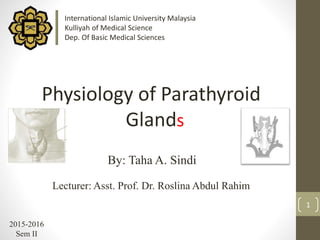
1. 1
By: Taha A. Sindi
Lecturer: Asst. Prof. Dr. Roslina Abdul Rahim
2015-2016
Sem II
Physiology of Parathyroid
Gland
International Islamic University Malaysia
Kulliyah of Medical Science
Dep. Of Basic Medical Sciences
s
2. Outline :
2
• Location of Parathyroid glands.
• Who discovered the glands.
• Some info. about it.
• Parathyroid hormone.
• Histology of the gland.
• PTH biosynthesis.
• The calcium-sensing receptors (CaSR)
• Why Calcium is so Important?
• Calcitonin
• 1,25-Dihyroxyvitamin D
• Metabolic bone diseases
(Hypercalcaemia and hypocalcaemia)
4. 4
Richard Owen, (1804 – 1892) who
discovered the parathyroid glands in 1850
a rhinoceros.
*Source: the Natural History Museum
Interesting Fact:
5. 5
• 4 tiny parathyroid glands, in the neck , on the posterior surface
of thyroid gland . Have two superiorly and 2 inferiorly
• Small in size(a grain of rice), each weighting around 30-50mg,
but may weight as much as 70 mg, and in diameter is 3-4 or till 8
millimeters.
• Woman usually have larger parathyroid gland than men
(Hinson, et al., 2010)
• It’s produce only one hormone (abbreviate: PTH)
• control the body's calcium levels
(why?)
Parathyroid Glands
*source: thyroid.com.au
6. 6
Parathyroid hormone (PTH)
Also known as parathormone or parathyrin
*source: wiki English
Produced by chief cells as a polypeptide
hormone containing 84 amino acids.
PTH half-life is approximately 4 minutes
PTH acts to increase the concentration of
ionic calcium (Ca2+) in the blood.
The major target end organs for parathyroid
hormone (PTH) action are the kidneys, skeletal
system, and intestine
8. 8
An image of the parathyroid gland reveals
two cell types:
*source: School of Anatomy and Human
Biology - The University of Western Australia
Histology
(Principal Cells): These cells release PTH.
are more numerous, smaller, with a slightly
eosinophilic cytoplasm
1- Chief cells
9. 9
2- Oxyphil cells
are less numerous, larger, and have a very
eosinophilic cytoplasm due to numerous
mitochondria. They are found individually, or
clustered in groups. The function of these cells
is unknown, yet their presence assists in
identifying this organ. Large numbers of
glycogen granules. Lack secretory vesicles.
Oxyphil cells increase in number in parathyroid
glands of patients with chronic kidney disease
(CKD) and are even more abundant in patients
receiving treatment for hyperparathyroidism
with calcitriol. (Ritter & et al., 2012)
*source: School of Anatomy and Human
Biology - The University of Western Australia
12. 12
The Role of the Parathyroid Glands
PTH raises the blood calcium level by:
1. breaking down the BONE (where most of the body's
calcium is stored) and causing calcium release.
2. increase the GIT ability to absorb calcium from food.
3. increasing the KIDNEY's ability to retain calcium that
would otherwise be lost in the urine.
14. 14
The calcium-sensing receptors (CaSR)
intracellular calcium-
sensing proteins
Extracellular calcium-
sensing proteins
Parathyroid cells respond to decreases in extracellular calcium
concentration by means of the calcium-sensing receptor, a cell surface
receptor that alters phosphatidylinositol turnover and intracellular
calcium, ultimately effecting an increase in parathyroid hormone secretion.
(Hendy & et al., 2000 )
CaSR is a plasma membrane or a cell surface receptor, structured as G
protein-coupled that is expressed in the parathyroid hormone-producing
chief cells of the parathyroid gland and the cells lining the kidney tubule.
(Hendy & et al., 2000 )
studies have demonstrated that the CaSR is expressed in the kidney, the
parathyroid glands (Riccardi & Brown,2010 ;Hendy & et al., 2000 ) brain
and gastrointestinal tract (Hebert, & et al., 1997).
CaSR: are receptors which senses levels of calcium ion
15. 15
A specific gland just to maintain Ca level !
Why Calcium is so Important?
Stimulate hormone secretion.
Muscle contraction.
Blood clotting.
Nerve function- transmit nerve impulse.
Necessary for the activation of some enzyme.
Transport ion across membrane.
Maintain regular heart beat (conduct electricity).
most of Calcium stored in the skeleton complexed
with phosphate, Ca++ has a wide range of functions:
16. 16
Calcium Homeostasis
in Calcium homeostasis process many key components are involving:
parathyroid hormone (PTH) : action on GIT, Kidneys and bones
1,25-dihydroxyvitamin D-3 (active form of vitamin D )
Calcitonin
Normal level of Ca in the blood is :
Around 10 mg/dL (Ganong’s review of med. Phy.)
17. 17
• hormone produced by the
parafollicular cells (also known as C-cells) of
the thyroid, that regulates calcium levels.
Calcitonin
• also known as thyrocalcitonin is a
32-amino acid linear polypeptide
• Action:
receptors for Calcitonin are found in
the bones and the kidneys.
• It inhibits the activity of osteoclasts
and also increase Ca++ in the urine.
18. 18
Vitamin D-3 formed in the skin when a cholesterol precursor,
is exposed to ultraviolet light. Activation occurs when the substance
undergoes 25-hydroxylation in the liver and 1-hydroxylation in the
kidney.
The primary action of 1,25-(OH)2 D3 is to promote gut absorption of
calcium by stimulating formation of calcium-binding protein within the
intestinal epithelial cells. Vitamin D also promotes intestinal absorption
of phosphate ion, although the exact mechanism is unclear. Negatively
charged phosphate ion may passively flow through the intestinal cell
because of flux of the positively charged calcium ion.
In bone, vitamin D may play a synergistic role with parathyroid hormone
(PTH) in stimulating osteoclast proliferation and bone resorption.
Compared to parathyroid hormone (PTH), vitamin D exerts a much
slower regulatory effect on calcium balance.
Vitamin D
19. 19
PTH increases blood calcium concentrations when
calcium ion levels fall below normal. First, PTH
enhances reabsorption of calcium by the kidneys; it
then stimulates osteoclast activity and inhibits
osteoblast activity. Finally, PTH stimulates synthesis
and secretion of calcitriol by the kidneys, which
enhances Ca2+ absorption by the digestive system.
Summary:
21. Hypercalcemia & Hypocalcemia
• Hypercalcemia is a disorder in which there is too
much calcium in the blood.
21
• On the other hand, if too little PTH is made,
hypocalcemia develops.
22. • The most common cause of hypercalcemia is
primary hyperparathyroidism. The problem is
usually a tumor, or adenoma. The tumor is
benign, which means that it doesn't spread
through the body. The parathyroid gland
enlarges and the additional cells produce extra
parathyroid hormone, causing an elevated
calcium level in the blood.
22
• The bones may weaken as PTH stimulates them
to release calcium, and extra calcium may
enter the kidneys, producing kidney stones.
The parathyroid gland tumor can be removed
by surgery.
Hypercalcemia
23. Other Causes of Hypercalcemia
• Adrenal gland failure
• Certain medications, especially thiazide diuretics (“water pills”)
or lithium
• Taking large amounts of calcium or vitamin D supplements for a
long time may also increase the calcium level in the blood.
• Dehydration can produce a temporary increase in the
concentration of calcium in the bloodstream.
• Kidney or adrenal gland problems can produce hypercalcemia.
23
24. Symptoms of Hypercalcemia
• Bone problems: pain, curvature of bones, fractures
• Muscle problems: weakness, twitches
• Gastrointestinal tract problems: pain, nausea, vomiting, loss
of appetite, constipation
• Kidney problems: back pain, thirst, frequent urination
• Nervous system problems: memory loss, confusion,
depression, fatigue
• hypertension
24
Treatment:
Medications that prevent bone breakdown such as
calcitonin
25. Hypocalcemia
25
In the blood calcium level is too low
Causes :
- Parathyroidectomy
- Renal failure
- Low levels of albumin
- Vitamin D deficiency
- Malabsorption
- Acute Pancreatitis (Precipitation of calcium )
26. Symptoms of Hypocalcemia
• brittle nails
• hair loss
• dry skin
• anxiety
• depression
• headaches
• memory loss
• muscle twitches
• weakness and fatigue
• muscle aches or cramps
• tingling in the lips, fingers or toes
26
Treatment :
Intravenous calcium chloride
27. References:
• Blaine, J., Chonchol, M., & Levi, M. (2014). Renal control of calcium, phosphate, and
magnesium homeostasis. Clinical Journal of the American Society of Nephrology, CJN-
09750913.
• Boundless. “Parathyroid Glands.” Accessed on 08 Jan. 2016. Retrieved 07 Apr. 2016
from boundless.com/biology/textbooks/boundless-biology-textbook/the-endocrine-system-37/endocrine-
glands-214/parathyroid-glands-806-12044
• Dr.Maizura’s lecture notes on Parathyroid glands. 2016
• Dr.Michael Barakate. How the Parathyroid Glands Work. Accessed on 17.3.2016.
thyroid.com.au/how-the-parathyroid-gland-works
• Essig, G. F.m Carron D, Carter W B, Jameson, M J , Talavera , F., Calhoun, K H Meyer,
A D ,Ramadan, H. H. (2016) Calcium Homeostasis 1.04.2016
emedicine.medscape.com/article/874690-overview
• Garmyan Yawar (2015) - Disorders of parathyroid gland
slideshare.net/GarmyanYawar/parathyroid-disorders
• Hinson, J. et al. (2010) The Endocrine System, Basic science and clinical conditions.
Elsevier Limited.
27
28. 28
• Introduction to Parathyroid Glands. Accessed on 30.03.2016
parathyroid.com/parathyroid.htm
• Histology of Thyroid and Parathyroid Glands. Accessed on 7.4.2016
quizlet.com/10459372/histology-lecture-3-thyroid-and-parathyroid-glands-flash-cards
• Hypercalcemia, Hypocalcemia and the Parathyroid Glands. Accessed on 7.4.2016 .
hubpages.com/health/Hypercalcemia-Hypocalcemia-and-the-Parathyroid-Gland
• Hypocalcemia vs. Hypercalcemia. Accessed on 7.4.2016. medbullets.com/step2-3-
renal/20703/hypocalcemia-vs-hypercalcemia .
• Kumar, R., & Thompson, J. R. (2011). The regulation of parathyroid hormone secretion
and synthesis. Journal of the American Society of Nephrology,22(2), 216-224.
• Parathyroid glands. Accessed on 17.3.2016
betterhealth.vic.gov.au/health/conditionsandtreatments/parathyroid-glands
• Hebert, S. C., Brown, E. M., & Harris, H. W. (1997). Role of the Ca (2+)-
sensing receptor in divalent mineral ion homeostasis. Journal of
Experimental Biology, 200(2), 295-302.
29. • Ritter, C. S., Haughey, B. H., Miller, B., & Brown, A. J. (2012). Differential gene expression by
oxyphil and chief cells of human parathyroid glands. The Journal of Clinical Endocrinology &
Metabolism, 97(8), E1499-E1505.
• School of Anatomy and Human Biology - The University of Western Australia.
Accessed on 7.4.2016 lab.anhb.uwa.edu.au/mb140/corepages/endocrines/endocrin.htm
• The American Association of Endocrine Surgeons (AAES). Parathyroid Glands: Function.
Accessed on 17.3.2016 endocrinediseases.org/parathyroid/parathyroid_background.shtml
• The Histology Guide – university of Leeds. Accessed on 7.4.2016
histology.leeds.ac.uk/glandular/thyroid.php
• National Library of Medicine. Accessed on 9.4.2016 nlm.nih.gov
• Riccardi, D., & Brown, E. M. (2010). Physiology and pathophysiology of
the calcium-sensing receptor in the kidney. American Journal of
Physiology-Renal Physiology, 298(3), F485-F499.
• Hendy, G. N., D'Souza-Li, L., Yang, B., Canaff, L., & Cole, D. E. (2000).
Mutations of the calcium-sensing receptor (CASR) in familial
hypocalciuric hypercalcemia, neonatal severe hyperparathyroidism, and
autosomal dominant hypocalcemia. Human mutation, 16(4), 281.
29