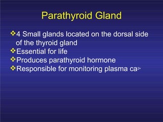
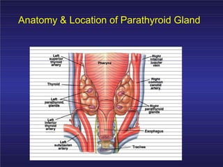
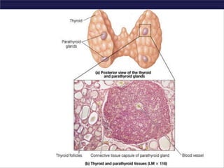
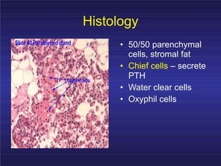
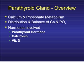
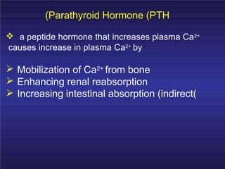
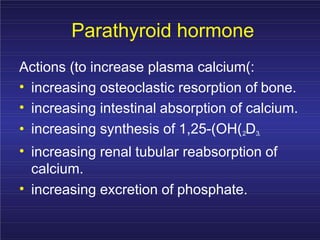
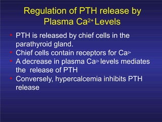
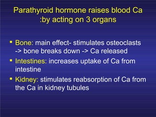
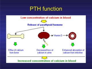
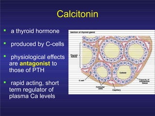
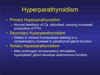
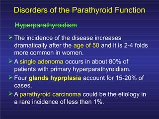
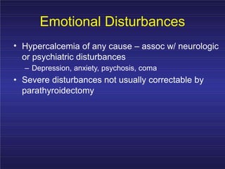
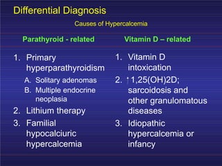
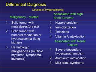
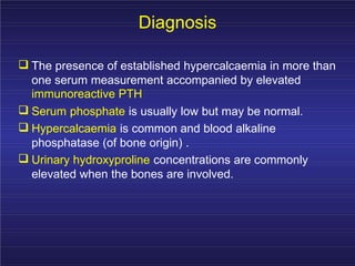
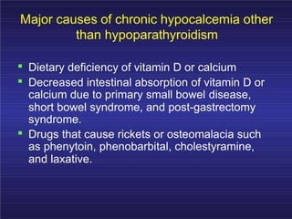
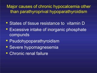
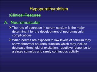
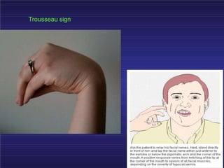
DISORDERS OFTHE
P
ARATHYROIDGLANDS
Obara thomas
Mclinmed Fh,bsc
clinmed,dip clinmed
Parathyroid Gland
4 Small glands located on the dorsal side
of the thyroid gland
Essential for life
Produces parathyroid hormone
Responsible for monitoring plasma ca2+
Anatomy & Location of Parathyroid Gland
Histology
• 50/50 parenchymal
cells, stromal fat
• Chief cells – secrete
PTH
• Water clear cells
• Oxyphil cells
Parathyroid Gland - Overview
. Calcium & Phosphate Metabolism
. Distribution & Balance of Ca & PO4
. Hormones involved
• Parathyroid Hormone
• Calcitonin
• Vit. D
(Parathyroid Hormone (PTH
a peptide hormone that increases plasma Ca2+
causes increase in plasma Ca2+ by
Mobilization of Ca2+ from bone
Enhancing renal reabsorption
Increasing intestinal absorption (indirect(
Parathyroid hormone
Actions (to increase plasma calcium(:
•
•
increasing osteoclastic resorption of bone.
increasing intestinal absorption of calcium.
• increasing synthesis of 1,25-(OH(2D3.
• increasing renal tubular reabsorption of
calcium.
• increasing excretion of phosphate.
Regulation of PTH release by
Plasma Ca2+ Levels
• PTH is released by chief cells in the
parathyroid gland.
•
•
Chief cells contain receptors for Ca2+
A decrease in plasma Ca2+ levels mediates
the release of PTH
• Conversely, hypercalcemia inhibits PTH
release
Parathyroid hormone raises blood Ca
:by acting on 3 organs
.
.
.
Bone: main effect- stimulates osteoclasts
-> bone breaks down -> Ca released
Intestines: increases uptake of Ca from
intestine
Kidney: stimulates reabsorption of Ca from
the Ca in kidney tubules
PTH function
Calcitonin
.
.
.
a thyroid hormone
produced by C-cells
physiological effects
are antagonist to
those of PTH
. rapid acting, short
term regulator of
plasma Ca levels
Calcitonin Lowers Blood Ca
Calcitonin is made by the C cells of the thyroid
gland
A large peptide "prohormone" is made and then
cut down to the 32 amino acid calcitonin
Stimulates osteoblasts, inhibits osteoclasts
Causes removal of Ca from plasma to calcify
new bone
• Lowers plasma Ca (opposes PTH(
•
•
Minor role in adult due to PTH feedback
Major role in children due to the rapid nature of
bone remodeling and its effect on osteoclastic
activity
Vit. D
• Vit. D family comprises of several diferent
compounds, all having similar functions.
• Most important is Vit. D3(cholecalciferol)
• Derived from irradiation of 7-dehydrocholesterol in
skin by UV rays
• Causes Ca absorption fron intestinal tract
• Active form of this hormone is 1,25-
dihydroxycholecalciferol (calcitriol)
Active Vitamin D (Calcitrol) is Made in 3
Steps by Different Organs
The skin uses ultraviolet sunlight to make vitamin D3
(cholecalciferol) from cholesterol
The vitamin D3 is converted to 25-Hydroxycholecalciferol in
the liver
Stimulated by PTH
The 25-Hydroxycholecalciferol is made into calcitriol (1, 25-
Dihydroxycholecalciferol) in the kidney
Stimulated by PTH
The main effect of calcitriol is to increase intestinal absorption
of Ca
Disorders of the Parathyroid Function
Hyperparathyroidism
Primary hyperparathyroidismis due to excessive
production of PTH by one or more of
hyperfunctioning parathyroid glands.
This leads to hyprcalcemia which fails to inhibit
the gland activity in the normal manner.
Hyperparathyroidism
• Primary Hyperparathyroidism
– Normal feedback of Ca disturbed, causing increased
production of PTH
• Secondary Hyperparathyroidism
– Defect in mineral homeostasis leading to a
compensatory increase in parathyroid gland function
• Tertiary Hyperparathyroidism
– After prolonged compensatory stimulation,
hyperplastic gland develops autonomous function
Disorders of the Parathyroid Function
Hyperparathyroidism
The cause of primary hyperparathyroidism is
unknown.
A genetic factor may be involved.
The clonal origin of most parathyroid adenomas
suggests a defect at the level of the gene
controlling the regulation and/or expression of
parathyroid hormone.
Disorders of the Parathyroid Function
Hyperparathyroidism
The incidence of the disease increases
dramatically after the age of 50 and it is 2-4 folds
more common in women.
A single adenoma occurs in about 80% of
patients with primary hyperparathyroidism.
Four glands hyprplasia account for 15-20% of
cases.
A parathyroid carcinoma could be the etiology in
a rare incidence of less then 1%.
Disorders of the Parathyroid Function
:Clinical Features
The two major sites of potential complications
are the bones and the kidneys.
The kidneys may have renal stones
(nephrolithiasis) or diffuse deposition of calcium-
phosphate complexes in the parachyma
(nephrocalcinosis).
Such complications are seen less
commonly and around 20% of patients or less
show such complications.
Disorders of the Parathyroid Function
:Clinical Features
In skeleton a condition called osteitis fibrosa
cystica could occur with subperiosteal resorption
of the distal phalanges, distal tappering of the
clavicles,
a “salt and pepper” appearance of the skull as
well as bone cysts and brown tumors of the long
bones.
Such overt bone disease even though typical of
primary hyperparathyroidism is very rarely
encountered.
Disorders of the Parathyroid Function
:Clinical Features
Other symptoms include muscle weakness,
easy fatigability, peptic ulcer disease,
pancreatitis, hypertension, gout and pseudogout
as well as anemia and depression have been
associated with primary hyperparathyroidism.
Primary HPT: Clinical Features
Symptomatic:
– Osteitis fibrosa cystica
– Nephrolithiasis
– Pathologic fractures
– Neuromuscular disease
– Life-threatening
hypercalcemia
– Peptic Ulcer Disease?
Asymptomatic:
– Fatigue
– Subjective muscle
weakness
– Depression
– Increased thirst
– Polyuria
– Constipation
– Musculoskeletal aches and
pains
Emotional Disturbances
• Hypercalcemia of any cause – assoc w/ neurologic
or psychiatric disturbances
– Depression, anxiety, psychosis, coma
• Severe disturbances not usually correctable by
parathyroidectomy
Differential Diagnosis
Causes of Hypercalcemia
Parathyroid - related Vitamin D – related
1. Vitamin D
intoxication
1. Primary
hyperparathyroidism
A. Solitary adenomas 2. 1,25(OH)2D;
sarcoidosis and
other granulomatous
diseases
B. Multiple endocrine
neoplasia
2. Lithium therapy
3. Familial 3. Idiopathic
hypocalciuric
hypercalcemia
hypercalcemia or
infancy
Differential Diagnosis
Causes of Hypercalcemia
Associated with high
bone turnover
Malignancy - related
1. Solid tumor with
metastases(breast)
1. Hyperthyroidism
2. Immobilization
3. Thiazides
2. Solid tumor with
humoral mediation of
hypercalcemia (lung
kidney)
4. Vitamin A intoxication
Assocated with Renal
Failure:
3. Hematologic
malignancies (multiple
myeloma, lymphoma,
leukemia)
1. Severe secondary
hyperparathyroidism
2. Aluminum intoxication
3. Milk alkali syndrome
Diagnosis
The presence of established hypercalcaemia in more than
one serum measurement accompanied by elevated
immunoreactive PTH
Serum phosphate is usually low but may be normal.
Hypercalcaemia is common and blood alkaline
phosphatase (of bone origin) .
Urinary hydroxyproline concentrations are commonly
elevated when the bones are involved.
Other Diagnostic tests
:The Glucocortisoid suppression test
The heypercalcaemic of non-parathyroid origin e.g.,
vitamin D intoxication, sarcoidosis and lymphoproliferative
syndromes
generally respond to the administration of prednisolone in
a dose of 40-60 mg daily for 10 days by a decrease in
serum calcium level.
A positive test result i.e. significant decrease in serum
calcium is a contraindication to neck exploration and
signals the need for investigation for a non-parathyroid
cause of the hypercalcaemia.
Other Diagnostic tests
:Radiograph
Plain X-ray of hands can be diagnostic showing
subperiosteal bone resorption usually on the radial surfacy
of the distal phalanx with distal phalangeal tufting as well
as cysts formation and generalzed osteopenia.
Ultrasonography
MRI
CT
Treatment
A large proportion of patients have “biochemical”
hyperparathyroidism but with prolonged follow up
they progress to overt clinical presentation.
In acute severe forms the main stay of therapy is
adequate hydration with saline .
Forced diuresis by diuretics to increase the urinary
excretion of calcium rapidly along with sodium and
prevent its reabsorption by the renal tubules.
Other agents
• Glucocostiroids
– In hypercalcaemia associated the hematological
malignant neoplasms
• Mythramycin
– A toxic antibiotics which inhibit bone resorption and is
used in hematological and solid neoplasms causing
hypercalcaemia.
•
•
Calcitonin
– Also inhibit osteoclast activity and prevent bone
resorption
Bisphosphonates
– They are given intravenously or orally to prevent bone
resorption.
Other agents
•
•
Phosphate
– Oral phosphate can be used as an
antihypercalcaemic agent and is commonly
used as a temporary measure during
diagnostic workup.
Estrogen
– It also decrease bone resorption and can be
given to postmenopausal women with primary
hyperparathyroidism using medical therapy
Surgery
Surgical treatment should be considered in all
cases with established diagnosis of primary
hyperparthyroidism.
During surgery the surgeon identifies all four
parathyroid glands (using biopsy if necessary)
followed by the removal of enlarged parathyroid
or 3 ½ glands in multiple glandular disease.
Other Complications
.
.
Deterioration of renal function
Metabolic disturbance e.g. hypomagnesia,
pancreatitis, gout or pseudogout
Hypoparathyroidism
Deficient secretion of PTH which manifests itself
biochemically by hypocalcemia, hyperphospatemia
diminished or absent circulating iPTH and clinically
the symptoms of neuromuscular hyperactivity.
Hypoparathyroidism
:Causes
• Surgical hypoparathyroidism – the
commonest
. After anterior neck exploration for thyroidectomy,
abnormal parathyroid gland removal, excision of a
neck lesion.
. It could be due to the removal of the parathyroid
glands or due to interruption of blood supply to the
glands.
Hypoparathyroidism
:Causes
. Idiopathic hypoparathyroidism
– A form occuring at an early age (genetic origin) with
autosomal recessive mode of transmission “multiple
endocrine deficiency –autoimmune-candidiasis
(MEDAC) syndrome”
– “Juvenile familial endocrinopathy”
– “Hypoparathyroidism – Addisson’s disease –
mucocutaneous candidiasis (HAM) syndrome”
Hypoparathyroidism
:Causes
. Idiopathic hypoparathyroidism
– Circulating antibodies for the parathyroid
glands and the adrenals are frequently
present.
– Other associated disease:
• Pernicious anemia
• Ovarian failure
• Autoimmune thyroiditis
• Diabetes mellitus
Hypoparathyroidism
:Causes
Functional hypoparathyroidism
– In patients who has chronic hypomagesaemia
of various causes.
– Magnesium is necessary for the PTH release
from the glands and also for the peripheral
action of the PTH.
Major causes of chronic hypocalcemia other
than hypoparathyroidism
. Dietary deficiency of vitamin D or calcium
. Decreased intestinal absorption of vitamin D or
calcium due to primary small bowel disease,
short bowel syndrome, and post-gastrectomy
syndrome.
. Drugs that cause rickets or osteomalacia such
as phenytoin, phenobarbital, cholestyramine,
and laxative.
Major causes of chronic hypocalcemia other
than parathyroprival hypoparathyroidism
. States of tissue resistance to vitamin D
. Excessive intake of inorganic phosphate
compunds
.
.
.
Psudohypoparathyroidism
Severe hypomagnesemia
Chronic renal failure
Hypoparathyroidism
:Clinical Features
A. Neuromuscular
The rate of decrease in serum calcium is the major
determinant for the development of neuromuscular
complications.
When nerves are exposed to low levels of calcium they
show abnormal neuronal function which may include
decrease threshold of excitation, repetitive response to
a single stimulus and rarely continuous activity.
Hypoparathyroidism
:Clinical Features
A. Neuromuscular
– Parathesia (sensation of numbness or tingling on the skin)
– Tetany
– Hyperventilation
– Adrenergic symptoms
– Convulsion (More common in young people and it can
take the form of either generalized tetany followed by
prolonged tonic spasms or the typical epileptiform
seizures.
– Signs of latent tetany
• Chvostek sign (nilateral spasm of the oris muscle is initiated
by a slight tap over the facial)
• Trousseau sign
• Extrapyramidal signs (due to basal ganglia calcification)
Involuntary movements, Tremors and rigidity
Trousseau sign
Hypoparathyroidism
:Clinical Features
B. Other clinical manifestation
1. Posterio lenticular cataract
2. Cardiac manifestation:
3. Prolonged QT interval in the ECG
4. Resistance to digitalis
5. Hypotension
6. Refractory heart failure with cardiomegally can
occur.
Hypoparathyroidism
:Clinical Features
B. Other clinical manifestation
3. Dental Manifestation
Abnormal enamel formation with delayed or absent
dental eruption and defective dental root formation.
4. Malabsorption syndrome
Presumably secondary to decreased calcium level
and may lead to steatorrhoea with long standing
untreated disease.
Hypoparathyroidism
:Diagnosis
In the absence of renal failure the presence of
hypocalcaemia with hyperphosphataemia is
virtually diagnostic of hypoparathyroidism.
Undetectable serum iPTH confirms the diagnosis or
it can be detectable if the assay is very sensitive.
Hypoparathyroidism
:Treatment
The mainstay of treatment is a combination of oral
calcium with pharmacological doses of vitamin D or
its potent analogues.
Phosphate restriction in diet may also be useful
with or without aluminum hydroxide gel to lower
serum phosphate level.
Emergency Treatment for Hypocalcaemic
:Tetany
Calcium should be given parenterally till
adequate serum calcium level is
obtained and then vitamin D
supplementation with oral calcium should
be initiated.
Emergency Treatment for Hypocalcaemic
:Hungry bone syndrome
In patients with hypoparathyroidism and severe
bone disease who undergo successful
parathyroidectomy hypocalcaemia may be
severe and parenteral calcium infusion with later
supplementation with oral calcium and vitamin D.
Parathyroid disorders.pptx

Parathyroid disorders.pptx

  • 1.
  • 2.
    Parathyroid Gland 4 Smallglands located on the dorsal side of the thyroid gland Essential for life Produces parathyroid hormone Responsible for monitoring plasma ca2+
  • 3.
    Anatomy & Locationof Parathyroid Gland
  • 5.
    Histology • 50/50 parenchymal cells,stromal fat • Chief cells – secrete PTH • Water clear cells • Oxyphil cells
  • 6.
    Parathyroid Gland -Overview . Calcium & Phosphate Metabolism . Distribution & Balance of Ca & PO4 . Hormones involved • Parathyroid Hormone • Calcitonin • Vit. D
  • 7.
    (Parathyroid Hormone (PTH apeptide hormone that increases plasma Ca2+ causes increase in plasma Ca2+ by Mobilization of Ca2+ from bone Enhancing renal reabsorption Increasing intestinal absorption (indirect(
  • 8.
    Parathyroid hormone Actions (toincrease plasma calcium(: • • increasing osteoclastic resorption of bone. increasing intestinal absorption of calcium. • increasing synthesis of 1,25-(OH(2D3. • increasing renal tubular reabsorption of calcium. • increasing excretion of phosphate.
  • 9.
    Regulation of PTHrelease by Plasma Ca2+ Levels • PTH is released by chief cells in the parathyroid gland. • • Chief cells contain receptors for Ca2+ A decrease in plasma Ca2+ levels mediates the release of PTH • Conversely, hypercalcemia inhibits PTH release
  • 10.
    Parathyroid hormone raisesblood Ca :by acting on 3 organs . . . Bone: main effect- stimulates osteoclasts -> bone breaks down -> Ca released Intestines: increases uptake of Ca from intestine Kidney: stimulates reabsorption of Ca from the Ca in kidney tubules
  • 11.
  • 12.
    Calcitonin . . . a thyroid hormone producedby C-cells physiological effects are antagonist to those of PTH . rapid acting, short term regulator of plasma Ca levels
  • 13.
    Calcitonin Lowers BloodCa Calcitonin is made by the C cells of the thyroid gland A large peptide "prohormone" is made and then cut down to the 32 amino acid calcitonin Stimulates osteoblasts, inhibits osteoclasts Causes removal of Ca from plasma to calcify new bone • Lowers plasma Ca (opposes PTH( • • Minor role in adult due to PTH feedback Major role in children due to the rapid nature of bone remodeling and its effect on osteoclastic activity
  • 14.
    Vit. D • Vit.D family comprises of several diferent compounds, all having similar functions. • Most important is Vit. D3(cholecalciferol) • Derived from irradiation of 7-dehydrocholesterol in skin by UV rays • Causes Ca absorption fron intestinal tract • Active form of this hormone is 1,25- dihydroxycholecalciferol (calcitriol)
  • 15.
    Active Vitamin D(Calcitrol) is Made in 3 Steps by Different Organs The skin uses ultraviolet sunlight to make vitamin D3 (cholecalciferol) from cholesterol The vitamin D3 is converted to 25-Hydroxycholecalciferol in the liver Stimulated by PTH The 25-Hydroxycholecalciferol is made into calcitriol (1, 25- Dihydroxycholecalciferol) in the kidney Stimulated by PTH The main effect of calcitriol is to increase intestinal absorption of Ca
  • 16.
    Disorders of theParathyroid Function Hyperparathyroidism Primary hyperparathyroidismis due to excessive production of PTH by one or more of hyperfunctioning parathyroid glands. This leads to hyprcalcemia which fails to inhibit the gland activity in the normal manner.
  • 17.
    Hyperparathyroidism • Primary Hyperparathyroidism –Normal feedback of Ca disturbed, causing increased production of PTH • Secondary Hyperparathyroidism – Defect in mineral homeostasis leading to a compensatory increase in parathyroid gland function • Tertiary Hyperparathyroidism – After prolonged compensatory stimulation, hyperplastic gland develops autonomous function
  • 18.
    Disorders of theParathyroid Function Hyperparathyroidism The cause of primary hyperparathyroidism is unknown. A genetic factor may be involved. The clonal origin of most parathyroid adenomas suggests a defect at the level of the gene controlling the regulation and/or expression of parathyroid hormone.
  • 19.
    Disorders of theParathyroid Function Hyperparathyroidism The incidence of the disease increases dramatically after the age of 50 and it is 2-4 folds more common in women. A single adenoma occurs in about 80% of patients with primary hyperparathyroidism. Four glands hyprplasia account for 15-20% of cases. A parathyroid carcinoma could be the etiology in a rare incidence of less then 1%.
  • 20.
    Disorders of theParathyroid Function :Clinical Features The two major sites of potential complications are the bones and the kidneys. The kidneys may have renal stones (nephrolithiasis) or diffuse deposition of calcium- phosphate complexes in the parachyma (nephrocalcinosis). Such complications are seen less commonly and around 20% of patients or less show such complications.
  • 21.
    Disorders of theParathyroid Function :Clinical Features In skeleton a condition called osteitis fibrosa cystica could occur with subperiosteal resorption of the distal phalanges, distal tappering of the clavicles, a “salt and pepper” appearance of the skull as well as bone cysts and brown tumors of the long bones. Such overt bone disease even though typical of primary hyperparathyroidism is very rarely encountered.
  • 22.
    Disorders of theParathyroid Function :Clinical Features Other symptoms include muscle weakness, easy fatigability, peptic ulcer disease, pancreatitis, hypertension, gout and pseudogout as well as anemia and depression have been associated with primary hyperparathyroidism.
  • 23.
    Primary HPT: ClinicalFeatures Symptomatic: – Osteitis fibrosa cystica – Nephrolithiasis – Pathologic fractures – Neuromuscular disease – Life-threatening hypercalcemia – Peptic Ulcer Disease? Asymptomatic: – Fatigue – Subjective muscle weakness – Depression – Increased thirst – Polyuria – Constipation – Musculoskeletal aches and pains
  • 24.
    Emotional Disturbances • Hypercalcemiaof any cause – assoc w/ neurologic or psychiatric disturbances – Depression, anxiety, psychosis, coma • Severe disturbances not usually correctable by parathyroidectomy
  • 25.
    Differential Diagnosis Causes ofHypercalcemia Parathyroid - related Vitamin D – related 1. Vitamin D intoxication 1. Primary hyperparathyroidism A. Solitary adenomas 2. 1,25(OH)2D; sarcoidosis and other granulomatous diseases B. Multiple endocrine neoplasia 2. Lithium therapy 3. Familial 3. Idiopathic hypocalciuric hypercalcemia hypercalcemia or infancy
  • 26.
    Differential Diagnosis Causes ofHypercalcemia Associated with high bone turnover Malignancy - related 1. Solid tumor with metastases(breast) 1. Hyperthyroidism 2. Immobilization 3. Thiazides 2. Solid tumor with humoral mediation of hypercalcemia (lung kidney) 4. Vitamin A intoxication Assocated with Renal Failure: 3. Hematologic malignancies (multiple myeloma, lymphoma, leukemia) 1. Severe secondary hyperparathyroidism 2. Aluminum intoxication 3. Milk alkali syndrome
  • 27.
    Diagnosis The presence ofestablished hypercalcaemia in more than one serum measurement accompanied by elevated immunoreactive PTH Serum phosphate is usually low but may be normal. Hypercalcaemia is common and blood alkaline phosphatase (of bone origin) . Urinary hydroxyproline concentrations are commonly elevated when the bones are involved.
  • 28.
    Other Diagnostic tests :TheGlucocortisoid suppression test The heypercalcaemic of non-parathyroid origin e.g., vitamin D intoxication, sarcoidosis and lymphoproliferative syndromes generally respond to the administration of prednisolone in a dose of 40-60 mg daily for 10 days by a decrease in serum calcium level. A positive test result i.e. significant decrease in serum calcium is a contraindication to neck exploration and signals the need for investigation for a non-parathyroid cause of the hypercalcaemia.
  • 29.
    Other Diagnostic tests :Radiograph PlainX-ray of hands can be diagnostic showing subperiosteal bone resorption usually on the radial surfacy of the distal phalanx with distal phalangeal tufting as well as cysts formation and generalzed osteopenia. Ultrasonography MRI CT
  • 30.
    Treatment A large proportionof patients have “biochemical” hyperparathyroidism but with prolonged follow up they progress to overt clinical presentation. In acute severe forms the main stay of therapy is adequate hydration with saline . Forced diuresis by diuretics to increase the urinary excretion of calcium rapidly along with sodium and prevent its reabsorption by the renal tubules.
  • 31.
    Other agents • Glucocostiroids –In hypercalcaemia associated the hematological malignant neoplasms • Mythramycin – A toxic antibiotics which inhibit bone resorption and is used in hematological and solid neoplasms causing hypercalcaemia. • • Calcitonin – Also inhibit osteoclast activity and prevent bone resorption Bisphosphonates – They are given intravenously or orally to prevent bone resorption.
  • 32.
    Other agents • • Phosphate – Oralphosphate can be used as an antihypercalcaemic agent and is commonly used as a temporary measure during diagnostic workup. Estrogen – It also decrease bone resorption and can be given to postmenopausal women with primary hyperparathyroidism using medical therapy
  • 33.
    Surgery Surgical treatment shouldbe considered in all cases with established diagnosis of primary hyperparthyroidism. During surgery the surgeon identifies all four parathyroid glands (using biopsy if necessary) followed by the removal of enlarged parathyroid or 3 ½ glands in multiple glandular disease.
  • 34.
    Other Complications . . Deterioration ofrenal function Metabolic disturbance e.g. hypomagnesia, pancreatitis, gout or pseudogout
  • 35.
    Hypoparathyroidism Deficient secretion ofPTH which manifests itself biochemically by hypocalcemia, hyperphospatemia diminished or absent circulating iPTH and clinically the symptoms of neuromuscular hyperactivity.
  • 36.
    Hypoparathyroidism :Causes • Surgical hypoparathyroidism– the commonest . After anterior neck exploration for thyroidectomy, abnormal parathyroid gland removal, excision of a neck lesion. . It could be due to the removal of the parathyroid glands or due to interruption of blood supply to the glands.
  • 37.
    Hypoparathyroidism :Causes . Idiopathic hypoparathyroidism –A form occuring at an early age (genetic origin) with autosomal recessive mode of transmission “multiple endocrine deficiency –autoimmune-candidiasis (MEDAC) syndrome” – “Juvenile familial endocrinopathy” – “Hypoparathyroidism – Addisson’s disease – mucocutaneous candidiasis (HAM) syndrome”
  • 38.
    Hypoparathyroidism :Causes . Idiopathic hypoparathyroidism –Circulating antibodies for the parathyroid glands and the adrenals are frequently present. – Other associated disease: • Pernicious anemia • Ovarian failure • Autoimmune thyroiditis • Diabetes mellitus
  • 39.
    Hypoparathyroidism :Causes Functional hypoparathyroidism – Inpatients who has chronic hypomagesaemia of various causes. – Magnesium is necessary for the PTH release from the glands and also for the peripheral action of the PTH.
  • 40.
    Major causes ofchronic hypocalcemia other than hypoparathyroidism . Dietary deficiency of vitamin D or calcium . Decreased intestinal absorption of vitamin D or calcium due to primary small bowel disease, short bowel syndrome, and post-gastrectomy syndrome. . Drugs that cause rickets or osteomalacia such as phenytoin, phenobarbital, cholestyramine, and laxative.
  • 41.
    Major causes ofchronic hypocalcemia other than parathyroprival hypoparathyroidism . States of tissue resistance to vitamin D . Excessive intake of inorganic phosphate compunds . . . Psudohypoparathyroidism Severe hypomagnesemia Chronic renal failure
  • 42.
    Hypoparathyroidism :Clinical Features A. Neuromuscular Therate of decrease in serum calcium is the major determinant for the development of neuromuscular complications. When nerves are exposed to low levels of calcium they show abnormal neuronal function which may include decrease threshold of excitation, repetitive response to a single stimulus and rarely continuous activity.
  • 43.
    Hypoparathyroidism :Clinical Features A. Neuromuscular –Parathesia (sensation of numbness or tingling on the skin) – Tetany – Hyperventilation – Adrenergic symptoms – Convulsion (More common in young people and it can take the form of either generalized tetany followed by prolonged tonic spasms or the typical epileptiform seizures. – Signs of latent tetany • Chvostek sign (nilateral spasm of the oris muscle is initiated by a slight tap over the facial) • Trousseau sign • Extrapyramidal signs (due to basal ganglia calcification) Involuntary movements, Tremors and rigidity
  • 44.
  • 45.
    Hypoparathyroidism :Clinical Features B. Otherclinical manifestation 1. Posterio lenticular cataract 2. Cardiac manifestation: 3. Prolonged QT interval in the ECG 4. Resistance to digitalis 5. Hypotension 6. Refractory heart failure with cardiomegally can occur.
  • 46.
    Hypoparathyroidism :Clinical Features B. Otherclinical manifestation 3. Dental Manifestation Abnormal enamel formation with delayed or absent dental eruption and defective dental root formation. 4. Malabsorption syndrome Presumably secondary to decreased calcium level and may lead to steatorrhoea with long standing untreated disease.
  • 47.
    Hypoparathyroidism :Diagnosis In the absenceof renal failure the presence of hypocalcaemia with hyperphosphataemia is virtually diagnostic of hypoparathyroidism. Undetectable serum iPTH confirms the diagnosis or it can be detectable if the assay is very sensitive.
  • 48.
    Hypoparathyroidism :Treatment The mainstay oftreatment is a combination of oral calcium with pharmacological doses of vitamin D or its potent analogues. Phosphate restriction in diet may also be useful with or without aluminum hydroxide gel to lower serum phosphate level.
  • 49.
    Emergency Treatment forHypocalcaemic :Tetany Calcium should be given parenterally till adequate serum calcium level is obtained and then vitamin D supplementation with oral calcium should be initiated.
  • 50.
    Emergency Treatment forHypocalcaemic :Hungry bone syndrome In patients with hypoparathyroidism and severe bone disease who undergo successful parathyroidectomy hypocalcaemia may be severe and parenteral calcium infusion with later supplementation with oral calcium and vitamin D.