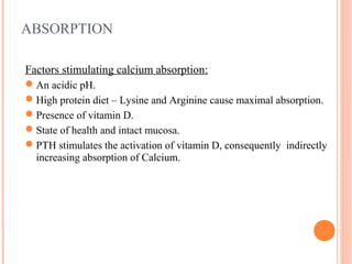
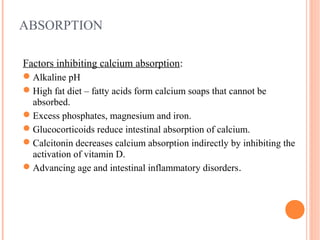
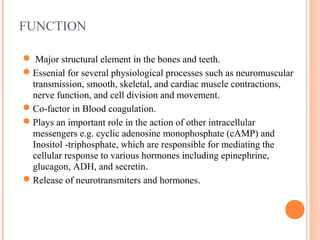
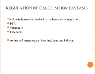
Calcium homeostasis involves absorption of calcium from the intestine, regulation by parathyroid hormone (PTH), vitamin D, and calcitonin, and storage in bone. PTH increases calcium levels by stimulating bone resorption and renal reabsorption and vitamin D absorption. Vitamin D increases intestinal calcium absorption. Calcitonin decreases calcium by inhibiting bone resorption. Hypocalcemia causes neurological symptoms and hypercalcemia causes gastrointestinal and renal issues. Conditions are diagnosed by calcium levels and other tests and treated by calcium supplementation or intravenous calcium for hypocalcemia and hydration and medications for hypercalcemia.

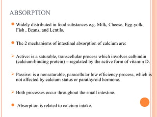
![DIAGNOSIS
Total (amended) Calcium
amended[Ca]=measured total[Ca]+0.02(40-[albumin])
Albumin
PTH ( high hypocalcaemia, low hypercalcaemia)
Vitamin D (high hypocalcaemia, low hypercalcaemia)
LFT (ALP is raised hypercalcaemia and normal in hypocalcaemia)
(Phos raised in hypocalcaemia and low in hypercalcaemia)
U&E (urea and creatinine increased in hypercalcaemia)
Iron (raised in hypocalcaemia)
PH (Acidosis increases calcium and alkalosis decreases calicum)
24-hour urinary calcium (elevated in hyperparathyroidism, renal
failure and decreased in hypoparathyroidism, malabsorption disorders)](https://image.slidesharecdn.com/6fa218da-9495-4dea-9c73-37f6acac8331-161103215019/85/Calcium-homeostasis-16-320.jpg)
![References
Baynes J. (1999) Medical Biochemistry. Basildon.Harcourt Brace and
Company Limited.
Baker S. et al.(2002) The essentials of Calcium, Magnesium and Phosphate
Metabolism. Critical Care and resuscitation. 4(4) pp. 301-306.[online]
Nessar A. (2010) Clinical Biochemistry. New York. Oxford University Press.
Mundy G. et al. (1999) Hormonal Control of Calcium Homeostasis, Clinical
Chemistry, 45 (8), pp.1347-1352.
Warrel D. et al.(2003) Oxford Textbook of Medicine. London.Oxford University
Press.[online]
Peacock M. (2010) Calcium Metabolism in Health and Disease, Clinical
Journal of American Society of Nephrology,5(1), pp. 23-30.[online]](https://image.slidesharecdn.com/6fa218da-9495-4dea-9c73-37f6acac8331-161103215019/85/Calcium-homeostasis-18-320.jpg)


