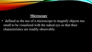
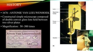
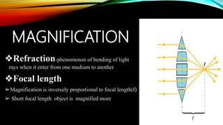
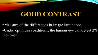
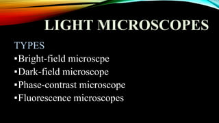
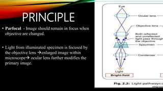
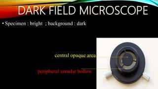
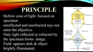
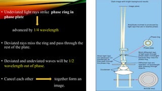
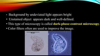
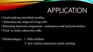
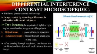
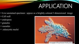
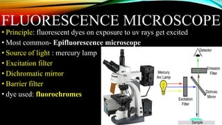
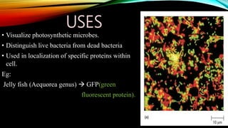
MEDICAL MICROSCOPY
Dr. ANNA KURIAN
Microscopy
• defined as the use of a microscope to magnify objects too
small to be visualized with the naked eye so that their
characteristics are readily observable.
HISTORY
• 1676 –ANTONIE VAN LEEUWENHOEK
• Constructed simple microscope composed
of double convex glass lens held between
two silver plates
• Magnification: 50 -300 times
GENERAL PROPERTIES
1. GOOD MAGNIFICATION
2. GOOD RESOLUTION
3. GOOD CONTRAST
MAGNIFICATION
❖Refraction-phenomenon of bending of light
rays when it enter from one medium to another
❖Focal length
➢Magnification is inversely proportional to focal length(f)
➢ Short focal length object is magnified more
RESOLUTION
❖- Described by Ernst Abbe , Ability of lens to separate or distinguish between small
objects that are close together.
.
Abbé equation : Minimal distance (d) between two objects that reveals them as separate
entities depends on
d = 0.5 λ
n sin ϴ
( λ) : the wavelength of light used to illuminate the specimen
(n sin ϴ ) : the numerical aperture of the lens
NUMERICALAPERTURE (NSIN ϴ)
defined by two components:
• RI of the medium(n)
• Angular aperture(ϴ) : 1/2 the
angle of the cone of light entering
an objective
• Maximum NA of lens obtained in
air =1 as,
RI (air) =1
sinϴ =1 ( sin90)
GOOD RESOLUTION
1. increasing the numerical aperture
 increase the refractive index with
immersion oil
(light rays that did not enter the objective
due to reflection and refraction at the surfaces
will now do so.)
2. decreasing the wavelength
light at the blue end of the visible spectrum in the range of 450 to 500
nm
Working distance
➢Characteristic of an objective lens.
➢Distance between front surface of the lens and surface of the
cover glass or the specimen when in focus.
➢Objectives with large NA and great resolving power  short
working distance
GOOD CONTRAST
•Measure of the differences in image luminance.
•Under optimum conditions, the human eye can detect 2%
contrast .
LIGHT MICROSCOPES
TYPES
▪Bright-field microscpe
▪Dark-field microscope
▪Phase-contrast microscope
▪Fluorescence microscopes
BRIGHT FIELD MICROSCOPE
• Routinely used in microbiology labs.
• Employed to examine both stained and unstained specimens.
• forms dark image against brighter background.
• 2 types:
1. Simple Microscope
✓ contains single magnifying glass
2. Compound Microscope
✓ uses series of lenses for magnification.
✓ Total magnification =mag of objective lens * mag of ocular lens
STRUCTURE
1. base: Light source
2. arm.
• Two focusing knobs  on the arm.
• Stage at halfway up arm with slide
clip or stage clip.
• Sub stage condenser
3. metal body or stand
• Connects the nosepiece and ocular
lens or eyepiece
• Nose piece - 3 to 5 objective lenses of
different magnifying power.
PRINCIPLE
• Parfocal - Image should remain in focus when
objective are changed.
• Light from illuminated specimen is focused by
the objective lens enlarged image within
microscope ocular lens further modifies the
primary image.
APPLICATION
•Observation of morphology of microorganisms.
• Detection of cell structures.
•Observation of intracellular structures.
•Observation of motility.
• Measurement of size.
•Observation of blood smears
DARK FIELD MICROSCOPE
• Specimen : bright ; background : dark
central opaque area
peripheral annular hollow
PRINCIPLE
• Hollow cone of light focused on
specimen
• unreflected and unrefracted rays not
enter the objective.
• Only light reflected or refracted by
the specimen forms image.
• Field appears dark & object
brightly illuminated.
APPLICATION
➢Allows to observe living unstained cell
and organisms
➢Reveal internal structure in larger
eukaryotic microorganism.
➢Identify thin and distinctively shaped
Treponema Pallidum.
DISADVANTAGE :
• Not only the specimen, but dust and
other particles scatter the light
PHASE CONTRAST MICROSCOPE
• Invented by Frits Zernite 1930s, received
the Nobel prize in physics in 1953
• Excellent way to observe unstained living
cells.
• Contrast enhanced
• Object: dark ; background :bright
• Converts slight differences in refractive
index and cell density into easily detected
variations in light intensity.
COMPONENTS • Condenser annulus
• Phase plate
PRINCIPLE
• Light waves through a cell structure is slowed more than those in surrounding
medium
• Deviated light rays – cell structures
• Undeviated light rays –through and
around the cell
• Deviated light rays slowed more
than undeviated
• Crests and troughs do not align
• Deviated rays retarded by ¼ th the wavelength of undeviated
• Undeviated light rays strike phase ring in
phase plate
advanced by 1/4 wavelength
• Deviated rays miss the ring and pass through the
rest of the plate.
• Deviated and undeviated waves will be 1/2
wavelength out of phase.
• Cancel each other together form an
image.
• Background by undeviated light appears bright
• Unstained object appears dark and well-defined.
• This type of microscopy is called dark-phase-contrast microscopy.
• Color filters often are used to improve the image.
APPLICATION
• Used studying microbial motility,
• Determine the shape of living cells
• Detecting bacterial components - endospores and inclusion bodies.
• Used to study eukaryotic cells.
• Disadvantages : 1. Halo artifacts
2. low contrast specimens needs staining
.
DIFFERENTIAL INTERFERENCE
CONTRAST MICROSCOPE(DIC)
➢Similar to phase-contrast microscope
➢Image created by detecting differences in
refractive indices and thickness.
➢Two beams(plane-polarized light) at right
angles to each other are generated by prisms.
1. Object beam : passes through specimen
2. Reference beam : passes through clear area
of the slide.
➢ After passing through specimen, two beams are
combined and interfere with each other to form an
image.
APPLICATION
• Live unstained specimen : appear as a brightly colored 3 dimensional image
• Cell wall
• endospore
• granules
• vacuoles
• eukaryotic nuclei
FLUORESCENCE MICROSCOPE
• Principle: fluorescent dyes on exposure to uv rays get excited
• Most common- Epifluorescence microscope
• Source of light : mercury lamp
• Excitation filter
• Dichromatic mirror
• Barrier filter
• dye used: fluorochromes
FLUROCHROME:
FLUOROCHROMES USES
Acridine orange Stains DNA
Diamidino-2-phenyl indole(DAPI) Stains DNA
Fluorescein isothiocyanate(FITC) Attached to antibodies that bind specific
cellular components or to DNA probes.
Tetra methyl rhodamine
isothiocyanate(TRITC or rhodamine)
Attached to antibodies that bind specific
cellular components.
USES
• Visualize photosynthetic microbes.
• Distinguish live bacteria from dead bacteria
• Used in localization of specific proteins within
cell.
Eg:
Jelly fish (Aequorea genus)  GFP(green
fluorescent protein).
CONFOCAL MICROSCOPY
• Uses optical sectioning for better resolution
• When 3D objects are viewed  light enters from all areas of the
object not just the plane of focus blurred image.
• confocal scanning laser microscope (CSLM) or confocal
microscope.
• Uses a laser beam to illuminate a
fluorescently stained specimen
• Major component is aperture placed
above objective lens
• Eliminates stray light from parts of the
specimen that lie above and below the
plane of focus.
• Light used to create the image is from
the plane of focus clearer sharper
image formed.
• A computer attached to confocal
microscope receives digitized
information from each plane in the
specimen.
• Used to create clear composite image
• 3D reconstruction of the specimen.
BENEFITIS OF CONFOCAL MICROSCOPY
• Reduce blurring of image from light scattering.
• Increased & effective resolution.
• Clear examination of thick specimen.
• Z- axis scanning.
• Depth perception in Z- sectioned images.
• Magnification can be adjusted electronically.
USES
✓Study of biofilm
✓DNA hybridization
✓Epitope tagging
✓Stem cell research
THANK YOU

Microscopy 4 anna

  • 1.
  • 2.
    Microscopy • defined asthe use of a microscope to magnify objects too small to be visualized with the naked eye so that their characteristics are readily observable.
  • 3.
    HISTORY • 1676 –ANTONIEVAN LEEUWENHOEK • Constructed simple microscope composed of double convex glass lens held between two silver plates • Magnification: 50 -300 times
  • 4.
    GENERAL PROPERTIES 1. GOODMAGNIFICATION 2. GOOD RESOLUTION 3. GOOD CONTRAST
  • 5.
    MAGNIFICATION ❖Refraction-phenomenon of bendingof light rays when it enter from one medium to another ❖Focal length ➢Magnification is inversely proportional to focal length(f) ➢ Short focal length object is magnified more
  • 6.
    RESOLUTION ❖- Described byErnst Abbe , Ability of lens to separate or distinguish between small objects that are close together. . Abbé equation : Minimal distance (d) between two objects that reveals them as separate entities depends on d = 0.5 λ n sin ϴ ( λ) : the wavelength of light used to illuminate the specimen (n sin ϴ ) : the numerical aperture of the lens
  • 7.
    NUMERICALAPERTURE (NSIN ϴ) definedby two components: • RI of the medium(n) • Angular aperture(ϴ) : 1/2 the angle of the cone of light entering an objective • Maximum NA of lens obtained in air =1 as, RI (air) =1 sinϴ =1 ( sin90)
  • 8.
    GOOD RESOLUTION 1. increasingthe numerical aperture  increase the refractive index with immersion oil (light rays that did not enter the objective due to reflection and refraction at the surfaces will now do so.) 2. decreasing the wavelength light at the blue end of the visible spectrum in the range of 450 to 500 nm
  • 9.
    Working distance ➢Characteristic ofan objective lens. ➢Distance between front surface of the lens and surface of the cover glass or the specimen when in focus. ➢Objectives with large NA and great resolving power  short working distance
  • 10.
    GOOD CONTRAST •Measure ofthe differences in image luminance. •Under optimum conditions, the human eye can detect 2% contrast .
  • 11.
    LIGHT MICROSCOPES TYPES ▪Bright-field microscpe ▪Dark-fieldmicroscope ▪Phase-contrast microscope ▪Fluorescence microscopes
  • 12.
    BRIGHT FIELD MICROSCOPE •Routinely used in microbiology labs. • Employed to examine both stained and unstained specimens. • forms dark image against brighter background. • 2 types: 1. Simple Microscope ✓ contains single magnifying glass 2. Compound Microscope ✓ uses series of lenses for magnification. ✓ Total magnification =mag of objective lens * mag of ocular lens
  • 13.
    STRUCTURE 1. base: Lightsource 2. arm. • Two focusing knobs  on the arm. • Stage at halfway up arm with slide clip or stage clip. • Sub stage condenser 3. metal body or stand • Connects the nosepiece and ocular lens or eyepiece • Nose piece - 3 to 5 objective lenses of different magnifying power.
  • 14.
    PRINCIPLE • Parfocal -Image should remain in focus when objective are changed. • Light from illuminated specimen is focused by the objective lens enlarged image within microscope ocular lens further modifies the primary image.
  • 15.
    APPLICATION •Observation of morphologyof microorganisms. • Detection of cell structures. •Observation of intracellular structures. •Observation of motility. • Measurement of size. •Observation of blood smears
  • 16.
    DARK FIELD MICROSCOPE •Specimen : bright ; background : dark central opaque area peripheral annular hollow
  • 17.
    PRINCIPLE • Hollow coneof light focused on specimen • unreflected and unrefracted rays not enter the objective. • Only light reflected or refracted by the specimen forms image. • Field appears dark & object brightly illuminated.
  • 18.
    APPLICATION ➢Allows to observeliving unstained cell and organisms ➢Reveal internal structure in larger eukaryotic microorganism. ➢Identify thin and distinctively shaped Treponema Pallidum. DISADVANTAGE : • Not only the specimen, but dust and other particles scatter the light
  • 19.
    PHASE CONTRAST MICROSCOPE •Invented by Frits Zernite 1930s, received the Nobel prize in physics in 1953 • Excellent way to observe unstained living cells. • Contrast enhanced • Object: dark ; background :bright • Converts slight differences in refractive index and cell density into easily detected variations in light intensity.
  • 20.
    COMPONENTS • Condenserannulus • Phase plate
  • 21.
    PRINCIPLE • Light wavesthrough a cell structure is slowed more than those in surrounding medium • Deviated light rays – cell structures • Undeviated light rays –through and around the cell • Deviated light rays slowed more than undeviated • Crests and troughs do not align • Deviated rays retarded by ¼ th the wavelength of undeviated
  • 22.
    • Undeviated lightrays strike phase ring in phase plate advanced by 1/4 wavelength • Deviated rays miss the ring and pass through the rest of the plate. • Deviated and undeviated waves will be 1/2 wavelength out of phase. • Cancel each other together form an image.
  • 23.
    • Background byundeviated light appears bright • Unstained object appears dark and well-defined. • This type of microscopy is called dark-phase-contrast microscopy. • Color filters often are used to improve the image.
  • 24.
    APPLICATION • Used studyingmicrobial motility, • Determine the shape of living cells • Detecting bacterial components - endospores and inclusion bodies. • Used to study eukaryotic cells. • Disadvantages : 1. Halo artifacts 2. low contrast specimens needs staining .
  • 25.
    DIFFERENTIAL INTERFERENCE CONTRAST MICROSCOPE(DIC) ➢Similarto phase-contrast microscope ➢Image created by detecting differences in refractive indices and thickness. ➢Two beams(plane-polarized light) at right angles to each other are generated by prisms. 1. Object beam : passes through specimen 2. Reference beam : passes through clear area of the slide. ➢ After passing through specimen, two beams are combined and interfere with each other to form an image.
  • 26.
    APPLICATION • Live unstainedspecimen : appear as a brightly colored 3 dimensional image • Cell wall • endospore • granules • vacuoles • eukaryotic nuclei
  • 27.
    FLUORESCENCE MICROSCOPE • Principle:fluorescent dyes on exposure to uv rays get excited • Most common- Epifluorescence microscope • Source of light : mercury lamp • Excitation filter • Dichromatic mirror • Barrier filter • dye used: fluorochromes
  • 28.
    FLUROCHROME: FLUOROCHROMES USES Acridine orangeStains DNA Diamidino-2-phenyl indole(DAPI) Stains DNA Fluorescein isothiocyanate(FITC) Attached to antibodies that bind specific cellular components or to DNA probes. Tetra methyl rhodamine isothiocyanate(TRITC or rhodamine) Attached to antibodies that bind specific cellular components.
  • 29.
    USES • Visualize photosyntheticmicrobes. • Distinguish live bacteria from dead bacteria • Used in localization of specific proteins within cell. Eg: Jelly fish (Aequorea genus)  GFP(green fluorescent protein).
  • 30.
    CONFOCAL MICROSCOPY • Usesoptical sectioning for better resolution • When 3D objects are viewed  light enters from all areas of the object not just the plane of focus blurred image. • confocal scanning laser microscope (CSLM) or confocal microscope.
  • 31.
    • Uses alaser beam to illuminate a fluorescently stained specimen • Major component is aperture placed above objective lens • Eliminates stray light from parts of the specimen that lie above and below the plane of focus. • Light used to create the image is from the plane of focus clearer sharper image formed. • A computer attached to confocal microscope receives digitized information from each plane in the specimen. • Used to create clear composite image • 3D reconstruction of the specimen.
  • 32.
    BENEFITIS OF CONFOCALMICROSCOPY • Reduce blurring of image from light scattering. • Increased & effective resolution. • Clear examination of thick specimen. • Z- axis scanning. • Depth perception in Z- sectioned images. • Magnification can be adjusted electronically.
  • 33.
    USES ✓Study of biofilm ✓DNAhybridization ✓Epitope tagging ✓Stem cell research
  • 34.

Editor's Notes

  • #3 Microbiology concerned with organism so small that cannot be seen with unaided eyes. Microscope is of crucial importance for seeing microorganism and understanding their function. It provides valuble information about functions of structures. Therefore its important to understand how microscopes work and how specimens are prepared for examinations
  • #6 Lenses act like a collection of prisms operating as a unit. When the light source is distant so that parallel rays of light strike the lens, a convex lens will focus these rays at a specific point, the focal point (F in figure 2.2). The distance between the center of the lens and the focal point is called the focal length ( f in figure 2.2).
  • #7 optical theory underlying microscope design
  • #8 When this cone has a narrow angle and tapers to a sharp point, it does not spread out much after leaving the slide and therefore does not adequately separate images of closely packed objects. If the cone of light has a very wide angle and spreads out rapidly after passing through a specimen, closely packed objects appear widely separated and are resolved
  • #10 No lens working in air have a numerical aperture > 1.00
  • #15 These lenses can be rotated to position any objective beneath the body assembly
  • #20 Unpigmented living cells are not clearly visible in the brightfield microscope because there is little difference in contrast between the cells and water. to increase contrast and create variations in color between cell structures you haveto stain or kill the cells. But what if an investigator must view living cellsin order to observe a dynamic process such as movement or phagocytosis? Phase-contrast microscopy can be used in this situation.
  • #23 Phase ring is special optical disk located in the objective.
  • #25 *that contain poly--hydroxyalkanoates (e.g., poly--hydroxybutyrate), polymetaphosphate, sulfur, or other substances These are clearly visible because they have refractive indices markedly different from that of water Most imp brk through is the ability to observe living particle in natural state. Major disadv of phase contrast microscope – produce halo artifacts
  • #28 exposes a specimen to ultraviolet,violet, or blue light and forms an image of the object with the resulting fluorescent light.
  • #29 The fluorochrome absorbs light energy and fluoresces brightly
  • #30 Live – green Red – dead Their pigments naturally fluoresce when excited by light of specific wavelengths.
  • #34 Computers are integral to the process of creating confocal images