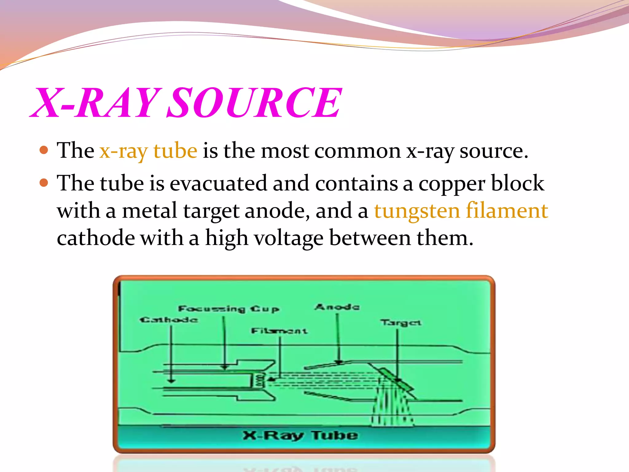
contents
1) Introduction about X-ray crystallography
2) X-ray and its properties
3) History
4) Principle
5) X-ray diffraction
6) Instrumentation
7) Applications
8) Conclusion
INTRODUCTION
 X-ray crystallography is the technique of determining
the arrangement of atoms within a crystal, in which a
beam of x-rays strikes a crystal and causes the beam of
light to spread into many specific direction.
 From the angles and intensities of these diffracted
beams, a crystallographer can produce the 3-
dimensional picture of density of the electron within
the crystal.
 Here , we see about history, principle,
instrumentation, uses and limitations of x-ray
crystallography.
X-RAY AND ITS PROPERTIES
 An electromagnetic wave of high energy and very
short wavelength (between UV light and gamma ray).
PROPERTIES:
 X-rays travels in a straight line.
 They are electrically neutral
 They are invisible ray.
 They can pass through any materials which are opaque
to light.
history
 X-ray crystallography was discovered by JOHN
KAPLAE in the 17th century.
 With the evidence of x-ray crystallography only, Dr.
JAMES WATSON and Dr. FRANCIS CRICK discovered
the double helix structure of DNA.
PRINCIPLE
 The vital principle of x-ray crystallography is the
DIFFRACTION.
 The crystal is made to strike against x-ray beam.
 Due to striking the atoms present in the crystal
diffracts the x-ray beam into different diffractions.
 The angle and intensity of this diffraction rays is along
to spatial arrangement of atom in crystal.
 By studying these angle, the 3D structure of any crystal
can be determined.
 X-rays are generated by bombarding electron on a
metallic anode.
X-RAY DIFFRACTION
 X-ray crystallography uses
the uniformity of light
diffraction of crystals to
determine the structure of
a molecule or atom.
 They use an x-ray beam to
hit the crystallized
molecule .
 The electron surrounding
the molecule diffract as x-
rays hits them. This forms
a pattern , this type of
pattern is called X-RAY
DIFFRACTION PATTERN.
instrumentation
x-ray crystallography uses
the instrument called the x-
ray diffractometer. It has
the following parts:
 X-ray source
 Mono chromator
 Calorimeter
 Goniometer
 Photographic film plate
 Detector.
X-RAY SOURCE
 The x-ray tube is the most common x-ray source.
 The tube is evacuated and contains a copper block
with a metal target anode, and a tungsten filament
cathode with a high voltage between them.
MONOCHROMATOR
 The word “mono” means
SINGLE and “chroma”
means COLOR.
 The device used to
convert the different color
radiation into single
color.
 It removes the unwanted
rays other than x-rays.
 It produces the x-rays
about 12-24 A*.
There is 2 closely packed
metal plate which are
0.3mm apart from each
other.
The x-ray beam
originate from x-ray tube
passes through this gap
and follow single line
path.
COLLIMATOR
GONIOMETER
 It is a device on which
crystal structure is to
be determined and
mounted.
 The device spin slowly
in according to crystal
which to rotate on
constant speed.
 It is placed between
the collimator and
photo plate.
PHOTO PLATE FILM
 The film captures the
diffraction rays of
crystal.
 The light-sensitive
emulsion of silver salts
was coated on a glass
plate, typically thinner
than common window
glass, instead of a
clear plastic film.
DETECTOR
The captured data has
been send to computer
for further processing by
detector where 3D
structure of crystal gets
developed.
WORKING OF THE X-RAY
DIFFRACTO METER
APPLICATIONS
MEDICINE
HIV:
scientists discovered the x-ray crystallo -graphic
structure of HIV PROTEASE, a viral enzyme in 1989
and to prevent the spread of virus in the body.
ARTHRITIS:
To create an effective pain killer that does not cause
ulcers.
Atomic science
 The arrangement of various atomic structure of
crystals like salts, metals, semiconductors and many
minerals are identified and studied.
 The atomic arrangement of diamond, graphite like
carbon compounds also studied by x-ray diffraction.
Molecular study
 The lattice structure of crystal revealed by diffraction
of the x-rays.
 The bonds like ionic and covalent bonds between
the molecules can be elucidated and studied.
 Molecular structures of the penicillin, vitamin-B12,
insulin, and other proteins can be easily revealed
and studied by x-ray diffraction.
DAIRY SCIENCE
LIQUID MILK:
The arrangement of lactose and other mineral
constituents of liquid milk are easily provided by
the x-ray analysis.
MILK STONE:
The atomic arrangement and chemical
composition of milk stone by x-ray diffraction
through photographic film.
DAIRY SCIENCE
MILK POWDER:
The study of milk powder is given by analysis of the
x-rays to provide the structural group spacings within
the milk proteins.
Thus the x-ray crystallography is a widely used tool for
the elucidation of the compounds present in milk and
dairy products obtained through their structure and
functional relationship.
CONCLUSION
 Thus the x-ray crystallography is used to reveal the
structure of viruses, nucleic acids, and antibodies, etc.
 It is widely used in the atomic and molecular studies.
Hence , the x-ray crystallography has a vast applications
in the various scientific fields.
REFERENCE
 X-ray crystallography by JAN DREATH.
 Biochemistry by KUMERASAN.
 www.chem.ucla.edu.com
 www.blogspot.com
X ray crystallography

X ray crystallography

  • 2.
    contents 1) Introduction aboutX-ray crystallography 2) X-ray and its properties 3) History 4) Principle 5) X-ray diffraction 6) Instrumentation 7) Applications 8) Conclusion
  • 3.
    INTRODUCTION  X-ray crystallographyis the technique of determining the arrangement of atoms within a crystal, in which a beam of x-rays strikes a crystal and causes the beam of light to spread into many specific direction.  From the angles and intensities of these diffracted beams, a crystallographer can produce the 3- dimensional picture of density of the electron within the crystal.  Here , we see about history, principle, instrumentation, uses and limitations of x-ray crystallography.
  • 4.
    X-RAY AND ITSPROPERTIES  An electromagnetic wave of high energy and very short wavelength (between UV light and gamma ray). PROPERTIES:  X-rays travels in a straight line.  They are electrically neutral  They are invisible ray.  They can pass through any materials which are opaque to light.
  • 5.
    history  X-ray crystallographywas discovered by JOHN KAPLAE in the 17th century.  With the evidence of x-ray crystallography only, Dr. JAMES WATSON and Dr. FRANCIS CRICK discovered the double helix structure of DNA.
  • 6.
    PRINCIPLE  The vitalprinciple of x-ray crystallography is the DIFFRACTION.  The crystal is made to strike against x-ray beam.  Due to striking the atoms present in the crystal diffracts the x-ray beam into different diffractions.  The angle and intensity of this diffraction rays is along to spatial arrangement of atom in crystal.  By studying these angle, the 3D structure of any crystal can be determined.  X-rays are generated by bombarding electron on a metallic anode.
  • 7.
    X-RAY DIFFRACTION  X-raycrystallography uses the uniformity of light diffraction of crystals to determine the structure of a molecule or atom.  They use an x-ray beam to hit the crystallized molecule .  The electron surrounding the molecule diffract as x- rays hits them. This forms a pattern , this type of pattern is called X-RAY DIFFRACTION PATTERN.
  • 8.
    instrumentation x-ray crystallography uses theinstrument called the x- ray diffractometer. It has the following parts:  X-ray source  Mono chromator  Calorimeter  Goniometer  Photographic film plate  Detector.
  • 9.
    X-RAY SOURCE  Thex-ray tube is the most common x-ray source.  The tube is evacuated and contains a copper block with a metal target anode, and a tungsten filament cathode with a high voltage between them.
  • 10.
    MONOCHROMATOR  The word“mono” means SINGLE and “chroma” means COLOR.  The device used to convert the different color radiation into single color.  It removes the unwanted rays other than x-rays.  It produces the x-rays about 12-24 A*.
  • 11.
    There is 2closely packed metal plate which are 0.3mm apart from each other. The x-ray beam originate from x-ray tube passes through this gap and follow single line path. COLLIMATOR
  • 12.
    GONIOMETER  It isa device on which crystal structure is to be determined and mounted.  The device spin slowly in according to crystal which to rotate on constant speed.  It is placed between the collimator and photo plate.
  • 13.
    PHOTO PLATE FILM The film captures the diffraction rays of crystal.  The light-sensitive emulsion of silver salts was coated on a glass plate, typically thinner than common window glass, instead of a clear plastic film.
  • 14.
    DETECTOR The captured datahas been send to computer for further processing by detector where 3D structure of crystal gets developed.
  • 15.
    WORKING OF THEX-RAY DIFFRACTO METER
  • 16.
  • 17.
    MEDICINE HIV: scientists discovered thex-ray crystallo -graphic structure of HIV PROTEASE, a viral enzyme in 1989 and to prevent the spread of virus in the body. ARTHRITIS: To create an effective pain killer that does not cause ulcers.
  • 18.
    Atomic science  Thearrangement of various atomic structure of crystals like salts, metals, semiconductors and many minerals are identified and studied.  The atomic arrangement of diamond, graphite like carbon compounds also studied by x-ray diffraction.
  • 19.
    Molecular study  Thelattice structure of crystal revealed by diffraction of the x-rays.  The bonds like ionic and covalent bonds between the molecules can be elucidated and studied.  Molecular structures of the penicillin, vitamin-B12, insulin, and other proteins can be easily revealed and studied by x-ray diffraction.
  • 20.
    DAIRY SCIENCE LIQUID MILK: Thearrangement of lactose and other mineral constituents of liquid milk are easily provided by the x-ray analysis. MILK STONE: The atomic arrangement and chemical composition of milk stone by x-ray diffraction through photographic film.
  • 21.
    DAIRY SCIENCE MILK POWDER: Thestudy of milk powder is given by analysis of the x-rays to provide the structural group spacings within the milk proteins. Thus the x-ray crystallography is a widely used tool for the elucidation of the compounds present in milk and dairy products obtained through their structure and functional relationship.
  • 22.
    CONCLUSION  Thus thex-ray crystallography is used to reveal the structure of viruses, nucleic acids, and antibodies, etc.  It is widely used in the atomic and molecular studies. Hence , the x-ray crystallography has a vast applications in the various scientific fields.
  • 23.
    REFERENCE  X-ray crystallographyby JAN DREATH.  Biochemistry by KUMERASAN.  www.chem.ucla.edu.com  www.blogspot.com