SlideShare a Scribd company logo
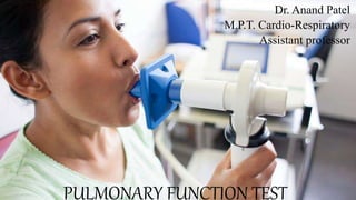
PULMONARY FUNCTION TEST
Dr. Anand Patel
M.P.T. Cardio-Respiratory
Assistant professor
INTRODUCTION
• Pulmonary function tests or lung function tests are useful in assessing
the functional status of the respiratory system both in physiological
and pathological conditions.
• MEASURE:
(1) Dynamic flow rates of gases through the airways,
(2) Lung volumes and capacities, and
(3) The ability of the lungs to diffuse gases
• Identify the presence and type of pulmonary impairments and the degree of
pulmonary disease present.
INDICATIONS
• To identify and quantify changes in pulmonary function
• To evaluate need and quantify therapeutic effectiveness
• To perform epidemiologic surveillance for pulmonary disease
• To assess patients for risk for postoperative pulmonary complications
• To determine pulmonary disability
Contraindications
• acute, unstable cardiopulmonary problems, such as
• hemoptysis,
• pneumothorax,
• myocardial infarction,
• pulmonary embolism, and
• patients with acute chest or abdominal pain should not be tested.
• Patients who have nausea and who have recently vomited
• Recent cataract removal surgery
Lung Volumes and Capacities
• 4 lung volumes and 4 lung capacities
Tidal volume (VT),
Inspiratory reserve volume
(IRV),
Expiratory reserve volume
(ERV),
Residual volume
Total lung capacity
Inspiratory capacity
Vital capacity
Functional Residual Capacity
LUNG VOLUMES
• TIDAL VOLUME: Tidal volume (TV) is the volume of air breathed in
and out of lungs in a single normal quiet respiration. Tidal volume
signifies the normal depth of breathing.
• Normal value: 500ml
• INSPIRATORY RESERVE VOLUME: Inspiratory reserve volume (IRV) is
an additional volume of air that can be inspired forcefully after the
end of normal inspiration.
• Normal value: 3,300 ml
• EXPIRATORY RESERVE VOLUME: Expiratory reserve volume is the
additional volume of air that can be expired out forcefully, after
normal expiration.
• Normal value: 1000 ml
• RESIDUAL VOLUME: Residual volume (RV) is the volume of air
remaining in lungs even after forced expiration. Normally, lungs
cannot be emptied completely even by forceful expiration. Some
quantity of air always remains in the lungs even after the forced
expiration.
• Normal value: 1200ml
LUNG CAPACITIES
• INSPIRATORY CAPACITY: Inspiratory capacity (IC) is the maximum
volume of air that is inspired after normal expiration. It includes tidal
volume and inspiratory reserve volume.
• IC = TV + IRV
500 + 3,300 = 3,800 mL
• VITAL CAPACITY: Vital capacity (VC) is the maximum volume of air that
can be expelled out forcefully after a deep inspiration. VC includes
inspiratory reserve volume, tidal volume and expiratory reserve
volume.
• VC = IRV + TV + ERV
• = 3,300 + 500 + 1,000 = 4,800 mL
• FUNCTIONAL RESIDUAL CAPACITY: Functional residual capacity (FRC)
is the volume of air remaining in lungs after normal expiration.
Functional residual capacity includes expiratory reserve volume and
residual volume.
• FRC = ERV + RV = 1,000 + 1,200 = 2,200 mL
• TOTAL LUNG CAPACITY:
• Total lung capacity (TLC) is the volume of air present in lungs after a deep
inspiration. It includes all the volumes.
• TLC = IRV + TV + ERV + RV
• = 3,300 + 500 + 1,000 + 1,200 = 6,000 mL
PFT
• INCLUDES:
• SPIROMETRY
• LUNG VOLUMES AND CAPACITIES
• GAS TRANSFER
SPIROMETRY
• SPIROMETRY: Measures of breathing
• Spiro means breathing
• Metry means measurement
• Spirometry is a method of assessing lung function by measuring the
total volumes of air the patient can expel from the lungs after a
maximal inhalation.
• It measure how much and how quickly air can be expelled following
deep breath
• Spirometry is an effort-dependent test that requires careful patient
instruction, understanding, coordination, and cooperation
• Measures:
• Forced vital capacity (FVC)
• Forced expiratory volume in one second. (FEV1)
• FEF25-75 - Forced Expiratory Flow between 25% to 75% of forced vital capacity
• peak expiratory flow rate (PEFR)
• Fev1/FVC
Forced vital capacity
• Volume of air that can be maximally and forcibly expelled from the
lung after a deep inhalation.
• Depends on:
• Lung elasticity
• Size of airways
• Resistance to flow along these airways
Fev1/FVC
• Expressed as percentage
• In normal adult, the ratio ranges from 75%-85%.
• Childrens may have higher flow.
• Obstructive condition: Decreased
• Restrictive condition: Normal or Increase
PEFR
• Maximum rate at which air can be expired after a deep inspiration.
• Normal values: 400L/min
• Measured by peak flow meter
• Pefr reduced in obstructive disease more than restrictive disease.
Before the test- avoid
• Alcohol – 4hours
• Large meal -2 hours
• Smoking – 1hr
• Vigorous exercise- 30 minutes
• Wear loose, comfortable clothing
• Avoid use of bronchodilators
Prior to test
• Gain verbal consent
• Check for contraindication
• Gain accurate height and weight
• Patient should sit comfortable and upright
• Technique in detail should be explained
Spirometry maneuver
• A few normal tidal respiration
• Then deep inspiration
• Breath holding,
• Very forced and fast expiration as hard as patient can
• Then deep, quick and full inspiration.
• Repeat atleast 3 times.
• Take the best readings.
Forced vital capacity
• Possible causes of a decrease in FVC:
• 1. The problem may be with the lung itself. There may have been a
resectional surgical procedure or areas of collapse. Various other
conditions can render the lung less expandable, such as fibrosis,
congestive heart failure, and thickened pleura. Obstructive lung
diseases may reduce the FVC by limiting deflation of the lung.
• 2. The problem may be in the pleural cavity, such as an enlarged
heart, pleural fluid, or a tumor encroaching on the lung.
• 3. Another possibility is restriction of the chest wall. The lung cannot
inflate and deflate normally if the motion of the chest wall (which
includes its abdominal components) is restricted.
• 4. Inflation and deflation of the system require normal function of the
respiratory muscles, primarily the diaphragm, the intercostal muscles,
and the abdominal muscles.
FEV1
• Fev1 is the volume of air exhaled in the first second of the FVC test.
• Normal value depends on patient age, size, sex and race.
• When flow rates are slowed by airway obstruction, as in emphysema,
the FEV1 is decreased by an amount that reflects the severity of the
disease.
• The FVC may also be reduced, although usually to a lesser degree.
FEV1 /FVC
• The FEV1 /FVC ratio is generally expressed as a percentage.
• The amount exhaled during the first second is a fairly constant
fraction of the FVC, irrespective of lung size.
• In the normal adult, the ratio ranges from 75% to 85%, but it
decreases somewhat with aging.
• Children have high flows for their size, and thus, their ratios are
higher, up to 90%.
• Significance:
• It aids in quickly identifying persons with airway obstruction
• The ratio is valuable for identifying the cause of a low FEV
• In pulmonary restriction, the FEV1 and FVC are decreased
proportionally; hence, the ratio is in the normal range, as in the case
of fibrosis.
• How to determine whether airway obstruction or a restrictive:
• A low FEV1 with a normal ratio usually indicates a restrictive process
• A low FEV1 and a decreased ratio signify a predominantly obstructive process.
MAXIMAL VOLUNTARY VENTILATION
• The test for maximal voluntary ventilation (MVV) is an athletic event.
• The subject is instructed to breathe as hard and fast as possible for 10
to 15 seconds.
• The result is extrapolated to 60 seconds and reported in liters per
minute.
• A low MVV can occur in obstructive disease, in restrictive disease, in
neuromuscular disease, in heart disease, in a patient who does not
try or who does not understand, or in a frail patient
MAXIMAL INSPIRATORY FLOW
• With spirometer systems that measure both expiratory and
inspiratory flows, the maximal inspiratory flow (MIF) can be
measured.
• The subject exhales maximally (the FVC test) and then immediately
inhales as rapidly and completely as possible, producing an
inspiratory curve.
• The combined expiratory and inspiratory FV curves form the FV loop.
• Increased airway resistance decreases both maximal expiratory flow
and MIF
Reversibility
• Based on the initial results of baseline spirometry, additional testing
of pulmonary mechanics is often desirable.
• If the baseline test indicates airway obstruction, determining the
reversibility of the obstruction is indicated.
• RTs also use the concept of reversibility when evaluating routine
therapy by performing spirometry before and after therapy.
• In the laboratory, the FVC maneuver is often repeated after the
patient has received a bronchodilator administered by small volume
nebulizer or metered dose inhaler.
• Reversibility of the airway obstruction indicates effective therapy.
• Although improvement in other measurements of pulmonary
function is sometimes used, reversibility is defined as a 15% or
greater improvement in FEV1 and at least a 200-ml increase in FEV1.
• % Improvement = PostFEV1 – PreFEV1/ PreFEV1 * 100
Bronchoprovocation
• When the patient’s history suggests episodic symptoms of
hyperreactive airways and airway obstruction, such as seasonal or
exercise-induced wheezing, and the results of baseline spirometry are
normal, performing a bronchial provocation may be indicated.
• Bronchial provocation testing uses an agent to stimulate a
hyperreactive airway response and to create airway obstruction.
• The procedure usually begins with the patient inhaling a normal
saline aerosol and then repeating the FVC maneuver.
• Some very sensitive patients exhibit hyperreactive airways with saline
alone; a positive response to saline is defined as a decrease in FEV1 of
10% or greater.
• The methacholine provocation protocol systematically exposes the patient
to increasing dosages of methacholine. Usually starting with a low dose of
0.03 mg/ml, patients inhale methacholine aerosol and then repeat the FVC
maneuver.
• A positive response to methacholine is defined as a decrease in FEV1 of
20% or greater.
• If a positive response does not occur, the methacholine dose is doubled to
0.06 mg/ml, and the FVC maneuver is repeated.
• The process of “double-dosing” and performing FVC maneuvers continues
until there is a positive response or until the full dose, 16 mg/ml, is given.
• If a positive response occurs, treatment with a fast-acting bronchodilator is
indicated, and sometimes administering O2 is helpful
Static lung volume
• The three most commonly used methods of measuring the FRC are
• Nitrogen washout,
• Inert gas dilution, and
• Plethysmography.
Helium Dilution
• The helium dilution technique for measuring lung volumes uses a closed,
rebreathing circuit.
• This technique is based on the assumptions that the patient has no He in
his or her lungs, and that an equilibration of He can occur between the
spirometer and the lungs.
• First, volume (V1) and concentration (C1) of He are measured at the
beginning of the test.
• Next, the valve is turned to connect the patient to the breathing circuit,
usually at the resting expiratory level of the FRC.
• The patient is connected to the He-air mixture, and the concentration of
He is diluted slowly by the patient’s lung volume.
• The patient is connected to the He-air mixture, and the concentration
of He is diluted slowly by the patient’s lung volume.
• Wearing nose clips, the patient breathes normally in the closed
circuit.
• Exhaled CO2 is absorbed with soda lime, and O2 is added at a rate
equal to the patient’s O2 consumption.
• A constant volume is maintained to ensure accurate He concentration
measurements.
• The patient rebreathes the gas in the system until equilibrium of He
concentration is established.
• In healthy patients and patients with a small FRC, equilibration occurs
in 2 to 5 minutes.
• Patients with obstructive lung disease may require 20 minutes to
equilibrate because of slow gas mixing in the lungs.
Nitrogen Washout Method
• The nitrogen washout technique uses a nonrebreathing or open
circuit.
• The technique is based on the assumptions that the N concentration
in the lungs is 78% and in equilibrium with the atmosphere, that the
patient inhales 100% O2, and that the O2 replaces all of the N in the
lungs.
• Similar to the He dilution technique, the patient is connected to the
system at FRC.
• The patient’s exhaled gas is monitored, and its volume and N
percentage are measured.
Plethysmography
• The principle of plethysmography is based on Boyle’s law, which
states that the product of the pressure (P) and volume (V) of a gas is
constant under constant temperature conditions.
• The whole-body plethysmograph consists of a sealed chamber in
which the patient sits.
• Pressure transducers (electronic manometers) measure pressure at
the mouth and in the chamber.
• The plethysmographic method measures essentially all the gas in the
lung, including that in poorly ventilated areas.
Diffusing Capacity
• The third major category of pulmonary function testing is measuring
the ability of the lungs to transfer gases across the alveolar-capillary
membrane.
• Vgas = DL× − ( P1-p2 )
• Vgas = Amount of the gas transferred into the lungs
• P1 = Partial pressure of the gas in the alveolus
• P2 = Partial pressure of the gas in the pulmonary capillary
• Carbon monoxide (CO) is the gas normally used to measure the DL.
• The diffusing capacity of the lung for carbon monoxide (DLCO) is expressed
in ml/min/mm Hg under standard temperature and pressure and dry
conditions.
• CO is used as the transfer gas because CO is similar to O2 in important
ways.
• CO and O2 have similar molecular weights and solubility coefficients.
Similar to O2, CO also chemically combines with hemoglobin (Hb).
• CO has a very high affinity for Hb and diffuses rapidly into the pulmonary
blood, keeping the pulmonary capillary partial pressure of CO near zero.
An average normal value is 20 to 30 mL/min/mm Hg; that is, 20 to 30
mL CO is transferred per minute per mm Hg difference in the driving
pressure of CO, namely, the difference between the partial pressure of
CO in alveolar air and blood.
The normal values depend on age, sex , and size.
Causes of Increased Dlco
• 1. Supine position: Rarely is the Dlco measured while the subject is supine,
but this position produces a higher value because of increased perfusion
and blood volume of the upper lobes.
• 2. Exercise: It is difficult to hold one’s breath for 10 seconds during exercise.
When this is done just after exercise, however, Dlco is increased because of
increased pulmonary blood volumes.
• 3. Asthma: Some patients with asthma, especially when symptom-free,
have an increased Dlco, possibly because of more uniform distribution of
pulmonary blood flow.
• 4. Obesity: The Dlco can be increased in obese persons, especially those
who are massively obese. This increase is thought to be due to an
increased pulmonary blood volume.
Pft
Pft
Pft

More Related Content

What's hot

Pulmonary function tests
Pulmonary function testsPulmonary function tests
Pulmonary function tests
logon2kingofkings
 
peak expiratory flow rate presentation
peak expiratory flow rate presentationpeak expiratory flow rate presentation
peak expiratory flow rate presentation
Rekha Marbate
 
Exercise induced asthma &bronchospasm
Exercise induced asthma &bronchospasmExercise induced asthma &bronchospasm
Exercise induced asthma &bronchospasmFawzia Abo-Ali
 
Peep & cpap
Peep & cpapPeep & cpap
Peep & cpap
Davis Kurian
 
Equal pressure point
Equal pressure pointEqual pressure point
Equal pressure point
Rekha Marbate
 
Pulmonary function test
Pulmonary function testPulmonary function test
Pulmonary function test
raghu srikanti
 
PEFR & mini peak flow meter
PEFR & mini peak flow meterPEFR & mini peak flow meter
PEFR & mini peak flow meter
Yapa
 
Bronchopleuralfistula
BronchopleuralfistulaBronchopleuralfistula
Bronchopleuralfistula
Neelu Aryal
 
Breathing patterns
Breathing patternsBreathing patterns
Breathing patterns
Dr.Priyanka Das
 
Flutter a device for clearance of airway
Flutter  a device for clearance of airwayFlutter  a device for clearance of airway
Flutter a device for clearance of airway
Shilpasree Saha
 
CLINICAL ASSESSMENT OF COPD
CLINICAL ASSESSMENT OF COPDCLINICAL ASSESSMENT OF COPD
CLINICAL ASSESSMENT OF COPD
SoM
 
Lung mechanics
Lung mechanicsLung mechanics
Lung mechanics
mauryaramgopal
 
Ats guidelines for the six minute walk test by dr kartik sood
Ats guidelines for the six minute walk test by dr kartik soodAts guidelines for the six minute walk test by dr kartik sood
Ats guidelines for the six minute walk test by dr kartik sood
Kartik Sood
 
Pep devices
Pep devicesPep devices
Pep devices
AragyaKhadka
 
Spirometry
SpirometrySpirometry
Spirometry
Tapeshwar Yadav
 
Interpretation OF PFT
Interpretation OF PFT		Interpretation OF PFT
Interpretation OF PFT Khalid
 
Pulmonary function testing
Pulmonary function testingPulmonary function testing
pneumonectomy
pneumonectomypneumonectomy
pneumonectomy
BPT4thyearJamiaMilli
 
Lung volumes capacities pfts
Lung volumes capacities pfts Lung volumes capacities pfts
Lung volumes capacities pfts
Rajesh Munigial
 
Physiotherapy
PhysiotherapyPhysiotherapy
Physiotherapy
Nursing Hi Nursing
 

What's hot (20)

Pulmonary function tests
Pulmonary function testsPulmonary function tests
Pulmonary function tests
 
peak expiratory flow rate presentation
peak expiratory flow rate presentationpeak expiratory flow rate presentation
peak expiratory flow rate presentation
 
Exercise induced asthma &bronchospasm
Exercise induced asthma &bronchospasmExercise induced asthma &bronchospasm
Exercise induced asthma &bronchospasm
 
Peep & cpap
Peep & cpapPeep & cpap
Peep & cpap
 
Equal pressure point
Equal pressure pointEqual pressure point
Equal pressure point
 
Pulmonary function test
Pulmonary function testPulmonary function test
Pulmonary function test
 
PEFR & mini peak flow meter
PEFR & mini peak flow meterPEFR & mini peak flow meter
PEFR & mini peak flow meter
 
Bronchopleuralfistula
BronchopleuralfistulaBronchopleuralfistula
Bronchopleuralfistula
 
Breathing patterns
Breathing patternsBreathing patterns
Breathing patterns
 
Flutter a device for clearance of airway
Flutter  a device for clearance of airwayFlutter  a device for clearance of airway
Flutter a device for clearance of airway
 
CLINICAL ASSESSMENT OF COPD
CLINICAL ASSESSMENT OF COPDCLINICAL ASSESSMENT OF COPD
CLINICAL ASSESSMENT OF COPD
 
Lung mechanics
Lung mechanicsLung mechanics
Lung mechanics
 
Ats guidelines for the six minute walk test by dr kartik sood
Ats guidelines for the six minute walk test by dr kartik soodAts guidelines for the six minute walk test by dr kartik sood
Ats guidelines for the six minute walk test by dr kartik sood
 
Pep devices
Pep devicesPep devices
Pep devices
 
Spirometry
SpirometrySpirometry
Spirometry
 
Interpretation OF PFT
Interpretation OF PFT		Interpretation OF PFT
Interpretation OF PFT
 
Pulmonary function testing
Pulmonary function testingPulmonary function testing
Pulmonary function testing
 
pneumonectomy
pneumonectomypneumonectomy
pneumonectomy
 
Lung volumes capacities pfts
Lung volumes capacities pfts Lung volumes capacities pfts
Lung volumes capacities pfts
 
Physiotherapy
PhysiotherapyPhysiotherapy
Physiotherapy
 

Similar to Pft

Pulmonary function test in children (spirometer)
Pulmonary function test in children (spirometer)Pulmonary function test in children (spirometer)
Pulmonary function test in children (spirometer)
Azad Haleem
 
Lung volumes and capacities.pptx
Lung volumes and capacities.pptxLung volumes and capacities.pptx
Lung volumes and capacities.pptx
Manoj Aryal
 
PFT
PFT PFT
Lung fuction tests
Lung fuction tests  Lung fuction tests
Lung fuction tests
Sid Kaithakkoden
 
pft6777777777777777777777hddgghjkkkk.pdf
pft6777777777777777777777hddgghjkkkk.pdfpft6777777777777777777777hddgghjkkkk.pdf
pft6777777777777777777777hddgghjkkkk.pdf
PTMAAbdelrahman
 
Pft
PftPft
Spirometry
SpirometrySpirometry
Spirometry
Navin Adhikari
 
Introduction to pulmonary function tests
Introduction to pulmonary function testsIntroduction to pulmonary function tests
Introduction to pulmonary function tests
Chanukya Vanam . Dr
 
Pulmonary Function Test's
Pulmonary Function Test's Pulmonary Function Test's
Pulmonary Function Test's
HSK College of Pharmacy
 
pulmonary function test
pulmonary function test pulmonary function test
pulmonary function test
imsurgeon
 
pulmonary function test
pulmonary function testpulmonary function test
pulmonary function test
mohamed abuelnaga
 
PULMONARY FUNCTION TESTS - LAB DATA INTERPRETATION
PULMONARY FUNCTION TESTS - LAB DATA INTERPRETATIONPULMONARY FUNCTION TESTS - LAB DATA INTERPRETATION
PULMONARY FUNCTION TESTS - LAB DATA INTERPRETATION
LincyAsha
 
Pulmonary function tests
Pulmonary function testsPulmonary function tests
Pulmonary function tests
Ranjeet Singha
 
Spirometry
SpirometrySpirometry
Spirometry
ArabilReang
 
spirometry functional lung test
spirometry functional lung test spirometry functional lung test
spirometry functional lung test
Harith Riyadh
 
pulmonary Function Test
pulmonary Function Testpulmonary Function Test
pulmonary Function Test
Rekha Marbate
 
PFT 3rd year FDPS.pptx
PFT 3rd year FDPS.pptxPFT 3rd year FDPS.pptx
PFT 3rd year FDPS.pptx
Rekha Marbate
 
PULMONARY FUNCTION TESTS.docx
PULMONARY FUNCTION TESTS.docxPULMONARY FUNCTION TESTS.docx
PULMONARY FUNCTION TESTS.docx
NbkKarim1
 
Lung volume and capacities 2022.pptx
Lung volume and capacities 2022.pptxLung volume and capacities 2022.pptx
Lung volume and capacities 2022.pptx
shama praveen
 
Lung function tests
Lung function testsLung function tests
Lung function tests
deka dada
 

Similar to Pft (20)

Pulmonary function test in children (spirometer)
Pulmonary function test in children (spirometer)Pulmonary function test in children (spirometer)
Pulmonary function test in children (spirometer)
 
Lung volumes and capacities.pptx
Lung volumes and capacities.pptxLung volumes and capacities.pptx
Lung volumes and capacities.pptx
 
PFT
PFT PFT
PFT
 
Lung fuction tests
Lung fuction tests  Lung fuction tests
Lung fuction tests
 
pft6777777777777777777777hddgghjkkkk.pdf
pft6777777777777777777777hddgghjkkkk.pdfpft6777777777777777777777hddgghjkkkk.pdf
pft6777777777777777777777hddgghjkkkk.pdf
 
Pft
PftPft
Pft
 
Spirometry
SpirometrySpirometry
Spirometry
 
Introduction to pulmonary function tests
Introduction to pulmonary function testsIntroduction to pulmonary function tests
Introduction to pulmonary function tests
 
Pulmonary Function Test's
Pulmonary Function Test's Pulmonary Function Test's
Pulmonary Function Test's
 
pulmonary function test
pulmonary function test pulmonary function test
pulmonary function test
 
pulmonary function test
pulmonary function testpulmonary function test
pulmonary function test
 
PULMONARY FUNCTION TESTS - LAB DATA INTERPRETATION
PULMONARY FUNCTION TESTS - LAB DATA INTERPRETATIONPULMONARY FUNCTION TESTS - LAB DATA INTERPRETATION
PULMONARY FUNCTION TESTS - LAB DATA INTERPRETATION
 
Pulmonary function tests
Pulmonary function testsPulmonary function tests
Pulmonary function tests
 
Spirometry
SpirometrySpirometry
Spirometry
 
spirometry functional lung test
spirometry functional lung test spirometry functional lung test
spirometry functional lung test
 
pulmonary Function Test
pulmonary Function Testpulmonary Function Test
pulmonary Function Test
 
PFT 3rd year FDPS.pptx
PFT 3rd year FDPS.pptxPFT 3rd year FDPS.pptx
PFT 3rd year FDPS.pptx
 
PULMONARY FUNCTION TESTS.docx
PULMONARY FUNCTION TESTS.docxPULMONARY FUNCTION TESTS.docx
PULMONARY FUNCTION TESTS.docx
 
Lung volume and capacities 2022.pptx
Lung volume and capacities 2022.pptxLung volume and capacities 2022.pptx
Lung volume and capacities 2022.pptx
 
Lung function tests
Lung function testsLung function tests
Lung function tests
 

More from Anand Patel

Joint Function.pptx
Joint Function.pptxJoint Function.pptx
Joint Function.pptx
Anand Patel
 
General properties of connective tissues.pptx
General properties of connective tissues.pptxGeneral properties of connective tissues.pptx
General properties of connective tissues.pptx
Anand Patel
 
Decortication of the lung.pptx
Decortication of the lung.pptxDecortication of the lung.pptx
Decortication of the lung.pptx
Anand Patel
 
General properties of connective tissues.pptx
General properties of connective tissues.pptxGeneral properties of connective tissues.pptx
General properties of connective tissues.pptx
Anand Patel
 
biomechanics of respiration.pptx
biomechanics of respiration.pptxbiomechanics of respiration.pptx
biomechanics of respiration.pptx
Anand Patel
 
6 mwt
6 mwt6 mwt
Vital sign examination
Vital sign examinationVital sign examination
Vital sign examination
Anand Patel
 
Electrocardiography
ElectrocardiographyElectrocardiography
Electrocardiography
Anand Patel
 

More from Anand Patel (8)

Joint Function.pptx
Joint Function.pptxJoint Function.pptx
Joint Function.pptx
 
General properties of connective tissues.pptx
General properties of connective tissues.pptxGeneral properties of connective tissues.pptx
General properties of connective tissues.pptx
 
Decortication of the lung.pptx
Decortication of the lung.pptxDecortication of the lung.pptx
Decortication of the lung.pptx
 
General properties of connective tissues.pptx
General properties of connective tissues.pptxGeneral properties of connective tissues.pptx
General properties of connective tissues.pptx
 
biomechanics of respiration.pptx
biomechanics of respiration.pptxbiomechanics of respiration.pptx
biomechanics of respiration.pptx
 
6 mwt
6 mwt6 mwt
6 mwt
 
Vital sign examination
Vital sign examinationVital sign examination
Vital sign examination
 
Electrocardiography
ElectrocardiographyElectrocardiography
Electrocardiography
 

Recently uploaded

Maxilla, Mandible & Hyoid Bone & Clinical Correlations by Dr. RIG.pptx
Maxilla, Mandible & Hyoid Bone & Clinical Correlations by Dr. RIG.pptxMaxilla, Mandible & Hyoid Bone & Clinical Correlations by Dr. RIG.pptx
Maxilla, Mandible & Hyoid Bone & Clinical Correlations by Dr. RIG.pptx
Dr. Rabia Inam Gandapore
 
KDIGO 2024 guidelines for diabetologists
KDIGO 2024 guidelines for diabetologistsKDIGO 2024 guidelines for diabetologists
KDIGO 2024 guidelines for diabetologists
د.محمود نجيب
 
Surat @ℂall @Girls ꧁❤8527049040❤꧂@ℂall @Girls Service Vip Top Model Safe
Surat @ℂall @Girls ꧁❤8527049040❤꧂@ℂall @Girls Service Vip Top Model SafeSurat @ℂall @Girls ꧁❤8527049040❤꧂@ℂall @Girls Service Vip Top Model Safe
Surat @ℂall @Girls ꧁❤8527049040❤꧂@ℂall @Girls Service Vip Top Model Safe
Savita Shen $i11
 
BRACHYTHERAPY OVERVIEW AND APPLICATORS
BRACHYTHERAPY OVERVIEW  AND  APPLICATORSBRACHYTHERAPY OVERVIEW  AND  APPLICATORS
BRACHYTHERAPY OVERVIEW AND APPLICATORS
Krishan Murari
 
Hemodialysis: Chapter 3, Dialysis Water Unit - Dr.Gawad
Hemodialysis: Chapter 3, Dialysis Water Unit - Dr.GawadHemodialysis: Chapter 3, Dialysis Water Unit - Dr.Gawad
Hemodialysis: Chapter 3, Dialysis Water Unit - Dr.Gawad
NephroTube - Dr.Gawad
 
ANATOMY AND PHYSIOLOGY OF URINARY SYSTEM.pptx
ANATOMY AND PHYSIOLOGY OF URINARY SYSTEM.pptxANATOMY AND PHYSIOLOGY OF URINARY SYSTEM.pptx
ANATOMY AND PHYSIOLOGY OF URINARY SYSTEM.pptx
Swetaba Besh
 
Charaka Samhita Sutra sthana Chapter 15 Upakalpaniyaadhyaya
Charaka Samhita Sutra sthana Chapter 15 UpakalpaniyaadhyayaCharaka Samhita Sutra sthana Chapter 15 Upakalpaniyaadhyaya
Charaka Samhita Sutra sthana Chapter 15 Upakalpaniyaadhyaya
Dr KHALID B.M
 
New Drug Discovery and Development .....
New Drug Discovery and Development .....New Drug Discovery and Development .....
New Drug Discovery and Development .....
NEHA GUPTA
 
BENIGN PROSTATIC HYPERPLASIA.BPH. BPHpdf
BENIGN PROSTATIC HYPERPLASIA.BPH. BPHpdfBENIGN PROSTATIC HYPERPLASIA.BPH. BPHpdf
BENIGN PROSTATIC HYPERPLASIA.BPH. BPHpdf
DR SETH JOTHAM
 
basicmodesofventilation2022-220313203758.pdf
basicmodesofventilation2022-220313203758.pdfbasicmodesofventilation2022-220313203758.pdf
basicmodesofventilation2022-220313203758.pdf
aljamhori teaching hospital
 
Lung Cancer: Artificial Intelligence, Synergetics, Complex System Analysis, S...
Lung Cancer: Artificial Intelligence, Synergetics, Complex System Analysis, S...Lung Cancer: Artificial Intelligence, Synergetics, Complex System Analysis, S...
Lung Cancer: Artificial Intelligence, Synergetics, Complex System Analysis, S...
Oleg Kshivets
 
For Better Surat #ℂall #Girl Service ❤85270-49040❤ Surat #ℂall #Girls
For Better Surat #ℂall #Girl Service ❤85270-49040❤ Surat #ℂall #GirlsFor Better Surat #ℂall #Girl Service ❤85270-49040❤ Surat #ℂall #Girls
For Better Surat #ℂall #Girl Service ❤85270-49040❤ Surat #ℂall #Girls
Savita Shen $i11
 
Knee anatomy and clinical tests 2024.pdf
Knee anatomy and clinical tests 2024.pdfKnee anatomy and clinical tests 2024.pdf
Knee anatomy and clinical tests 2024.pdf
vimalpl1234
 
24 Upakrama.pptx class ppt useful in all
24 Upakrama.pptx class ppt useful in all24 Upakrama.pptx class ppt useful in all
24 Upakrama.pptx class ppt useful in all
DrSathishMS1
 
Physiology of Special Chemical Sensation of Taste
Physiology of Special Chemical Sensation of TastePhysiology of Special Chemical Sensation of Taste
Physiology of Special Chemical Sensation of Taste
MedicoseAcademics
 
New Directions in Targeted Therapeutic Approaches for Older Adults With Mantl...
New Directions in Targeted Therapeutic Approaches for Older Adults With Mantl...New Directions in Targeted Therapeutic Approaches for Older Adults With Mantl...
New Directions in Targeted Therapeutic Approaches for Older Adults With Mantl...
i3 Health
 
Novas diretrizes da OMS para os cuidados perinatais de mais qualidade
Novas diretrizes da OMS para os cuidados perinatais de mais qualidadeNovas diretrizes da OMS para os cuidados perinatais de mais qualidade
Novas diretrizes da OMS para os cuidados perinatais de mais qualidade
Prof. Marcus Renato de Carvalho
 
NVBDCP.pptx Nation vector borne disease control program
NVBDCP.pptx Nation vector borne disease control programNVBDCP.pptx Nation vector borne disease control program
NVBDCP.pptx Nation vector borne disease control program
Sapna Thakur
 
POST OPERATIVE OLIGURIA and its management
POST OPERATIVE OLIGURIA and its managementPOST OPERATIVE OLIGURIA and its management
POST OPERATIVE OLIGURIA and its management
touseefaziz1
 
HOT NEW PRODUCT! BIG SALES FAST SHIPPING NOW FROM CHINA!! EU KU DB BK substit...
HOT NEW PRODUCT! BIG SALES FAST SHIPPING NOW FROM CHINA!! EU KU DB BK substit...HOT NEW PRODUCT! BIG SALES FAST SHIPPING NOW FROM CHINA!! EU KU DB BK substit...
HOT NEW PRODUCT! BIG SALES FAST SHIPPING NOW FROM CHINA!! EU KU DB BK substit...
GL Anaacs
 

Recently uploaded (20)

Maxilla, Mandible & Hyoid Bone & Clinical Correlations by Dr. RIG.pptx
Maxilla, Mandible & Hyoid Bone & Clinical Correlations by Dr. RIG.pptxMaxilla, Mandible & Hyoid Bone & Clinical Correlations by Dr. RIG.pptx
Maxilla, Mandible & Hyoid Bone & Clinical Correlations by Dr. RIG.pptx
 
KDIGO 2024 guidelines for diabetologists
KDIGO 2024 guidelines for diabetologistsKDIGO 2024 guidelines for diabetologists
KDIGO 2024 guidelines for diabetologists
 
Surat @ℂall @Girls ꧁❤8527049040❤꧂@ℂall @Girls Service Vip Top Model Safe
Surat @ℂall @Girls ꧁❤8527049040❤꧂@ℂall @Girls Service Vip Top Model SafeSurat @ℂall @Girls ꧁❤8527049040❤꧂@ℂall @Girls Service Vip Top Model Safe
Surat @ℂall @Girls ꧁❤8527049040❤꧂@ℂall @Girls Service Vip Top Model Safe
 
BRACHYTHERAPY OVERVIEW AND APPLICATORS
BRACHYTHERAPY OVERVIEW  AND  APPLICATORSBRACHYTHERAPY OVERVIEW  AND  APPLICATORS
BRACHYTHERAPY OVERVIEW AND APPLICATORS
 
Hemodialysis: Chapter 3, Dialysis Water Unit - Dr.Gawad
Hemodialysis: Chapter 3, Dialysis Water Unit - Dr.GawadHemodialysis: Chapter 3, Dialysis Water Unit - Dr.Gawad
Hemodialysis: Chapter 3, Dialysis Water Unit - Dr.Gawad
 
ANATOMY AND PHYSIOLOGY OF URINARY SYSTEM.pptx
ANATOMY AND PHYSIOLOGY OF URINARY SYSTEM.pptxANATOMY AND PHYSIOLOGY OF URINARY SYSTEM.pptx
ANATOMY AND PHYSIOLOGY OF URINARY SYSTEM.pptx
 
Charaka Samhita Sutra sthana Chapter 15 Upakalpaniyaadhyaya
Charaka Samhita Sutra sthana Chapter 15 UpakalpaniyaadhyayaCharaka Samhita Sutra sthana Chapter 15 Upakalpaniyaadhyaya
Charaka Samhita Sutra sthana Chapter 15 Upakalpaniyaadhyaya
 
New Drug Discovery and Development .....
New Drug Discovery and Development .....New Drug Discovery and Development .....
New Drug Discovery and Development .....
 
BENIGN PROSTATIC HYPERPLASIA.BPH. BPHpdf
BENIGN PROSTATIC HYPERPLASIA.BPH. BPHpdfBENIGN PROSTATIC HYPERPLASIA.BPH. BPHpdf
BENIGN PROSTATIC HYPERPLASIA.BPH. BPHpdf
 
basicmodesofventilation2022-220313203758.pdf
basicmodesofventilation2022-220313203758.pdfbasicmodesofventilation2022-220313203758.pdf
basicmodesofventilation2022-220313203758.pdf
 
Lung Cancer: Artificial Intelligence, Synergetics, Complex System Analysis, S...
Lung Cancer: Artificial Intelligence, Synergetics, Complex System Analysis, S...Lung Cancer: Artificial Intelligence, Synergetics, Complex System Analysis, S...
Lung Cancer: Artificial Intelligence, Synergetics, Complex System Analysis, S...
 
For Better Surat #ℂall #Girl Service ❤85270-49040❤ Surat #ℂall #Girls
For Better Surat #ℂall #Girl Service ❤85270-49040❤ Surat #ℂall #GirlsFor Better Surat #ℂall #Girl Service ❤85270-49040❤ Surat #ℂall #Girls
For Better Surat #ℂall #Girl Service ❤85270-49040❤ Surat #ℂall #Girls
 
Knee anatomy and clinical tests 2024.pdf
Knee anatomy and clinical tests 2024.pdfKnee anatomy and clinical tests 2024.pdf
Knee anatomy and clinical tests 2024.pdf
 
24 Upakrama.pptx class ppt useful in all
24 Upakrama.pptx class ppt useful in all24 Upakrama.pptx class ppt useful in all
24 Upakrama.pptx class ppt useful in all
 
Physiology of Special Chemical Sensation of Taste
Physiology of Special Chemical Sensation of TastePhysiology of Special Chemical Sensation of Taste
Physiology of Special Chemical Sensation of Taste
 
New Directions in Targeted Therapeutic Approaches for Older Adults With Mantl...
New Directions in Targeted Therapeutic Approaches for Older Adults With Mantl...New Directions in Targeted Therapeutic Approaches for Older Adults With Mantl...
New Directions in Targeted Therapeutic Approaches for Older Adults With Mantl...
 
Novas diretrizes da OMS para os cuidados perinatais de mais qualidade
Novas diretrizes da OMS para os cuidados perinatais de mais qualidadeNovas diretrizes da OMS para os cuidados perinatais de mais qualidade
Novas diretrizes da OMS para os cuidados perinatais de mais qualidade
 
NVBDCP.pptx Nation vector borne disease control program
NVBDCP.pptx Nation vector borne disease control programNVBDCP.pptx Nation vector borne disease control program
NVBDCP.pptx Nation vector borne disease control program
 
POST OPERATIVE OLIGURIA and its management
POST OPERATIVE OLIGURIA and its managementPOST OPERATIVE OLIGURIA and its management
POST OPERATIVE OLIGURIA and its management
 
HOT NEW PRODUCT! BIG SALES FAST SHIPPING NOW FROM CHINA!! EU KU DB BK substit...
HOT NEW PRODUCT! BIG SALES FAST SHIPPING NOW FROM CHINA!! EU KU DB BK substit...HOT NEW PRODUCT! BIG SALES FAST SHIPPING NOW FROM CHINA!! EU KU DB BK substit...
HOT NEW PRODUCT! BIG SALES FAST SHIPPING NOW FROM CHINA!! EU KU DB BK substit...
 

Pft

  • 1. PULMONARY FUNCTION TEST Dr. Anand Patel M.P.T. Cardio-Respiratory Assistant professor
  • 2. INTRODUCTION • Pulmonary function tests or lung function tests are useful in assessing the functional status of the respiratory system both in physiological and pathological conditions. • MEASURE: (1) Dynamic flow rates of gases through the airways, (2) Lung volumes and capacities, and (3) The ability of the lungs to diffuse gases • Identify the presence and type of pulmonary impairments and the degree of pulmonary disease present.
  • 3. INDICATIONS • To identify and quantify changes in pulmonary function • To evaluate need and quantify therapeutic effectiveness • To perform epidemiologic surveillance for pulmonary disease • To assess patients for risk for postoperative pulmonary complications • To determine pulmonary disability
  • 4. Contraindications • acute, unstable cardiopulmonary problems, such as • hemoptysis, • pneumothorax, • myocardial infarction, • pulmonary embolism, and • patients with acute chest or abdominal pain should not be tested. • Patients who have nausea and who have recently vomited • Recent cataract removal surgery
  • 5. Lung Volumes and Capacities • 4 lung volumes and 4 lung capacities Tidal volume (VT), Inspiratory reserve volume (IRV), Expiratory reserve volume (ERV), Residual volume Total lung capacity Inspiratory capacity Vital capacity Functional Residual Capacity
  • 6. LUNG VOLUMES • TIDAL VOLUME: Tidal volume (TV) is the volume of air breathed in and out of lungs in a single normal quiet respiration. Tidal volume signifies the normal depth of breathing. • Normal value: 500ml • INSPIRATORY RESERVE VOLUME: Inspiratory reserve volume (IRV) is an additional volume of air that can be inspired forcefully after the end of normal inspiration. • Normal value: 3,300 ml
  • 7. • EXPIRATORY RESERVE VOLUME: Expiratory reserve volume is the additional volume of air that can be expired out forcefully, after normal expiration. • Normal value: 1000 ml • RESIDUAL VOLUME: Residual volume (RV) is the volume of air remaining in lungs even after forced expiration. Normally, lungs cannot be emptied completely even by forceful expiration. Some quantity of air always remains in the lungs even after the forced expiration. • Normal value: 1200ml
  • 8. LUNG CAPACITIES • INSPIRATORY CAPACITY: Inspiratory capacity (IC) is the maximum volume of air that is inspired after normal expiration. It includes tidal volume and inspiratory reserve volume. • IC = TV + IRV 500 + 3,300 = 3,800 mL • VITAL CAPACITY: Vital capacity (VC) is the maximum volume of air that can be expelled out forcefully after a deep inspiration. VC includes inspiratory reserve volume, tidal volume and expiratory reserve volume. • VC = IRV + TV + ERV • = 3,300 + 500 + 1,000 = 4,800 mL
  • 9. • FUNCTIONAL RESIDUAL CAPACITY: Functional residual capacity (FRC) is the volume of air remaining in lungs after normal expiration. Functional residual capacity includes expiratory reserve volume and residual volume. • FRC = ERV + RV = 1,000 + 1,200 = 2,200 mL • TOTAL LUNG CAPACITY: • Total lung capacity (TLC) is the volume of air present in lungs after a deep inspiration. It includes all the volumes. • TLC = IRV + TV + ERV + RV • = 3,300 + 500 + 1,000 + 1,200 = 6,000 mL
  • 10.
  • 11.
  • 12. PFT • INCLUDES: • SPIROMETRY • LUNG VOLUMES AND CAPACITIES • GAS TRANSFER
  • 13. SPIROMETRY • SPIROMETRY: Measures of breathing • Spiro means breathing • Metry means measurement • Spirometry is a method of assessing lung function by measuring the total volumes of air the patient can expel from the lungs after a maximal inhalation. • It measure how much and how quickly air can be expelled following deep breath • Spirometry is an effort-dependent test that requires careful patient instruction, understanding, coordination, and cooperation
  • 14.
  • 15. • Measures: • Forced vital capacity (FVC) • Forced expiratory volume in one second. (FEV1) • FEF25-75 - Forced Expiratory Flow between 25% to 75% of forced vital capacity • peak expiratory flow rate (PEFR) • Fev1/FVC
  • 16. Forced vital capacity • Volume of air that can be maximally and forcibly expelled from the lung after a deep inhalation. • Depends on: • Lung elasticity • Size of airways • Resistance to flow along these airways
  • 17.
  • 18. Fev1/FVC • Expressed as percentage • In normal adult, the ratio ranges from 75%-85%. • Childrens may have higher flow. • Obstructive condition: Decreased • Restrictive condition: Normal or Increase
  • 19. PEFR • Maximum rate at which air can be expired after a deep inspiration. • Normal values: 400L/min • Measured by peak flow meter • Pefr reduced in obstructive disease more than restrictive disease.
  • 20. Before the test- avoid • Alcohol – 4hours • Large meal -2 hours • Smoking – 1hr • Vigorous exercise- 30 minutes • Wear loose, comfortable clothing • Avoid use of bronchodilators
  • 21. Prior to test • Gain verbal consent • Check for contraindication • Gain accurate height and weight • Patient should sit comfortable and upright • Technique in detail should be explained
  • 22. Spirometry maneuver • A few normal tidal respiration • Then deep inspiration • Breath holding, • Very forced and fast expiration as hard as patient can • Then deep, quick and full inspiration. • Repeat atleast 3 times. • Take the best readings.
  • 23. Forced vital capacity • Possible causes of a decrease in FVC: • 1. The problem may be with the lung itself. There may have been a resectional surgical procedure or areas of collapse. Various other conditions can render the lung less expandable, such as fibrosis, congestive heart failure, and thickened pleura. Obstructive lung diseases may reduce the FVC by limiting deflation of the lung. • 2. The problem may be in the pleural cavity, such as an enlarged heart, pleural fluid, or a tumor encroaching on the lung.
  • 24. • 3. Another possibility is restriction of the chest wall. The lung cannot inflate and deflate normally if the motion of the chest wall (which includes its abdominal components) is restricted. • 4. Inflation and deflation of the system require normal function of the respiratory muscles, primarily the diaphragm, the intercostal muscles, and the abdominal muscles.
  • 25.
  • 26. FEV1 • Fev1 is the volume of air exhaled in the first second of the FVC test. • Normal value depends on patient age, size, sex and race. • When flow rates are slowed by airway obstruction, as in emphysema, the FEV1 is decreased by an amount that reflects the severity of the disease. • The FVC may also be reduced, although usually to a lesser degree.
  • 27. FEV1 /FVC • The FEV1 /FVC ratio is generally expressed as a percentage. • The amount exhaled during the first second is a fairly constant fraction of the FVC, irrespective of lung size. • In the normal adult, the ratio ranges from 75% to 85%, but it decreases somewhat with aging. • Children have high flows for their size, and thus, their ratios are higher, up to 90%.
  • 28. • Significance: • It aids in quickly identifying persons with airway obstruction • The ratio is valuable for identifying the cause of a low FEV • In pulmonary restriction, the FEV1 and FVC are decreased proportionally; hence, the ratio is in the normal range, as in the case of fibrosis.
  • 29. • How to determine whether airway obstruction or a restrictive: • A low FEV1 with a normal ratio usually indicates a restrictive process • A low FEV1 and a decreased ratio signify a predominantly obstructive process.
  • 30.
  • 31.
  • 32. MAXIMAL VOLUNTARY VENTILATION • The test for maximal voluntary ventilation (MVV) is an athletic event. • The subject is instructed to breathe as hard and fast as possible for 10 to 15 seconds. • The result is extrapolated to 60 seconds and reported in liters per minute. • A low MVV can occur in obstructive disease, in restrictive disease, in neuromuscular disease, in heart disease, in a patient who does not try or who does not understand, or in a frail patient
  • 33.
  • 34. MAXIMAL INSPIRATORY FLOW • With spirometer systems that measure both expiratory and inspiratory flows, the maximal inspiratory flow (MIF) can be measured. • The subject exhales maximally (the FVC test) and then immediately inhales as rapidly and completely as possible, producing an inspiratory curve. • The combined expiratory and inspiratory FV curves form the FV loop. • Increased airway resistance decreases both maximal expiratory flow and MIF
  • 35.
  • 36.
  • 37.
  • 38. Reversibility • Based on the initial results of baseline spirometry, additional testing of pulmonary mechanics is often desirable. • If the baseline test indicates airway obstruction, determining the reversibility of the obstruction is indicated. • RTs also use the concept of reversibility when evaluating routine therapy by performing spirometry before and after therapy. • In the laboratory, the FVC maneuver is often repeated after the patient has received a bronchodilator administered by small volume nebulizer or metered dose inhaler.
  • 39. • Reversibility of the airway obstruction indicates effective therapy. • Although improvement in other measurements of pulmonary function is sometimes used, reversibility is defined as a 15% or greater improvement in FEV1 and at least a 200-ml increase in FEV1. • % Improvement = PostFEV1 – PreFEV1/ PreFEV1 * 100
  • 40. Bronchoprovocation • When the patient’s history suggests episodic symptoms of hyperreactive airways and airway obstruction, such as seasonal or exercise-induced wheezing, and the results of baseline spirometry are normal, performing a bronchial provocation may be indicated. • Bronchial provocation testing uses an agent to stimulate a hyperreactive airway response and to create airway obstruction. • The procedure usually begins with the patient inhaling a normal saline aerosol and then repeating the FVC maneuver. • Some very sensitive patients exhibit hyperreactive airways with saline alone; a positive response to saline is defined as a decrease in FEV1 of 10% or greater.
  • 41. • The methacholine provocation protocol systematically exposes the patient to increasing dosages of methacholine. Usually starting with a low dose of 0.03 mg/ml, patients inhale methacholine aerosol and then repeat the FVC maneuver. • A positive response to methacholine is defined as a decrease in FEV1 of 20% or greater. • If a positive response does not occur, the methacholine dose is doubled to 0.06 mg/ml, and the FVC maneuver is repeated. • The process of “double-dosing” and performing FVC maneuvers continues until there is a positive response or until the full dose, 16 mg/ml, is given. • If a positive response occurs, treatment with a fast-acting bronchodilator is indicated, and sometimes administering O2 is helpful
  • 42. Static lung volume • The three most commonly used methods of measuring the FRC are • Nitrogen washout, • Inert gas dilution, and • Plethysmography.
  • 43. Helium Dilution • The helium dilution technique for measuring lung volumes uses a closed, rebreathing circuit. • This technique is based on the assumptions that the patient has no He in his or her lungs, and that an equilibration of He can occur between the spirometer and the lungs. • First, volume (V1) and concentration (C1) of He are measured at the beginning of the test. • Next, the valve is turned to connect the patient to the breathing circuit, usually at the resting expiratory level of the FRC. • The patient is connected to the He-air mixture, and the concentration of He is diluted slowly by the patient’s lung volume.
  • 44. • The patient is connected to the He-air mixture, and the concentration of He is diluted slowly by the patient’s lung volume. • Wearing nose clips, the patient breathes normally in the closed circuit. • Exhaled CO2 is absorbed with soda lime, and O2 is added at a rate equal to the patient’s O2 consumption. • A constant volume is maintained to ensure accurate He concentration measurements. • The patient rebreathes the gas in the system until equilibrium of He concentration is established.
  • 45. • In healthy patients and patients with a small FRC, equilibration occurs in 2 to 5 minutes. • Patients with obstructive lung disease may require 20 minutes to equilibrate because of slow gas mixing in the lungs.
  • 46.
  • 47. Nitrogen Washout Method • The nitrogen washout technique uses a nonrebreathing or open circuit. • The technique is based on the assumptions that the N concentration in the lungs is 78% and in equilibrium with the atmosphere, that the patient inhales 100% O2, and that the O2 replaces all of the N in the lungs. • Similar to the He dilution technique, the patient is connected to the system at FRC. • The patient’s exhaled gas is monitored, and its volume and N percentage are measured.
  • 48.
  • 49. Plethysmography • The principle of plethysmography is based on Boyle’s law, which states that the product of the pressure (P) and volume (V) of a gas is constant under constant temperature conditions. • The whole-body plethysmograph consists of a sealed chamber in which the patient sits. • Pressure transducers (electronic manometers) measure pressure at the mouth and in the chamber. • The plethysmographic method measures essentially all the gas in the lung, including that in poorly ventilated areas.
  • 50.
  • 51. Diffusing Capacity • The third major category of pulmonary function testing is measuring the ability of the lungs to transfer gases across the alveolar-capillary membrane. • Vgas = DL× − ( P1-p2 ) • Vgas = Amount of the gas transferred into the lungs • P1 = Partial pressure of the gas in the alveolus • P2 = Partial pressure of the gas in the pulmonary capillary
  • 52. • Carbon monoxide (CO) is the gas normally used to measure the DL. • The diffusing capacity of the lung for carbon monoxide (DLCO) is expressed in ml/min/mm Hg under standard temperature and pressure and dry conditions. • CO is used as the transfer gas because CO is similar to O2 in important ways. • CO and O2 have similar molecular weights and solubility coefficients. Similar to O2, CO also chemically combines with hemoglobin (Hb). • CO has a very high affinity for Hb and diffuses rapidly into the pulmonary blood, keeping the pulmonary capillary partial pressure of CO near zero.
  • 53. An average normal value is 20 to 30 mL/min/mm Hg; that is, 20 to 30 mL CO is transferred per minute per mm Hg difference in the driving pressure of CO, namely, the difference between the partial pressure of CO in alveolar air and blood. The normal values depend on age, sex , and size.
  • 54. Causes of Increased Dlco • 1. Supine position: Rarely is the Dlco measured while the subject is supine, but this position produces a higher value because of increased perfusion and blood volume of the upper lobes. • 2. Exercise: It is difficult to hold one’s breath for 10 seconds during exercise. When this is done just after exercise, however, Dlco is increased because of increased pulmonary blood volumes. • 3. Asthma: Some patients with asthma, especially when symptom-free, have an increased Dlco, possibly because of more uniform distribution of pulmonary blood flow. • 4. Obesity: The Dlco can be increased in obese persons, especially those who are massively obese. This increase is thought to be due to an increased pulmonary blood volume.