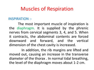
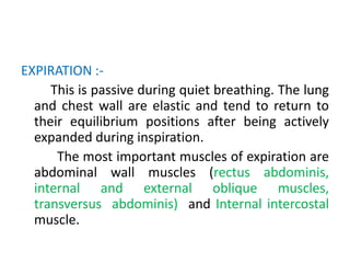
LUNG MECHANICS
PRESENTER :- DR RAVE RANJAN KUMAR
MODERATOR :- DR ZIA ARSHAD
Muscles of Respiration
INSPIRATION :-
The most important muscle of inspiration is
the diaphragm. It is supplied by the phrenic
nerves from cervical segments 3, 4, and 5. When
it contracts, the abdominal contents are forced
downward and forward, and the vertical
dimension of the chest cavity is increased.
In addition, the rib margins are lifted and
moved out, causing an increase in the transverse
diameter of the thorax . In normal tidal breathing,
the level of the diaphragm moves about 1-2 cm.
The external intercostal muscles connect
adjacent ribs and slope downwards and forward.
When they contract, the ribs are pulled upward
and forward, causing an increase in both the
lateral and the antero-posterior diameters of
the thorax. The lateral dimension increases
because of the “bucket-handle” movement of the
ribs.
The accessory muscles of inspiration include
the scalene muscles, which elevate the first two
ribs, and the sternomastoids, which raise the
sternum.
EXPIRATION :-
This is passive during quiet breathing. The lung
and chest wall are elastic and tend to return to
their equilibrium positions after being actively
expanded during inspiration.
The most important muscles of expiration are
abdominal wall muscles (rectus abdominis,
internal and external oblique muscles,
transversus abdominis) and Internal intercostal
muscle.
Spontaneous Ventilation
Alveolar pressure is normally atmospheric (zero
for reference) at end-inspiration and end-
expiration. The difference in pressure between the
inside and the outside of the lung is known as
transpulmonary pressure.
P transpulmonary = P alveolar − P intrapleural
The pressure surrounding the lung is less than
atmospheric, because of the elastic recoil of the lung.
At end-expiration, intrapleural pressure normally
averages about - 5 cm H2O, and because alveolar
pressure is 0 (no flow), transpulmonary pressure is +
5 cm H2O.
Diaphragmatic and intercostal muscle
activation during inspiration expands the chest
and decreases intrapleural pressure from –5
cm H2O to –8 or –9 cm H2O. As a result, alveolar
pressure also decreases (between –3 and –4 cm
H2O), and an alveolar–upper airway gradient is
established; gas flows from the upper airway
into alveoli.
At end-inspiration (when gas inflow has
ceased), alveolar pressure returns to zero, but
intrapleural pressure remains decreased; the
new transpulmonary pressure (8 cm H2O)
sustains lung expansion.
During expiration, diaphragmatic relaxation
returns intrapleural pressure to –5 cm H2O.
Now the transpulmonary pressure does not
support the new lung volume, and the elastic
recoil of the lung causes a reversal of the
previous alveolar–upper airway gradient; gas
flows out of alveoli, and original lung volume
is restored.
Regional Differences in Ventilation
Intrapleural pressure is higher (less negative) at the
bottom than the top of the lung due to weight of the
lung. Because the expanding pressure at the base
of the lung is small, this region has a small resting
volume. As the lung is easier to inflate at low
volumes than at high volumes, it expands well on
inspiration.
By contrast, the apex of the lung has a large
expanding pressure,hence, a big resting volume, and
thus a small change in volume occurs in inspiration.
Similarly, dependent lung is ventilated more in both
the supine and lateral positions.
The movement of the lungs is determined by :-
Elastic resistance of tissues
Gas–liquid interface
Nonelastic resistance to gas flow.
Elastic resistance governs lung volume and the
associated pressures under static conditions.
Resistance to gas flow relates to frictional resistance
to airflow and tissue deformation. The work
necessary to overcome elastic resistance is stored as
potential energy, but the work necessary to
overcome nonelastic resistance is lost as heat.
Elastic Resistance
Both the lungs and the chest have elastic
properties. The chest has a tendency to
expand outward at volumes, whereas the
lungs have a tendency to collapse.
The recoil properties of the chest are
due to chest wall muscle tone. The elastic
recoil of the lungs is due to their high content
of elastin fibers, and, even more important,
the surface tension forces acting at the air–
fluid interface in alveoli.
Surface Tension Forces
The gas–fluid interface lining the alveoli causes
them to behave as bubbles. Surface tension
forces tend to reduce the area of the interface
and favor alveolar collapse. Laplace’s law:-
Pressure =2 × Surface tension/Radius
Alveolar collapse is therefore directly
proportional to surface tension. Pulmonary
surfactant (secreted by type II alveolar cells) is a
phospholipid (dipalmitoyl phosphatidylcholine)
(DPPC), which decreases alveolar surface tension.
.
How does surfactant reduce the surface
tension so much?
Apparently the molecules of DPPC
are hydrophobic at one end and hydrophilic at
the other, and they align themselves in the
surface. When this occurs, their
intermolecular repulsive forces oppose the
normal attracting forces between the liquid
surface molecules that are responsible for
surface tension.
The ability of the surfactant to lower surface
tension is directly proportional to its
concentration within the alveolus.
As alveoli become smaller, the surfactant
within becomes more concentrated, and
surface tension is more effectively reduced.
Conversely, when alveoli are overdistended,
surfactant becomes less concentrated, and
surface tension increases. The net effect is to
stabilize alveoli; small alveoli are prevented
from getting smaller, whereas large alveoli
are prevented from getting larger.
A low surface tension in the alveoli increases the
compliance of the lung and reduces the work of
expanding it with each breath.
A third role of surfactant is to help to keep
the alveoli dry. Just as the surface tension forces
tend to collapse alveoli, they also tend to suck fluid
out of the capillaries. By reducing these surface
forces, surfactant prevents pulmonary edema to
develop.
Compliance
Elastic recoil is usually measured in terms of
compliance (C), which is defined as the change
in volume per unit change in the distending
pressure. Compliance measurements can be
obtained for either the chest, the lung, or
both together.
Lung compliance (CL) is defined as Change in
lung volume / Change in transpulmonary
pressure.
Various factors like lung volume, pulmonary
blood volume, extravascular lung water, and
pathological processes (eg, inflammation and
fibrosis) affect CL. An increased compliance
occurs in pulmonary emphysema and in the
normal aging lung.
Chest wall compliance (Cw)=Change in chest
volume /Change in transthoracic pressure, where
transthoracic pressure equals atmospheric
pressure minus intrapleural pressure.
Normal lung and chest wall compliance is
200 mL/cm H2O. Total compliance (lung and
chest wall together) is 100 mL/cm H2O.
Pressure-Volume Curve of the Lung
• Nonlinear, with the lung becoming stiffer at
high volumes
• Shows hysteresis ( curve that the lung follows
during inflation and deflation), the lung
volume at any given pressure during deflation
is larger than during inflation.
• Compliance is the slope of P-V curve (ΔV/ΔP)
• Behavior depends on both structural proteins
(collagen, elastin) and surface tension.
Functional Residual Capacity
The lung volume at the end of a normal
exhalation is called functional residual
capacity (FRC).
FRC =RV + ERV.
At this volume, the inward elastic recoil of the
lung approximates the outward elastic recoil
of the chest. Functional residual capacity can
be measured by nitrogen washout or helium
washin technique or body plethysmography.
Factors known to alter the FRC
• Body habitus: FRC is directly proportional to
height. Obesity markedly decreases FRC
(primarily as a result of reduced chest
compliance).
• Sex: FRC is reduced by about 10% in females
compared with males.
• Lung disease: Decreased compliance of the
lung, chest, or both is characteristic of
restrictive pulmonary disorders, all of which
are associated with a low FRC.
• Posture:- The supine position reduces the
FRC by 0.8–1.0 litre (alveolar collapse and
compression atelectasis due to loss of
inspiratory muscle tone, change in chest wall
rigidity, and upward shift of the diaphragm due
to upward push by abdominal contents).
The greatest change occurs between 0°
and 60° of inclination. No further decrease is
observed with a head-down position of up to 30°
Steep head-down (Trendelenburg) position
(>30°) may reduce FRC even further as
intrathoracic blood volume increases.
Closing Capacity
Small airways (lacking cartilaginous support)
depend on radial traction caused by the elastic
recoil of surrounding tissue to keep them open;
patency of these airways is highly dependent on
lung volume. The volume at which these airways
begin to close is called the closing capacity.
At lower lung volumes, alveoli in dependent
areas continue to be perfused, but are no longer
ventilated causing intrapulmonary shunting and
thus, hypoxemia .
Closing capacity is normally well below
FRC , but rises steadily with age. This increase is
probably responsible for the normal age-related
decline in arterial O2 tension.
At an average age of 44 years, closing
capacity equals FRC in the supine position; by
age 66, closing capacity equals or exceeds FRC in
the upright position in most individuals. Unlike
FRC, closing capacity is unaffected by posture.
Nonelastic Resistances
Airway resistance is the pressure difference between
the alveoli and the mouth divided by flow rate. Airway
Resistance to gas flow in the lung is a mixture of
laminar and turbulent flow.
Laminar flow can be thought of as consisting of
concentric cylinders of gas flowing at different
velocities; velocity is highest (2 times the average
velocity) in the center and decreases toward the
periphery. This changing velocity across the diameter
of the tube is known as the velocity profile.
During laminar flow,
Flow = Pressure gradient/ Raw, where Raw is
airway resistance.
Raw =8 × Length × Gas viscosity /π × (Radius)4
Turbulent flow is characterized by random movement
of the gas molecules down the air passages.
Pressure gradient ≈ Flow 2 × Gas density/Radius 5
Resistance is directly proportional to gas
density and inversely proportional to the fifth power of
the radius. As a result, turbulent gas flow is extremely
sensitive to airway caliber.
Turbulence generally occurs at high gas flows, at
sharp angles or branching points, and in response to
abrupt changes in airway diameter. Whether turbulent
or laminar flow occurs can be predicted by the
Reynolds number.
Reynolds number =Linear velocity × Diameter × Gas
density/Gas viscosity
Reynolds number < 1000 - laminar flow ,
> 2000 - turbulent flow.
Laminar flow normally occurs only distal to
small bronchioles ( < 1 mm). Flow in larger airways
is generally turbulent.
Of the gases used clinically, only helium has a
significantly lower density-to-viscosity ratio,
making it useful clinically during severe turbulent
flow (as in upper airway obstruction).
Normal total airway resistance is about 0.5–2
cm H2O/L/sec, with the largest contribution coming
from medium-sized bronchi (before the 7th generation).
Resistance in large bronchi is low because of
their large diameters, whereas resistance in small
bronchi is low because of their large total cross-
sectional area. Considerable small airway disease can
be present before the usual measurements of airway
resistance can detect an abnormality.
The most important causes of increased airway
resistance include bronchospasm, secretions, and
mucosal edema as well as volume-related and flow-
related airway collapse.
Volume-Related Airway Collapse
At low lung volumes, loss of radial
traction increases the contribution of small
airways to total resistance; airway resistance
becomes inversely proportional to lung
volume .
Increasing lung volume up to normal with
positive end-expiratory pressure (PEEP) can
reduce airway resistance.
Flow-Related Airway Collapse
During forced exhalation, reversal of the
normal transmural airway pressure can cause
collapse airways (dynamic airway compression).
Two factors are responsible:- generation of a
positive pleural pressure and a large pressure drop
across intrathoracic airways as a result of increased
airway resistance.
During dynamic compression, flow is
determined by alveolar pressure minus pleural
pressure (not mouth pressure).Hence, the
terminal portion of the flow/volume curve is
effort independent.
The point along the airways where
dynamic compression occurs is called the equal
pressure point. It is normally beyond the eleventh
to thirteenth generation of bronchioles where
cartilaginous support is absent. The equal
pressure point moves toward smaller airways as
lung volume decreases.
Emphysema or asthma predisposes
patients to dynamic airway compression.
Premature termination of exhalation may
increase FRC above normal, resulting in air
trapping and auto-PEEP.
Forced Vital Capacity
Forced Vital Capacity provides important
information about airway resistance. The ratio of
the forced expiratory volume in the first second
of exhalation (FEV1) to the total forced vital
capacity (FVC) is proportional to the degree of
airway obstruction. Normally, FEV1/FVC is ≥ 80%.
Whereas both FEV1 and FVC are effort
dependent, forced midexpiratory flow (FEF 25–
75%) is more effort independent and may be a
more reliable measurement of obstruction.
Tissue Resistance
This component of nonelastic
resistance may account for up to 20% of the
total (tissue + airway) resistance in young
normal subjects.
Tissue Resistance is primarily due to
viscoelastic (frictional) resistance of tissues to
gas flow, as they slide over each other during
respiration.
Work of Breathing
Because expiration is normally entirely
passive, both the inspiratory and the expiratory
work of breathing is performed by the inspiratory
muscles (primarily the diaphragm).
Three factors must be overcome during
ventilation:-
the elastic recoil of the chest and lung,
frictional resistance to gas flow in the airways,
and tissue frictional resistance.
During inhalation, nearly 50% of the energy
expended is stored as pulmonary elastic recoil. During
exhalation, the stored potential energy is released and
overcomes expiratory airway resistance. Increases in
either inspiratory or expiratory resistance are
compensated by increased inspiratory muscle effort.
When expiratory resistance increases, the normal
compensatory response is to increase lung volume
such that Vt breathing occurs at an abnormally high
FRC. The greater elastic recoil energy stored at a higher
lung volume overcomes the added expiratory
resistance.
The work required to overcome elastic
resistance increases as Vt increases, whereas
the work required to overcome airflow
resistance increases as respiratory rate (and,
necessarily, expiratory flow) increases. Faced
with either condition, patients minimize the
work of breathing by altering the respiratory
rate and Vt .
Patients with reduced compliance tend to
have rapid, shallow breaths, whereas those with
increased airflow resistance have a slow, deep
breathing pattern .
Respiratory muscles normally account for
only 2% to 3% of O2 consumption, but
operate at about 5-10% efficiency. Ninety
percent of the work is dissipated as heat (due
to elastic and airflow resistance).
Lung mechanics

Lung mechanics

  • 1.
    LUNG MECHANICS PRESENTER :-DR RAVE RANJAN KUMAR MODERATOR :- DR ZIA ARSHAD
  • 2.
    Muscles of Respiration INSPIRATION:- The most important muscle of inspiration is the diaphragm. It is supplied by the phrenic nerves from cervical segments 3, 4, and 5. When it contracts, the abdominal contents are forced downward and forward, and the vertical dimension of the chest cavity is increased. In addition, the rib margins are lifted and moved out, causing an increase in the transverse diameter of the thorax . In normal tidal breathing, the level of the diaphragm moves about 1-2 cm.
  • 3.
    The external intercostalmuscles connect adjacent ribs and slope downwards and forward. When they contract, the ribs are pulled upward and forward, causing an increase in both the lateral and the antero-posterior diameters of the thorax. The lateral dimension increases because of the “bucket-handle” movement of the ribs. The accessory muscles of inspiration include the scalene muscles, which elevate the first two ribs, and the sternomastoids, which raise the sternum.
  • 5.
    EXPIRATION :- This ispassive during quiet breathing. The lung and chest wall are elastic and tend to return to their equilibrium positions after being actively expanded during inspiration. The most important muscles of expiration are abdominal wall muscles (rectus abdominis, internal and external oblique muscles, transversus abdominis) and Internal intercostal muscle.
  • 6.
    Spontaneous Ventilation Alveolar pressureis normally atmospheric (zero for reference) at end-inspiration and end- expiration. The difference in pressure between the inside and the outside of the lung is known as transpulmonary pressure. P transpulmonary = P alveolar − P intrapleural The pressure surrounding the lung is less than atmospheric, because of the elastic recoil of the lung. At end-expiration, intrapleural pressure normally averages about - 5 cm H2O, and because alveolar pressure is 0 (no flow), transpulmonary pressure is + 5 cm H2O.
  • 7.
    Diaphragmatic and intercostalmuscle activation during inspiration expands the chest and decreases intrapleural pressure from –5 cm H2O to –8 or –9 cm H2O. As a result, alveolar pressure also decreases (between –3 and –4 cm H2O), and an alveolar–upper airway gradient is established; gas flows from the upper airway into alveoli. At end-inspiration (when gas inflow has ceased), alveolar pressure returns to zero, but intrapleural pressure remains decreased; the new transpulmonary pressure (8 cm H2O) sustains lung expansion.
  • 8.
    During expiration, diaphragmaticrelaxation returns intrapleural pressure to –5 cm H2O. Now the transpulmonary pressure does not support the new lung volume, and the elastic recoil of the lung causes a reversal of the previous alveolar–upper airway gradient; gas flows out of alveoli, and original lung volume is restored.
  • 10.
    Regional Differences inVentilation Intrapleural pressure is higher (less negative) at the bottom than the top of the lung due to weight of the lung. Because the expanding pressure at the base of the lung is small, this region has a small resting volume. As the lung is easier to inflate at low volumes than at high volumes, it expands well on inspiration. By contrast, the apex of the lung has a large expanding pressure,hence, a big resting volume, and thus a small change in volume occurs in inspiration. Similarly, dependent lung is ventilated more in both the supine and lateral positions.
  • 11.
    The movement ofthe lungs is determined by :- Elastic resistance of tissues Gas–liquid interface Nonelastic resistance to gas flow. Elastic resistance governs lung volume and the associated pressures under static conditions. Resistance to gas flow relates to frictional resistance to airflow and tissue deformation. The work necessary to overcome elastic resistance is stored as potential energy, but the work necessary to overcome nonelastic resistance is lost as heat.
  • 12.
    Elastic Resistance Both thelungs and the chest have elastic properties. The chest has a tendency to expand outward at volumes, whereas the lungs have a tendency to collapse. The recoil properties of the chest are due to chest wall muscle tone. The elastic recoil of the lungs is due to their high content of elastin fibers, and, even more important, the surface tension forces acting at the air– fluid interface in alveoli.
  • 13.
    Surface Tension Forces Thegas–fluid interface lining the alveoli causes them to behave as bubbles. Surface tension forces tend to reduce the area of the interface and favor alveolar collapse. Laplace’s law:- Pressure =2 × Surface tension/Radius Alveolar collapse is therefore directly proportional to surface tension. Pulmonary surfactant (secreted by type II alveolar cells) is a phospholipid (dipalmitoyl phosphatidylcholine) (DPPC), which decreases alveolar surface tension. .
  • 14.
    How does surfactantreduce the surface tension so much? Apparently the molecules of DPPC are hydrophobic at one end and hydrophilic at the other, and they align themselves in the surface. When this occurs, their intermolecular repulsive forces oppose the normal attracting forces between the liquid surface molecules that are responsible for surface tension.
  • 16.
    The ability ofthe surfactant to lower surface tension is directly proportional to its concentration within the alveolus. As alveoli become smaller, the surfactant within becomes more concentrated, and surface tension is more effectively reduced. Conversely, when alveoli are overdistended, surfactant becomes less concentrated, and surface tension increases. The net effect is to stabilize alveoli; small alveoli are prevented from getting smaller, whereas large alveoli are prevented from getting larger.
  • 17.
    A low surfacetension in the alveoli increases the compliance of the lung and reduces the work of expanding it with each breath. A third role of surfactant is to help to keep the alveoli dry. Just as the surface tension forces tend to collapse alveoli, they also tend to suck fluid out of the capillaries. By reducing these surface forces, surfactant prevents pulmonary edema to develop.
  • 18.
    Compliance Elastic recoil isusually measured in terms of compliance (C), which is defined as the change in volume per unit change in the distending pressure. Compliance measurements can be obtained for either the chest, the lung, or both together. Lung compliance (CL) is defined as Change in lung volume / Change in transpulmonary pressure.
  • 19.
    Various factors likelung volume, pulmonary blood volume, extravascular lung water, and pathological processes (eg, inflammation and fibrosis) affect CL. An increased compliance occurs in pulmonary emphysema and in the normal aging lung. Chest wall compliance (Cw)=Change in chest volume /Change in transthoracic pressure, where transthoracic pressure equals atmospheric pressure minus intrapleural pressure. Normal lung and chest wall compliance is 200 mL/cm H2O. Total compliance (lung and chest wall together) is 100 mL/cm H2O.
  • 20.
    Pressure-Volume Curve ofthe Lung • Nonlinear, with the lung becoming stiffer at high volumes • Shows hysteresis ( curve that the lung follows during inflation and deflation), the lung volume at any given pressure during deflation is larger than during inflation. • Compliance is the slope of P-V curve (ΔV/ΔP) • Behavior depends on both structural proteins (collagen, elastin) and surface tension.
  • 21.
    Functional Residual Capacity Thelung volume at the end of a normal exhalation is called functional residual capacity (FRC). FRC =RV + ERV. At this volume, the inward elastic recoil of the lung approximates the outward elastic recoil of the chest. Functional residual capacity can be measured by nitrogen washout or helium washin technique or body plethysmography.
  • 23.
    Factors known toalter the FRC • Body habitus: FRC is directly proportional to height. Obesity markedly decreases FRC (primarily as a result of reduced chest compliance). • Sex: FRC is reduced by about 10% in females compared with males. • Lung disease: Decreased compliance of the lung, chest, or both is characteristic of restrictive pulmonary disorders, all of which are associated with a low FRC.
  • 24.
    • Posture:- Thesupine position reduces the FRC by 0.8–1.0 litre (alveolar collapse and compression atelectasis due to loss of inspiratory muscle tone, change in chest wall rigidity, and upward shift of the diaphragm due to upward push by abdominal contents). The greatest change occurs between 0° and 60° of inclination. No further decrease is observed with a head-down position of up to 30° Steep head-down (Trendelenburg) position (>30°) may reduce FRC even further as intrathoracic blood volume increases.
  • 25.
    Closing Capacity Small airways(lacking cartilaginous support) depend on radial traction caused by the elastic recoil of surrounding tissue to keep them open; patency of these airways is highly dependent on lung volume. The volume at which these airways begin to close is called the closing capacity. At lower lung volumes, alveoli in dependent areas continue to be perfused, but are no longer ventilated causing intrapulmonary shunting and thus, hypoxemia .
  • 26.
    Closing capacity isnormally well below FRC , but rises steadily with age. This increase is probably responsible for the normal age-related decline in arterial O2 tension. At an average age of 44 years, closing capacity equals FRC in the supine position; by age 66, closing capacity equals or exceeds FRC in the upright position in most individuals. Unlike FRC, closing capacity is unaffected by posture.
  • 27.
    Nonelastic Resistances Airway resistanceis the pressure difference between the alveoli and the mouth divided by flow rate. Airway Resistance to gas flow in the lung is a mixture of laminar and turbulent flow. Laminar flow can be thought of as consisting of concentric cylinders of gas flowing at different velocities; velocity is highest (2 times the average velocity) in the center and decreases toward the periphery. This changing velocity across the diameter of the tube is known as the velocity profile. During laminar flow, Flow = Pressure gradient/ Raw, where Raw is airway resistance. Raw =8 × Length × Gas viscosity /π × (Radius)4
  • 29.
    Turbulent flow ischaracterized by random movement of the gas molecules down the air passages. Pressure gradient ≈ Flow 2 × Gas density/Radius 5 Resistance is directly proportional to gas density and inversely proportional to the fifth power of the radius. As a result, turbulent gas flow is extremely sensitive to airway caliber. Turbulence generally occurs at high gas flows, at sharp angles or branching points, and in response to abrupt changes in airway diameter. Whether turbulent or laminar flow occurs can be predicted by the Reynolds number. Reynolds number =Linear velocity × Diameter × Gas density/Gas viscosity
  • 31.
    Reynolds number <1000 - laminar flow , > 2000 - turbulent flow. Laminar flow normally occurs only distal to small bronchioles ( < 1 mm). Flow in larger airways is generally turbulent. Of the gases used clinically, only helium has a significantly lower density-to-viscosity ratio, making it useful clinically during severe turbulent flow (as in upper airway obstruction).
  • 32.
    Normal total airwayresistance is about 0.5–2 cm H2O/L/sec, with the largest contribution coming from medium-sized bronchi (before the 7th generation). Resistance in large bronchi is low because of their large diameters, whereas resistance in small bronchi is low because of their large total cross- sectional area. Considerable small airway disease can be present before the usual measurements of airway resistance can detect an abnormality. The most important causes of increased airway resistance include bronchospasm, secretions, and mucosal edema as well as volume-related and flow- related airway collapse.
  • 33.
    Volume-Related Airway Collapse Atlow lung volumes, loss of radial traction increases the contribution of small airways to total resistance; airway resistance becomes inversely proportional to lung volume . Increasing lung volume up to normal with positive end-expiratory pressure (PEEP) can reduce airway resistance.
  • 34.
    Flow-Related Airway Collapse Duringforced exhalation, reversal of the normal transmural airway pressure can cause collapse airways (dynamic airway compression). Two factors are responsible:- generation of a positive pleural pressure and a large pressure drop across intrathoracic airways as a result of increased airway resistance. During dynamic compression, flow is determined by alveolar pressure minus pleural pressure (not mouth pressure).Hence, the terminal portion of the flow/volume curve is effort independent.
  • 36.
    The point alongthe airways where dynamic compression occurs is called the equal pressure point. It is normally beyond the eleventh to thirteenth generation of bronchioles where cartilaginous support is absent. The equal pressure point moves toward smaller airways as lung volume decreases. Emphysema or asthma predisposes patients to dynamic airway compression. Premature termination of exhalation may increase FRC above normal, resulting in air trapping and auto-PEEP.
  • 37.
    Forced Vital Capacity ForcedVital Capacity provides important information about airway resistance. The ratio of the forced expiratory volume in the first second of exhalation (FEV1) to the total forced vital capacity (FVC) is proportional to the degree of airway obstruction. Normally, FEV1/FVC is ≥ 80%. Whereas both FEV1 and FVC are effort dependent, forced midexpiratory flow (FEF 25– 75%) is more effort independent and may be a more reliable measurement of obstruction.
  • 38.
    Tissue Resistance This componentof nonelastic resistance may account for up to 20% of the total (tissue + airway) resistance in young normal subjects. Tissue Resistance is primarily due to viscoelastic (frictional) resistance of tissues to gas flow, as they slide over each other during respiration.
  • 39.
    Work of Breathing Becauseexpiration is normally entirely passive, both the inspiratory and the expiratory work of breathing is performed by the inspiratory muscles (primarily the diaphragm). Three factors must be overcome during ventilation:- the elastic recoil of the chest and lung, frictional resistance to gas flow in the airways, and tissue frictional resistance.
  • 40.
    During inhalation, nearly50% of the energy expended is stored as pulmonary elastic recoil. During exhalation, the stored potential energy is released and overcomes expiratory airway resistance. Increases in either inspiratory or expiratory resistance are compensated by increased inspiratory muscle effort. When expiratory resistance increases, the normal compensatory response is to increase lung volume such that Vt breathing occurs at an abnormally high FRC. The greater elastic recoil energy stored at a higher lung volume overcomes the added expiratory resistance.
  • 41.
    The work requiredto overcome elastic resistance increases as Vt increases, whereas the work required to overcome airflow resistance increases as respiratory rate (and, necessarily, expiratory flow) increases. Faced with either condition, patients minimize the work of breathing by altering the respiratory rate and Vt . Patients with reduced compliance tend to have rapid, shallow breaths, whereas those with increased airflow resistance have a slow, deep breathing pattern .
  • 42.
    Respiratory muscles normallyaccount for only 2% to 3% of O2 consumption, but operate at about 5-10% efficiency. Ninety percent of the work is dissipated as heat (due to elastic and airflow resistance).