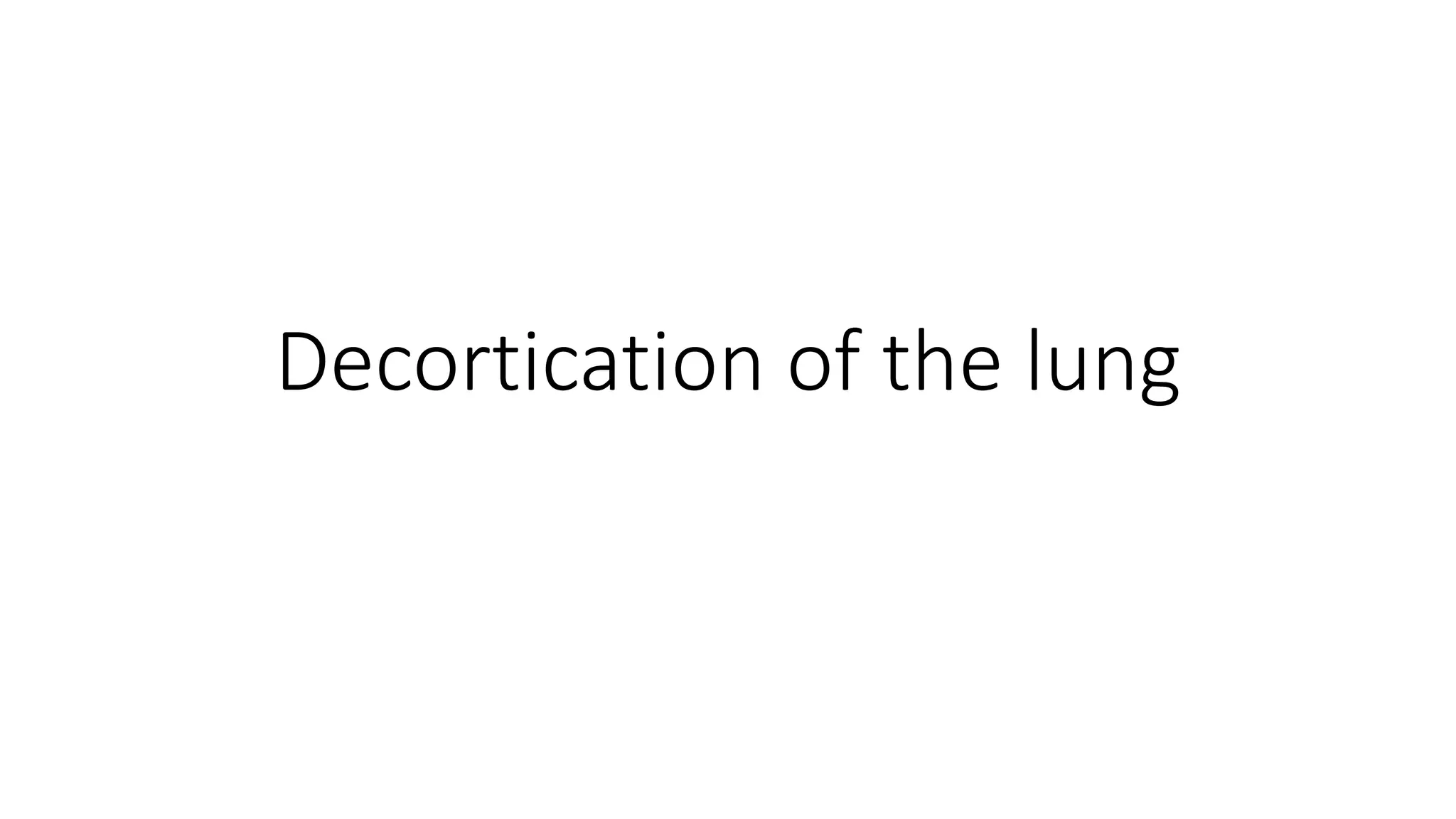
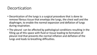
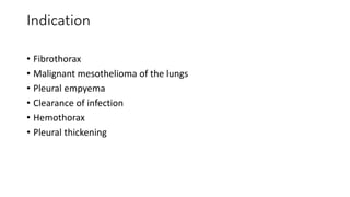
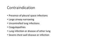
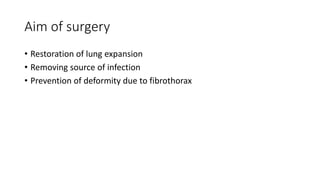
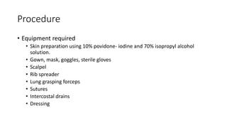
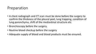
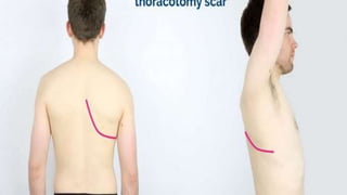
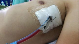
Decortication of the lung
Decortication
• Decortication of the lungs is a surgical procedure that is done to
remove fibrous tissue that envelops the lungs, the chest wall and the
diaphragm, to enable the normal expansion and deflation of lungs
during respiration.
• The pleural can be affected by pathological conditions resulting in the
filling up of this space with fluid or tissue leading to formation of
pleural rind that prevents the normal inflation and deflation of the
lungs and leads to breathing difficulties.
Indication
• Fibrothorax
• Malignant mesothelioma of the lungs
• Pleural empyema
• Clearance of infection
• Hemothorax
• Pleural thickening
Contraindication
• Presence of pleural space infections
• Large airway narrowing
• Uncontrolled lung infections
• Coagulopathies
• Lung infection or disease of other lung
• Severe chest wall disease or infection
Aim of surgery
• Restoration of lung expansion
• Removing source of infection
• Prevention of deformity due to fibrothorax
Procedure
• Equipment required
• Skin preparation using 10% povidone- iodine and 70% isopropyl alcohol
solution.
• Gown, mask, goggles, sterile gloves
• Scalpel
• Rib spreader
• Lung grasping forceps
• Sutures
• Intercostal drains
• Dressing
Preparation
• A chest radiograph and CT scan must be done before the surgery to
confirm the thickness of the pleural peel, lung trapping, condition of
lung parenchyma, shift of the mediastinal structure etc.
• Bronchoscopy before the surgery.
• Routine blood checkup before the surgery
• Adequate supply of blood and blood products must be ensured.
Pre procedure positioning
• The patient is placed in the lateral decubitus position with the
diseased side up.
• A folded towel or a roll is placed below the dependent side.
• The down leg is flexed to 90 degrees and a pillow is placed between
the legs.
Technique
• Postero-lateral thoracotomy is done.
• Entry into the thoracic cavity is established via fifth or sixth intercostal
space.
• A rib resection might be required if there is excessive crowding of
ribs.
• After the division of intercostal muscles, the extrapleural space is
entered.
• The mediastinum is generally not involved in inflammatory process,
therefore care must be taken to avoid injury to mediastinal structures.
• The apex of the lung must be freed carefully because injury to
subclavian vessels may occur during apical dissection and can cause
hemorrhage.
• Care taken to avoid injury to oesophagus or venacava during medial
dissection and diaphragm during inferior dissection.
• The pleural peel must be removed from the lung parenchyma,
including the fissures.
• After removal of thick peel, the lung is inflated to locate the air leaks.
• All the major air leaks must be closed with suture.
• The intercostal drain is inserted in the thoracic interspace.
• Some surgeons insert two drains one in the base and one in the apex.
• Subsequently chest wall closure is done.
Post op care
• Adequate analgesia.
• Antibiotic therapy.
• Hydration’
• Nutritional support.
• Patient may often require mechanical ventilation.
• Chest tube care must be done.
Complication
• Hemorrhage
• Persistent air leaks and bronchopleural fistula
• Persistent lung collapse
• Injury to vital structures
• Infection and sepsis
• Severe postoperative pain
• Chest wall deformity and scoliosis
Decortication of the lung.pptx

Decortication of the lung.pptx

  • 1.
  • 3.
    Decortication • Decortication ofthe lungs is a surgical procedure that is done to remove fibrous tissue that envelops the lungs, the chest wall and the diaphragm, to enable the normal expansion and deflation of lungs during respiration. • The pleural can be affected by pathological conditions resulting in the filling up of this space with fluid or tissue leading to formation of pleural rind that prevents the normal inflation and deflation of the lungs and leads to breathing difficulties.
  • 4.
    Indication • Fibrothorax • Malignantmesothelioma of the lungs • Pleural empyema • Clearance of infection • Hemothorax • Pleural thickening
  • 5.
    Contraindication • Presence ofpleural space infections • Large airway narrowing • Uncontrolled lung infections • Coagulopathies • Lung infection or disease of other lung • Severe chest wall disease or infection
  • 6.
    Aim of surgery •Restoration of lung expansion • Removing source of infection • Prevention of deformity due to fibrothorax
  • 7.
    Procedure • Equipment required •Skin preparation using 10% povidone- iodine and 70% isopropyl alcohol solution. • Gown, mask, goggles, sterile gloves • Scalpel • Rib spreader • Lung grasping forceps • Sutures • Intercostal drains • Dressing
  • 9.
    Preparation • A chestradiograph and CT scan must be done before the surgery to confirm the thickness of the pleural peel, lung trapping, condition of lung parenchyma, shift of the mediastinal structure etc. • Bronchoscopy before the surgery. • Routine blood checkup before the surgery • Adequate supply of blood and blood products must be ensured.
  • 10.
    Pre procedure positioning •The patient is placed in the lateral decubitus position with the diseased side up. • A folded towel or a roll is placed below the dependent side. • The down leg is flexed to 90 degrees and a pillow is placed between the legs.
  • 12.
    Technique • Postero-lateral thoracotomyis done. • Entry into the thoracic cavity is established via fifth or sixth intercostal space. • A rib resection might be required if there is excessive crowding of ribs. • After the division of intercostal muscles, the extrapleural space is entered. • The mediastinum is generally not involved in inflammatory process, therefore care must be taken to avoid injury to mediastinal structures.
  • 14.
    • The apexof the lung must be freed carefully because injury to subclavian vessels may occur during apical dissection and can cause hemorrhage. • Care taken to avoid injury to oesophagus or venacava during medial dissection and diaphragm during inferior dissection. • The pleural peel must be removed from the lung parenchyma, including the fissures.
  • 15.
    • After removalof thick peel, the lung is inflated to locate the air leaks. • All the major air leaks must be closed with suture. • The intercostal drain is inserted in the thoracic interspace. • Some surgeons insert two drains one in the base and one in the apex. • Subsequently chest wall closure is done.
  • 16.
    Post op care •Adequate analgesia. • Antibiotic therapy. • Hydration’ • Nutritional support. • Patient may often require mechanical ventilation. • Chest tube care must be done.
  • 19.
    Complication • Hemorrhage • Persistentair leaks and bronchopleural fistula • Persistent lung collapse • Injury to vital structures • Infection and sepsis • Severe postoperative pain • Chest wall deformity and scoliosis