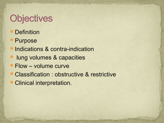
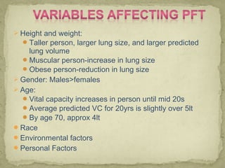
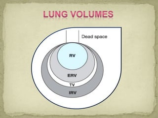
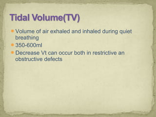
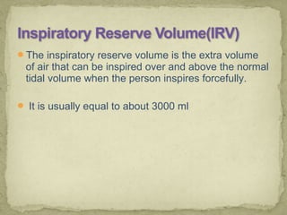
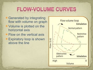
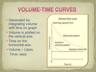
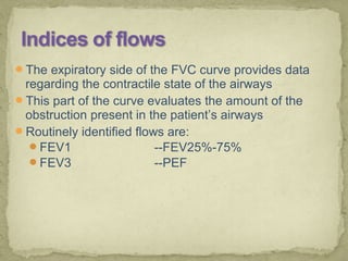
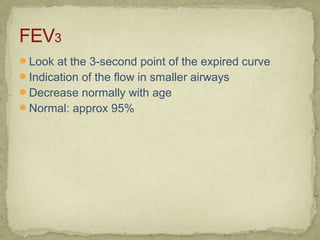
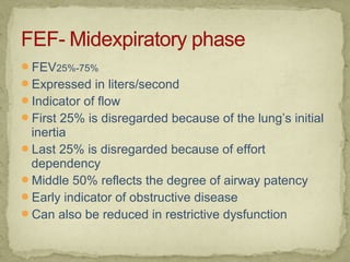
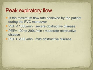
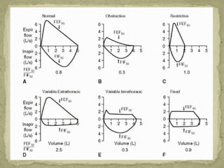
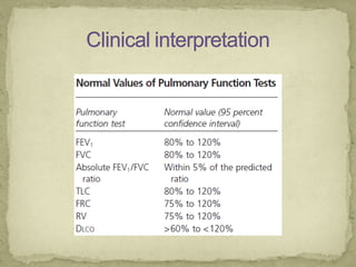
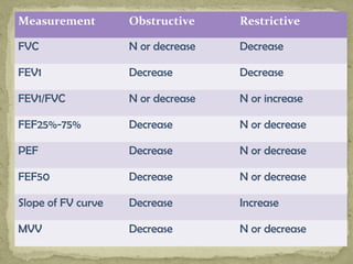
Dr.Nidhi Ahya
Assistant Professor
Cardio-Vascular & Respiratory PTDVVPF
College of Physiotherapy,
Ahmednagar 414111
Definition
Purpose
Indications & contra-indication
 lung volumes & capacities
Flow – volume curve
Classification : obstructive & restrictive
Clinical interpretation.
Pulmonary function testing is the process of
having the patient perform specific inspiratory
and expiratory maneuvers while breathing in and
out of tubing attached to the equipment that
measure a variety of variables
To determine the functional status of the
lungs
How much gas can be moved in and out of the lungs
How fast gas can be moved
The stiffness of the lung and chest wall
The diffusion characteristics of the alveolar-capillary
membrane
How well lung responds to the therapy
• To evaluate symptoms, signs or abnormal
laboratory tests
• To measure the effect of disease on pulmonary
function
• To differentiate between obstructive and
restrictive disease
• To screen individuals at risk of having pulmonary
disease
• To assess pre-operative risk
• To assess prognosis, response to therapy
• Hemoptysis of unknown origin
• Pneumothorax
• Unstable cardiovascular status or recent MI or
pulmonary embolus
• Thoracic, abdominal or cerebral aneurysms
• Presence of an acute disease process that might
interfere with test performance
• Recent surgery of thorax and abdomen
Nosocomial infection contracted from improperly
cleaned tubing, mouthpiece
 Height and weight:
Taller person, larger lung size, and larger predicted
lung volume
Muscular person-increase in lung size
Obese person-reduction in lung size
 Gender: Males>females
 Age:
Vital capacity increases in person until mid 20s
Average predicted VC for 20yrs is slightly over 5lt
By age 70, approx 4lt
Race
Environmental factors
Personal Factors
Volume of air exhaled and inhaled during quiet
breathing
350-600ml
Decrease Vt can occur both in restrictive an
obstructive defects
The inspiratory reserve volume is the extra volume
of air that can be inspired over and above the normal
tidal volume when the person inspires forcefully.
 It is usually equal to about 3000 ml
The expiratory reserve volume is the maximum
extra volume of air that can be expired by forceful
expiration after the end of a normal tidal expiration
 It normally amounts to about 1100 ml
The residual volume is the volume of air remaining in
the lungs after the most forceful expiration
 This volume averages about 1200 Ml
Is reduced in restrictive defects
Increased in obstructive defects
Expressed as ratio to TLC and VC
The inspiratory capacity equals the tidal volume
plus the inspiratory reserve volume.
It’s the amount of air a person can breathe in,
beginning at the normal expiratory level and
distending the lungs to the maximum amount.
about 3500 milliliters
 The vital capacity equals the inspiratory reserve
volume plus the tidal volume plus the expiratory
reserve volume.
This is the maximum amount of air a person can
expel from the lungs after first filling the lungs to
their maximum extent and then expiring to the
maximum extent
About 4600 ml
If the person forcefully exhales the volume, it is
called Forced vital capacity(FVC)
Three phases of FVC maneuver:
Maximal inspiratory effort
Initial expiratory blast
Forceful emptying of the lungs
Both restrictive and obstructive defects can cause
decrease in VC
Important preoperative assessment factor useful in
evaluating the patient’s need for mechanical
ventilation
FRC = ERV+RV
Resting volume of the lungs after exhalation of Vt
breath
This is the amount of air that remains in the lungs at
the end of normal expiration
About 2300 ml
Represents balance between the expanding chest
wall forces and the contractile rebound forces of
lung tissue
Sum of VC and RV
Is increased with most obstructive lung defects and
decreased with restrictive lung defects
To measure TLC, RV must be determined
The lung volumes that can be directly measured
using a spirometer include- Tidal Volume, Inspiratory
Reserve Volume, Expiratory Reserve Volume and
thus Vital Capacity
Total Lung Capacity can be measured if RV and
FRC are known.
RV and FRC is obtained in one of indirect methods:
Body plethysmograph (body box)
Open-circuit nitrogen washout
Closed-circuit helium dilution
Generated by integrating
flow with volume on graph
Volume is plotted on the
horizontal axis
Flow on the vertical axis
Expiratory loop is shown
above the line
Generated by
integrating volume
with time on graph
Volume is plotted on
the vertical axis
Time on the
horizontal axis
Volume – Liters
Time- secs
The expiratory side of the FVC curve provides data
regarding the contractile state of the airways
This part of the curve evaluates the amount of the
obstruction present in the patient’s airways
Routinely identified flows are:
FEV1 --FEV25%-75%
FEV3 --PEF
Measures the maximal volume of air exhaled during
the first second of expiration
Reflects the flow characteristics in the larger airways
Best indicator of obstructive diseases
Expressed as percentage of the observed FVC
(FEV1/FVC)
Normal: 75% of VC in 1 second
Decreased: acute and chronic obstructive pulmonary
disease
Normal in restrictive disorders
Look at the 3-second point of the expired curve
Indication of the flow in smaller airways
Decrease normally with age
Normal: approx 95%
FEV25%-75%
Expressed in liters/second
Indicator of flow
First 25% is disregarded because of the lung’s initial
inertia
Last 25% is disregarded because of effort
dependency
Middle 50% reflects the degree of airway patency
Early indicator of obstructive disease
Can also be reduced in restrictive dysfunction
Is the maximum flow rate achieved by the patient
during the FVC maneuver
PEF < 100L/min : severe obstructive disease
PEF= 100 to 200L/min : moderate obstructive
disease
PEF > 200L/min : mild obstructive disease
Indicates the respiratory muscle endurance
Requires the patient to inhale and exhale as quickly
as possible for a period of 12 secs
Normal: 170L/min for healthy young adult
Pulmonary function abnormalities can be
grouped into two main categories-
• Obstructive
• Restrictive
 Obstructive defects:
• Can occur in upper and larger airways or in smaller
airways(less than 2mm)
• Upper airways obstruction will reduce flow rates in the
initial 25% of a FVC maneuver
• Smaller airway obstruction will reduce flow rates in the
later portion of the exhaled volume
 Restrictive defects:
• Present when the lung volume are reduced to less
than 80% of the predicted levels
• Includes chest wall dysfunction, neurologic disorders,
scarring of lung, obesity
Measurement Obstructive Restrictive
FVC N or decrease Decrease
FEV1 Decrease Decrease
FEV1/FVC N or decrease N or increase
FEF25%-75% Decrease N or decrease
PEF Decrease N or decrease
FEF50 Decrease N or decrease
Slope of FV curve Decrease Increase
MVV Decrease N or decrease
Based on the initial results of a baseline spirometry,
additional testing of pulmonary mechanics is often
desirable.
If the baseline test indicates airway obstruction,
determining the reversibility of obstruction is
indicated.
In the laboratory , FVC maneuver is often repeated
after the patient has received a bronchodilator
Reversibility is defined as 15% or greater
improvement in FEV1
What is PFT & purpose of PFT.
Indications & contra-indication.
 lung volumes & capacities.
Flow – volume curve .
Classification : obstructive & restrictive.
Clinical interpretation.
1. WHAT IS PFT AND ITS PURPOSE? 5MARKS
2. WRITE THE INDICATION AND
CONTRAINDICATIONS OF THE PFT. 5MARKS
Pulmonary function testing

Pulmonary function testing

  • 1.
    Dr.Nidhi Ahya Assistant Professor Cardio-Vascular& Respiratory PTDVVPF College of Physiotherapy, Ahmednagar 414111
  • 2.
    Definition Purpose Indications & contra-indication lung volumes & capacities Flow – volume curve Classification : obstructive & restrictive Clinical interpretation.
  • 3.
    Pulmonary function testingis the process of having the patient perform specific inspiratory and expiratory maneuvers while breathing in and out of tubing attached to the equipment that measure a variety of variables
  • 4.
    To determine thefunctional status of the lungs How much gas can be moved in and out of the lungs How fast gas can be moved The stiffness of the lung and chest wall The diffusion characteristics of the alveolar-capillary membrane How well lung responds to the therapy
  • 6.
    • To evaluatesymptoms, signs or abnormal laboratory tests • To measure the effect of disease on pulmonary function • To differentiate between obstructive and restrictive disease • To screen individuals at risk of having pulmonary disease • To assess pre-operative risk • To assess prognosis, response to therapy
  • 7.
    • Hemoptysis ofunknown origin • Pneumothorax • Unstable cardiovascular status or recent MI or pulmonary embolus • Thoracic, abdominal or cerebral aneurysms • Presence of an acute disease process that might interfere with test performance • Recent surgery of thorax and abdomen
  • 8.
    Nosocomial infection contractedfrom improperly cleaned tubing, mouthpiece
  • 9.
     Height andweight: Taller person, larger lung size, and larger predicted lung volume Muscular person-increase in lung size Obese person-reduction in lung size  Gender: Males>females  Age: Vital capacity increases in person until mid 20s Average predicted VC for 20yrs is slightly over 5lt By age 70, approx 4lt Race Environmental factors Personal Factors
  • 11.
    Volume of airexhaled and inhaled during quiet breathing 350-600ml Decrease Vt can occur both in restrictive an obstructive defects
  • 12.
    The inspiratory reservevolume is the extra volume of air that can be inspired over and above the normal tidal volume when the person inspires forcefully.  It is usually equal to about 3000 ml
  • 13.
    The expiratory reservevolume is the maximum extra volume of air that can be expired by forceful expiration after the end of a normal tidal expiration  It normally amounts to about 1100 ml
  • 14.
    The residual volumeis the volume of air remaining in the lungs after the most forceful expiration  This volume averages about 1200 Ml Is reduced in restrictive defects Increased in obstructive defects Expressed as ratio to TLC and VC
  • 17.
    The inspiratory capacityequals the tidal volume plus the inspiratory reserve volume. It’s the amount of air a person can breathe in, beginning at the normal expiratory level and distending the lungs to the maximum amount. about 3500 milliliters
  • 18.
     The vitalcapacity equals the inspiratory reserve volume plus the tidal volume plus the expiratory reserve volume. This is the maximum amount of air a person can expel from the lungs after first filling the lungs to their maximum extent and then expiring to the maximum extent About 4600 ml If the person forcefully exhales the volume, it is called Forced vital capacity(FVC)
  • 19.
    Three phases ofFVC maneuver: Maximal inspiratory effort Initial expiratory blast Forceful emptying of the lungs Both restrictive and obstructive defects can cause decrease in VC Important preoperative assessment factor useful in evaluating the patient’s need for mechanical ventilation
  • 20.
    FRC = ERV+RV Restingvolume of the lungs after exhalation of Vt breath This is the amount of air that remains in the lungs at the end of normal expiration About 2300 ml Represents balance between the expanding chest wall forces and the contractile rebound forces of lung tissue
  • 21.
    Sum of VCand RV Is increased with most obstructive lung defects and decreased with restrictive lung defects To measure TLC, RV must be determined
  • 22.
    The lung volumesthat can be directly measured using a spirometer include- Tidal Volume, Inspiratory Reserve Volume, Expiratory Reserve Volume and thus Vital Capacity Total Lung Capacity can be measured if RV and FRC are known. RV and FRC is obtained in one of indirect methods: Body plethysmograph (body box) Open-circuit nitrogen washout Closed-circuit helium dilution
  • 23.
    Generated by integrating flowwith volume on graph Volume is plotted on the horizontal axis Flow on the vertical axis Expiratory loop is shown above the line
  • 24.
    Generated by integrating volume withtime on graph Volume is plotted on the vertical axis Time on the horizontal axis Volume – Liters Time- secs
  • 25.
    The expiratory sideof the FVC curve provides data regarding the contractile state of the airways This part of the curve evaluates the amount of the obstruction present in the patient’s airways Routinely identified flows are: FEV1 --FEV25%-75% FEV3 --PEF
  • 26.
    Measures the maximalvolume of air exhaled during the first second of expiration Reflects the flow characteristics in the larger airways Best indicator of obstructive diseases Expressed as percentage of the observed FVC (FEV1/FVC) Normal: 75% of VC in 1 second Decreased: acute and chronic obstructive pulmonary disease Normal in restrictive disorders
  • 27.
    Look at the3-second point of the expired curve Indication of the flow in smaller airways Decrease normally with age Normal: approx 95%
  • 28.
    FEV25%-75% Expressed in liters/second Indicatorof flow First 25% is disregarded because of the lung’s initial inertia Last 25% is disregarded because of effort dependency Middle 50% reflects the degree of airway patency Early indicator of obstructive disease Can also be reduced in restrictive dysfunction
  • 29.
    Is the maximumflow rate achieved by the patient during the FVC maneuver PEF < 100L/min : severe obstructive disease PEF= 100 to 200L/min : moderate obstructive disease PEF > 200L/min : mild obstructive disease
  • 30.
    Indicates the respiratorymuscle endurance Requires the patient to inhale and exhale as quickly as possible for a period of 12 secs Normal: 170L/min for healthy young adult
  • 31.
    Pulmonary function abnormalitiescan be grouped into two main categories- • Obstructive • Restrictive
  • 32.
     Obstructive defects: •Can occur in upper and larger airways or in smaller airways(less than 2mm) • Upper airways obstruction will reduce flow rates in the initial 25% of a FVC maneuver • Smaller airway obstruction will reduce flow rates in the later portion of the exhaled volume  Restrictive defects: • Present when the lung volume are reduced to less than 80% of the predicted levels • Includes chest wall dysfunction, neurologic disorders, scarring of lung, obesity
  • 36.
    Measurement Obstructive Restrictive FVCN or decrease Decrease FEV1 Decrease Decrease FEV1/FVC N or decrease N or increase FEF25%-75% Decrease N or decrease PEF Decrease N or decrease FEF50 Decrease N or decrease Slope of FV curve Decrease Increase MVV Decrease N or decrease
  • 38.
    Based on theinitial results of a baseline spirometry, additional testing of pulmonary mechanics is often desirable. If the baseline test indicates airway obstruction, determining the reversibility of obstruction is indicated. In the laboratory , FVC maneuver is often repeated after the patient has received a bronchodilator Reversibility is defined as 15% or greater improvement in FEV1
  • 39.
    What is PFT& purpose of PFT. Indications & contra-indication.  lung volumes & capacities. Flow – volume curve . Classification : obstructive & restrictive. Clinical interpretation.
  • 40.
    1. WHAT ISPFT AND ITS PURPOSE? 5MARKS 2. WRITE THE INDICATION AND CONTRAINDICATIONS OF THE PFT. 5MARKS