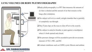
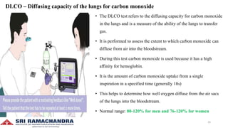
PULMONARY
FUNCTION TESTS
PRESENTED BY:
LINCY ASHA.S
M.PHARM (PHARMACY PRACTICE)
I SEMESTER
1
PULMONARY FUNCTION TEST
Pulmonary function tests are a series of tests performed to examine a patient’s respiratory system and identify the
severity of pulmonary impairment.
These tests are performed to measure a patient’s lung volume, capacity, flow rate and gas exchange.
This allows medical professionals to obtain an accurate diagnosis and determine the best course of medical
intervention for the patient.
In general there are two types of lung disorders that these tests can be used to assess
1. Obstructive lung diseases
2. Restrictive lung diseases
2
1.OBSTRUCTIVE LUNG DISEASES
• It include conditions that make it difficult to exhale air out of the lungs
• This results in shortness of breath that occurs from narrowing and
constriction of the airways and causes the patient to have decreased flow
rates. Eg. COPD, Asthma
2.RESTRICTIVE LUNG DISEASES
• It include conditions that make it difficult to fully fill the lungs with air
during inhalation.
• When the lungs aren’t fully able to expand it causes the patient to have
decreased lung volumes. Eg. Pulmonary fibrosis, interstitial lung disease
3
Pulmonary function tests would be indicated for the following:
• On healthy patients as part of a routine physical exam
• Evaluate signs and symptoms of lung disease
• Diagnosis of certain medical conditions
• Measure current stage of disease and evaluate its progress
• Assess how a patient is responding to different treatments
• Determine patient’s condition before surgery to assess the risk of respiratory complications
• Screen people who are at risk of pulmonary disease
• Determine how much a patient’s airways have narrowed due to disorders
• In certain types of work environments to assess the health of employees.
4
Additionally PFTs may be indicated for the following
1. Chronic lung conditions
2. Restrictive airway problems
3. Asthma
4. COPD
5. Shortness of breath
6. Impairment or disability
7. Early morning wheezing
8. Chest muscle weakness
9. Lung cancer
10. Respiratory infections
5
STATIC LUNG VOLUMES
Lung volume is the amount of air breathed by an individual under a specific condition.
1.Tidal Volume (TV)
It is the volume of air inspired or expired during normal breathing at rest.
2.Inspiratory Reserve Volume (IRV)
It is the volume of air inspired with maximum effort over and above the normal
tidal volume.
3.Expiratory Reserve Volume (ERV)
It is the volume of air expired forcefully after a normal respiration.
4.Residual Volume (RV)
It is the volume of air remaining in the lungs after a forceful expiration
6
LUNG
VOLUMES
NORMAL
VALUE (ml)
TV 500
IRV 3000
ERV 1100
RV 1200
STATIC LUNG CAPACITIES
1.Inspiratory capacity (IC)
 It is the amount of air a person can inspire forcefully after a normal respiration.
IC = TV+IRV
2.Functional Residual Capacity (FRC)
 It is the amount of air that remains in the lungs at the end of normal respiration.
FRC = ERV+RV
3.Vital Capacity (VC)
 It is the maximum volume of air exhaled forcefully from the lungs after a maximum inspiration.
VC = TV+IRV+ERV
4.Total Lung Capacity (TLC)
 It is the volume of air present in the lungs after maximum inspiration.
TLC = VC+RV
7
LUNG
CAPACITIES
NORMAL
VALUE (ml)
IC 3500
FRC 2300
VC 4600
TLC 5800
STATIC LUNG VOLUMES AND CAPACITIES
8
DYNAMIC LUNG VOLUMES AND CAPACITIES
TIMED VITAL CAPACITY (FORCED VITAL CAPACITY)
It is the measurement of vital capacity in relation to time.
FORCED EXPIRATORY VOLUME (FEV)
It is the volume of air expired forcefully in unit time.
It is expressed as percentage of FVC.
Timed vital capacity is expressed as:
FEV1= Volume of air expired forcefully in 1 second and the normal value is 83%
FEV2= Volume of air expired forcefully in 2 seconds and the normal value is 94%
FEV3= Volume of air expired forcefully in 3 seconds and the normal value is 97-100%
9
PEAK EXPIRATORY FLOW RATE (PEFR)
The maximum rate at which air can be exhaled forcefully after a deep inspiration
The normal value is 300-400 L/min.
Useful in assessing obstructive airway diseases such as asthma, cystic fibrosis and emphysema.
FORCED EXPIRATORY FLOW (FEF 25-75%)
It is the maximum air flow during the middle 50% of timed vital capacity
The normal value is 300 L/min.
FLOW VOLUME CURVE
Dynamic lung functions are measured clinically using Flow volume curves (loops).
They are graphical representations of the relationship between maximal flow rates and volume of gas during forced
maneuver.
Used to measure FEF, PEFR and TVC
10
TYPES OF
PULMONARY
FUNCTION TESTS
11
SPIROMETRY
• Spirometry is a PFT test that measures airflow and volume as it moves
into and out of the patient’s lungs.
• This means that spirometry testing occurs during both the inspiratory and
expiratory phases of the breathing cycle.
• It is measured by a simple instrument called spirometer.
• This test is done by having the patient take a deep breath into a spirometer
followed by a fast, maximum exhalation.
• When a patient breathes into and out of a spirometer, it measures the
volume of air and the flow rate during the ventilatory cycle.
• Since it measures both volume and flow, this means that it’s useful in
identifying both obstructive and restrictive respiratory conditions.
12
OVERVIEW OF THE PROCEDURE
• subject should exhale for atleast 6 seconds, stop when no volume change for one second.
• For patients with severe COPD, this can take upto 15 seconds
• Should obtain 3 spirometry readings
• The best two should be within 100 ml (or 5%) of each other.
SPIROMETRY MEASURES
FEV1
FVC
13
CALCULATIONS
FEV1/FVC
• Used to determine whether there is any evidence of restrictive or obstructive lung disease.
• Individual measurements of FEV1 and FVC need to be corrected for the patient’s sex, ethnicity, height and age.
• Expressed as a percentage of predicted for matched, healthy individuals with no lung disease.
• Many spirometers calculate the FEV1 and FVC as a percentage of predicted.
• FEV1 or FVC of <80% predicted indicates abnormality.
• A ratio of FEV1/FVC of 70% or above is considered normal.
OBSTRUCTIVE SPIROMETRY
• Obstructive lung disease is characterised by airway obstruction.
• Airflow obstruction causes a limitation of flow on forced expiration.
14
FEV1 reduced (<80% predicted)
FVC reduced to a lesser extent than FEV1 (<80%)
FEV1/FVC ratio of <70% (or values below LLN)
CAUSES
• Asthma
• COPD
RESTRICTIVE SPIROMETRY
• It is characterised by decreased lung volumes
• There is little or no airflow obstruction.
FEV1 reduced (<80% predicted)
FVC reduced (< 80% predicted)
FEV1 and FVC are reduced with preservation of the FEV1/FVC ratio ≥70%
15
CAUSES
• Idiopathic pulmonary fibrosis
• Obesity
• Deformities of the chest wall
• Neuromuscular disease
OTHER MEASUREMENTS
FEF25%-75%
• Measured in L/Sec
• Most sensitive marker for obstruction
• Useful in tracking asthma or slight progression of obstructive disease.
16
PEFR
<80% predicted = obstruction
Forced expiratory time (FET)
It is the number of seconds required to complete a forceful exhalation
after maximal inspiration.
Normal is <5 seconds
Severely obstructed patients may take upto 15 seconds.
CONTRAINDICATIONS
• Short acting bronchodilator within 6hr
• Long acting bronchodilator within 12hr
• Acutely ill or recently hospitalized
• Current use of steroids
17
• INTERPRETATION DEGREE OF OBSTRUCTION
• If the patient has abnormal spirometry indicating obstruction the next stage is to do a bronchodilator treatment.
Bronchodilator
• Short acting
18
• Sit for 15-20 mins
• Repeat spirometry
• Compare the outcomes
DEGREE OF REVERSIBILITY
• If FEV1 and FVC fully reversible = consider asthma
• If FEV1 increases by 12% or >200 ml = May have asthma or combination of asthma and COPD.
• In COPD, FEV1/FVC remains <70% even after treatment.
A simple spirometer can record all volumes and capacities except FRC, RV and TLC.
FRC, RV and TLC are determined by 3 methods
1. Helium dilution method
2. Nitrogen washout method
3. Body plethysmography
19
HELIUM DILUTION METHOD
The spirometer is filled with air mixed with helium of known concentration.
The subject inhales air from the spirometer commencing from the end of
normal respiration.
FRC can be determined by the degree of dilution of helium.
NITROGEN WASHOUT METHOD
At the end of normal expiration, the subject inspires pure oxygen and later
exhales into a Douglas bag.
This procedure is repeated for 7 minutes till the nitrogen in the lungs is
displaced by oxygen.
FRC calculated by knowing the volume of air in douglas bag, N2 conc in
atmosphere and air sample in douglas bag.
RV = FRC-ERV
TLC = FRC+IC 20
LUNG VOLUMES OR BODY PLETHYSMOGRAPHY
Body plethysmography is a PFT that measures the amount of
air that is inhaled and the amount of air that remains in the
lungs after exhalation.
The subject will sit in a small, airtight chamber that is partially
or completely see-through.
They’ll put clips on the nose to shut off air to the nostrils.
The subject is asked to breathe or pant against a mouthpiece
when it’s both opened and closed.
The pressure changes will be recorded to provide an accurate
measure of TLC, FRC, and RC.
Evaluate conditions such as COPD, cystic fibrosis and asthma.
21
TLC, RV and FRC
80% < Normal TLC < 120%
• <80% defines a restrictive ventilatory
defect
• >120% suggests Hyperinflation often seen
with airflow obstruction
RV/TLC ratio
20-35% in healthy adults
• RV/TLC% >35% + Normal TLC = Air
trapping
• RV/TLC% >35% + >Normal TLC =
Hyperinflation
22
PEAK FLOW METER
A peak flow meter is a portable device that measures the peak
expiratory flow rate (PEFR) i.e, how fast air comes out of the lungs
when exhaled forcefully.
Readings from the meter can help recognize early signs of worsening
asthma.
Keeping track of peak flow numbers is part of Asthma Action Plan.
Asthma Action Plan has three zones
• Green zone: Asthma is well-controlled. Peak flow is 80% to 100% of
personal best.
• Yellow zone: Asthma is getting worse or is poorly controlled. Peak flow
is 50% to 80% of personal best.
• Red zone: Asthma is severe. It requires emergency care. Peak flow is
less than 50% of personal best.
23
DLCO – Diffusing capacity of the lungs for carbon monoxide
• The DLCO test refers to the diffusing capacity for carbon monoxide
in the lungs and is a measure of the ability of the lungs to transfer
gas.
• It is performed to assess the extent to which carbon monoxide can
diffuse from air into the bloodstream.
• During this test carbon monoxide is used because it has a high
affinity for hemoglobin.
• It is the amount of carbon monoxide uptake from a single
inspiration in a specified time (generally 10s)
• This helps to determine how well oxygen diffuse from the air sacs
of the lungs into the bloodstream.
• Normal range: 80-120% for men and 76-120% for women
24
Obstructive pattern
(FEV1/FVC <70%
Predicted)
Restrictive pattern
(FEV1/FVC >70%
Predicted, FVC <80%
predicted)
Normal spirometry
Low DLCO • Emphysema • Interstitial lung
diseases
• Sarcoidosis
• Asbestosis
• Heart failure
• Anemia
• Pulmonary embolism
• Pulmonary
hypertension
Normal • Chronic bronchitis • Musculoskeletal
deformity
• Neuromuscular disease
Increased DLCO • Asthma • Morbid obesity • Pulmonary hemorrhage
• polycythemia
25
BRONCHOPROVOCATION TEST
• A bronchoprovocation test is used to diagnose asthma.
• It involves inhaling either aerosolized chemicals or cold air, or
performing exercises, to see if it induces asthma symptoms.
• By measuring the lung function after exposure to these triggers,
healthcare provider may be able to determine if the patient have
asthma.
• A baseline FEV1 is taken before bronchoprovocation and a
second FEV1 reading is taken after the test.
• A decline in FEV1 of 20% or more from your baseline is
considered a positive diagnosis for asthma.
26
CARDIOPULMONARY EXERCISE TESTING
This test monitors the patient's heart and lung function during
controlled exercise and rest periods.
Performed to assess exercise capacity, risk for a planned surgical
procedure and to determine cause of unexplained shortness of
breath.
Measurements include BP, heart rate, ECG changes, etc
SIX-MINUTE WALK TEST
This test measures the maximum distance the patient is able to
walk in six minutes.
It measures how far a COPD patient can walk in 6 mins and
provides an assessment of their functional status or ability to
perform daily activities.
Score range for healthy adults = 400-700m
27
TO SUMMARISE
28
REFERENCES
• Textbook of Medical Physiology by prof G K Pal, 2nd edition.
• Comprehensive textbook of Medical physiology, Pravati Pal, Nivedita Nanda, 2nd edition.
• Textbook of Medical physiology by Geetha N, 2nd edition.
• https://copd.net/clinical/diffusion-test
• https://youtu.be/jSkwBoed6Tw
• https://youtu.be/Vk_vEvC1aVI
• https://youtu.be/QbG8QFooO3U
• https://youtu.be/AZxyM27xV-U
• https://www.sciencedirect.com/science/article/pii/S0954611111000552
• https://www.lung.org/lung-health-diseases/lung-procedures-and-tests/six-minute-walk-test
• https://www.physio-pedia.com/Cardiopulmonary_Exercise_Testing_(CPET)_In_Adults
• https://asthma.net/diagnosis/peak-flow-meter
29
THANK YOU
30

PULMONARY FUNCTION TESTS - LAB DATA INTERPRETATION

  • 1.
    PULMONARY FUNCTION TESTS PRESENTED BY: LINCYASHA.S M.PHARM (PHARMACY PRACTICE) I SEMESTER 1
  • 2.
    PULMONARY FUNCTION TEST Pulmonaryfunction tests are a series of tests performed to examine a patient’s respiratory system and identify the severity of pulmonary impairment. These tests are performed to measure a patient’s lung volume, capacity, flow rate and gas exchange. This allows medical professionals to obtain an accurate diagnosis and determine the best course of medical intervention for the patient. In general there are two types of lung disorders that these tests can be used to assess 1. Obstructive lung diseases 2. Restrictive lung diseases 2
  • 3.
    1.OBSTRUCTIVE LUNG DISEASES •It include conditions that make it difficult to exhale air out of the lungs • This results in shortness of breath that occurs from narrowing and constriction of the airways and causes the patient to have decreased flow rates. Eg. COPD, Asthma 2.RESTRICTIVE LUNG DISEASES • It include conditions that make it difficult to fully fill the lungs with air during inhalation. • When the lungs aren’t fully able to expand it causes the patient to have decreased lung volumes. Eg. Pulmonary fibrosis, interstitial lung disease 3
  • 4.
    Pulmonary function testswould be indicated for the following: • On healthy patients as part of a routine physical exam • Evaluate signs and symptoms of lung disease • Diagnosis of certain medical conditions • Measure current stage of disease and evaluate its progress • Assess how a patient is responding to different treatments • Determine patient’s condition before surgery to assess the risk of respiratory complications • Screen people who are at risk of pulmonary disease • Determine how much a patient’s airways have narrowed due to disorders • In certain types of work environments to assess the health of employees. 4
  • 5.
    Additionally PFTs maybe indicated for the following 1. Chronic lung conditions 2. Restrictive airway problems 3. Asthma 4. COPD 5. Shortness of breath 6. Impairment or disability 7. Early morning wheezing 8. Chest muscle weakness 9. Lung cancer 10. Respiratory infections 5
  • 6.
    STATIC LUNG VOLUMES Lungvolume is the amount of air breathed by an individual under a specific condition. 1.Tidal Volume (TV) It is the volume of air inspired or expired during normal breathing at rest. 2.Inspiratory Reserve Volume (IRV) It is the volume of air inspired with maximum effort over and above the normal tidal volume. 3.Expiratory Reserve Volume (ERV) It is the volume of air expired forcefully after a normal respiration. 4.Residual Volume (RV) It is the volume of air remaining in the lungs after a forceful expiration 6 LUNG VOLUMES NORMAL VALUE (ml) TV 500 IRV 3000 ERV 1100 RV 1200
  • 7.
    STATIC LUNG CAPACITIES 1.Inspiratorycapacity (IC)  It is the amount of air a person can inspire forcefully after a normal respiration. IC = TV+IRV 2.Functional Residual Capacity (FRC)  It is the amount of air that remains in the lungs at the end of normal respiration. FRC = ERV+RV 3.Vital Capacity (VC)  It is the maximum volume of air exhaled forcefully from the lungs after a maximum inspiration. VC = TV+IRV+ERV 4.Total Lung Capacity (TLC)  It is the volume of air present in the lungs after maximum inspiration. TLC = VC+RV 7 LUNG CAPACITIES NORMAL VALUE (ml) IC 3500 FRC 2300 VC 4600 TLC 5800
  • 8.
    STATIC LUNG VOLUMESAND CAPACITIES 8
  • 9.
    DYNAMIC LUNG VOLUMESAND CAPACITIES TIMED VITAL CAPACITY (FORCED VITAL CAPACITY) It is the measurement of vital capacity in relation to time. FORCED EXPIRATORY VOLUME (FEV) It is the volume of air expired forcefully in unit time. It is expressed as percentage of FVC. Timed vital capacity is expressed as: FEV1= Volume of air expired forcefully in 1 second and the normal value is 83% FEV2= Volume of air expired forcefully in 2 seconds and the normal value is 94% FEV3= Volume of air expired forcefully in 3 seconds and the normal value is 97-100% 9
  • 10.
    PEAK EXPIRATORY FLOWRATE (PEFR) The maximum rate at which air can be exhaled forcefully after a deep inspiration The normal value is 300-400 L/min. Useful in assessing obstructive airway diseases such as asthma, cystic fibrosis and emphysema. FORCED EXPIRATORY FLOW (FEF 25-75%) It is the maximum air flow during the middle 50% of timed vital capacity The normal value is 300 L/min. FLOW VOLUME CURVE Dynamic lung functions are measured clinically using Flow volume curves (loops). They are graphical representations of the relationship between maximal flow rates and volume of gas during forced maneuver. Used to measure FEF, PEFR and TVC 10
  • 11.
  • 12.
    SPIROMETRY • Spirometry isa PFT test that measures airflow and volume as it moves into and out of the patient’s lungs. • This means that spirometry testing occurs during both the inspiratory and expiratory phases of the breathing cycle. • It is measured by a simple instrument called spirometer. • This test is done by having the patient take a deep breath into a spirometer followed by a fast, maximum exhalation. • When a patient breathes into and out of a spirometer, it measures the volume of air and the flow rate during the ventilatory cycle. • Since it measures both volume and flow, this means that it’s useful in identifying both obstructive and restrictive respiratory conditions. 12
  • 13.
    OVERVIEW OF THEPROCEDURE • subject should exhale for atleast 6 seconds, stop when no volume change for one second. • For patients with severe COPD, this can take upto 15 seconds • Should obtain 3 spirometry readings • The best two should be within 100 ml (or 5%) of each other. SPIROMETRY MEASURES FEV1 FVC 13
  • 14.
    CALCULATIONS FEV1/FVC • Used todetermine whether there is any evidence of restrictive or obstructive lung disease. • Individual measurements of FEV1 and FVC need to be corrected for the patient’s sex, ethnicity, height and age. • Expressed as a percentage of predicted for matched, healthy individuals with no lung disease. • Many spirometers calculate the FEV1 and FVC as a percentage of predicted. • FEV1 or FVC of <80% predicted indicates abnormality. • A ratio of FEV1/FVC of 70% or above is considered normal. OBSTRUCTIVE SPIROMETRY • Obstructive lung disease is characterised by airway obstruction. • Airflow obstruction causes a limitation of flow on forced expiration. 14
  • 15.
    FEV1 reduced (<80%predicted) FVC reduced to a lesser extent than FEV1 (<80%) FEV1/FVC ratio of <70% (or values below LLN) CAUSES • Asthma • COPD RESTRICTIVE SPIROMETRY • It is characterised by decreased lung volumes • There is little or no airflow obstruction. FEV1 reduced (<80% predicted) FVC reduced (< 80% predicted) FEV1 and FVC are reduced with preservation of the FEV1/FVC ratio ≥70% 15
  • 16.
    CAUSES • Idiopathic pulmonaryfibrosis • Obesity • Deformities of the chest wall • Neuromuscular disease OTHER MEASUREMENTS FEF25%-75% • Measured in L/Sec • Most sensitive marker for obstruction • Useful in tracking asthma or slight progression of obstructive disease. 16
  • 17.
    PEFR <80% predicted =obstruction Forced expiratory time (FET) It is the number of seconds required to complete a forceful exhalation after maximal inspiration. Normal is <5 seconds Severely obstructed patients may take upto 15 seconds. CONTRAINDICATIONS • Short acting bronchodilator within 6hr • Long acting bronchodilator within 12hr • Acutely ill or recently hospitalized • Current use of steroids 17
  • 18.
    • INTERPRETATION DEGREEOF OBSTRUCTION • If the patient has abnormal spirometry indicating obstruction the next stage is to do a bronchodilator treatment. Bronchodilator • Short acting 18
  • 19.
    • Sit for15-20 mins • Repeat spirometry • Compare the outcomes DEGREE OF REVERSIBILITY • If FEV1 and FVC fully reversible = consider asthma • If FEV1 increases by 12% or >200 ml = May have asthma or combination of asthma and COPD. • In COPD, FEV1/FVC remains <70% even after treatment. A simple spirometer can record all volumes and capacities except FRC, RV and TLC. FRC, RV and TLC are determined by 3 methods 1. Helium dilution method 2. Nitrogen washout method 3. Body plethysmography 19
  • 20.
    HELIUM DILUTION METHOD Thespirometer is filled with air mixed with helium of known concentration. The subject inhales air from the spirometer commencing from the end of normal respiration. FRC can be determined by the degree of dilution of helium. NITROGEN WASHOUT METHOD At the end of normal expiration, the subject inspires pure oxygen and later exhales into a Douglas bag. This procedure is repeated for 7 minutes till the nitrogen in the lungs is displaced by oxygen. FRC calculated by knowing the volume of air in douglas bag, N2 conc in atmosphere and air sample in douglas bag. RV = FRC-ERV TLC = FRC+IC 20
  • 21.
    LUNG VOLUMES ORBODY PLETHYSMOGRAPHY Body plethysmography is a PFT that measures the amount of air that is inhaled and the amount of air that remains in the lungs after exhalation. The subject will sit in a small, airtight chamber that is partially or completely see-through. They’ll put clips on the nose to shut off air to the nostrils. The subject is asked to breathe or pant against a mouthpiece when it’s both opened and closed. The pressure changes will be recorded to provide an accurate measure of TLC, FRC, and RC. Evaluate conditions such as COPD, cystic fibrosis and asthma. 21
  • 22.
    TLC, RV andFRC 80% < Normal TLC < 120% • <80% defines a restrictive ventilatory defect • >120% suggests Hyperinflation often seen with airflow obstruction RV/TLC ratio 20-35% in healthy adults • RV/TLC% >35% + Normal TLC = Air trapping • RV/TLC% >35% + >Normal TLC = Hyperinflation 22
  • 23.
    PEAK FLOW METER Apeak flow meter is a portable device that measures the peak expiratory flow rate (PEFR) i.e, how fast air comes out of the lungs when exhaled forcefully. Readings from the meter can help recognize early signs of worsening asthma. Keeping track of peak flow numbers is part of Asthma Action Plan. Asthma Action Plan has three zones • Green zone: Asthma is well-controlled. Peak flow is 80% to 100% of personal best. • Yellow zone: Asthma is getting worse or is poorly controlled. Peak flow is 50% to 80% of personal best. • Red zone: Asthma is severe. It requires emergency care. Peak flow is less than 50% of personal best. 23
  • 24.
    DLCO – Diffusingcapacity of the lungs for carbon monoxide • The DLCO test refers to the diffusing capacity for carbon monoxide in the lungs and is a measure of the ability of the lungs to transfer gas. • It is performed to assess the extent to which carbon monoxide can diffuse from air into the bloodstream. • During this test carbon monoxide is used because it has a high affinity for hemoglobin. • It is the amount of carbon monoxide uptake from a single inspiration in a specified time (generally 10s) • This helps to determine how well oxygen diffuse from the air sacs of the lungs into the bloodstream. • Normal range: 80-120% for men and 76-120% for women 24
  • 25.
    Obstructive pattern (FEV1/FVC <70% Predicted) Restrictivepattern (FEV1/FVC >70% Predicted, FVC <80% predicted) Normal spirometry Low DLCO • Emphysema • Interstitial lung diseases • Sarcoidosis • Asbestosis • Heart failure • Anemia • Pulmonary embolism • Pulmonary hypertension Normal • Chronic bronchitis • Musculoskeletal deformity • Neuromuscular disease Increased DLCO • Asthma • Morbid obesity • Pulmonary hemorrhage • polycythemia 25
  • 26.
    BRONCHOPROVOCATION TEST • Abronchoprovocation test is used to diagnose asthma. • It involves inhaling either aerosolized chemicals or cold air, or performing exercises, to see if it induces asthma symptoms. • By measuring the lung function after exposure to these triggers, healthcare provider may be able to determine if the patient have asthma. • A baseline FEV1 is taken before bronchoprovocation and a second FEV1 reading is taken after the test. • A decline in FEV1 of 20% or more from your baseline is considered a positive diagnosis for asthma. 26
  • 27.
    CARDIOPULMONARY EXERCISE TESTING Thistest monitors the patient's heart and lung function during controlled exercise and rest periods. Performed to assess exercise capacity, risk for a planned surgical procedure and to determine cause of unexplained shortness of breath. Measurements include BP, heart rate, ECG changes, etc SIX-MINUTE WALK TEST This test measures the maximum distance the patient is able to walk in six minutes. It measures how far a COPD patient can walk in 6 mins and provides an assessment of their functional status or ability to perform daily activities. Score range for healthy adults = 400-700m 27
  • 28.
  • 29.
    REFERENCES • Textbook ofMedical Physiology by prof G K Pal, 2nd edition. • Comprehensive textbook of Medical physiology, Pravati Pal, Nivedita Nanda, 2nd edition. • Textbook of Medical physiology by Geetha N, 2nd edition. • https://copd.net/clinical/diffusion-test • https://youtu.be/jSkwBoed6Tw • https://youtu.be/Vk_vEvC1aVI • https://youtu.be/QbG8QFooO3U • https://youtu.be/AZxyM27xV-U • https://www.sciencedirect.com/science/article/pii/S0954611111000552 • https://www.lung.org/lung-health-diseases/lung-procedures-and-tests/six-minute-walk-test • https://www.physio-pedia.com/Cardiopulmonary_Exercise_Testing_(CPET)_In_Adults • https://asthma.net/diagnosis/peak-flow-meter 29
  • 30.