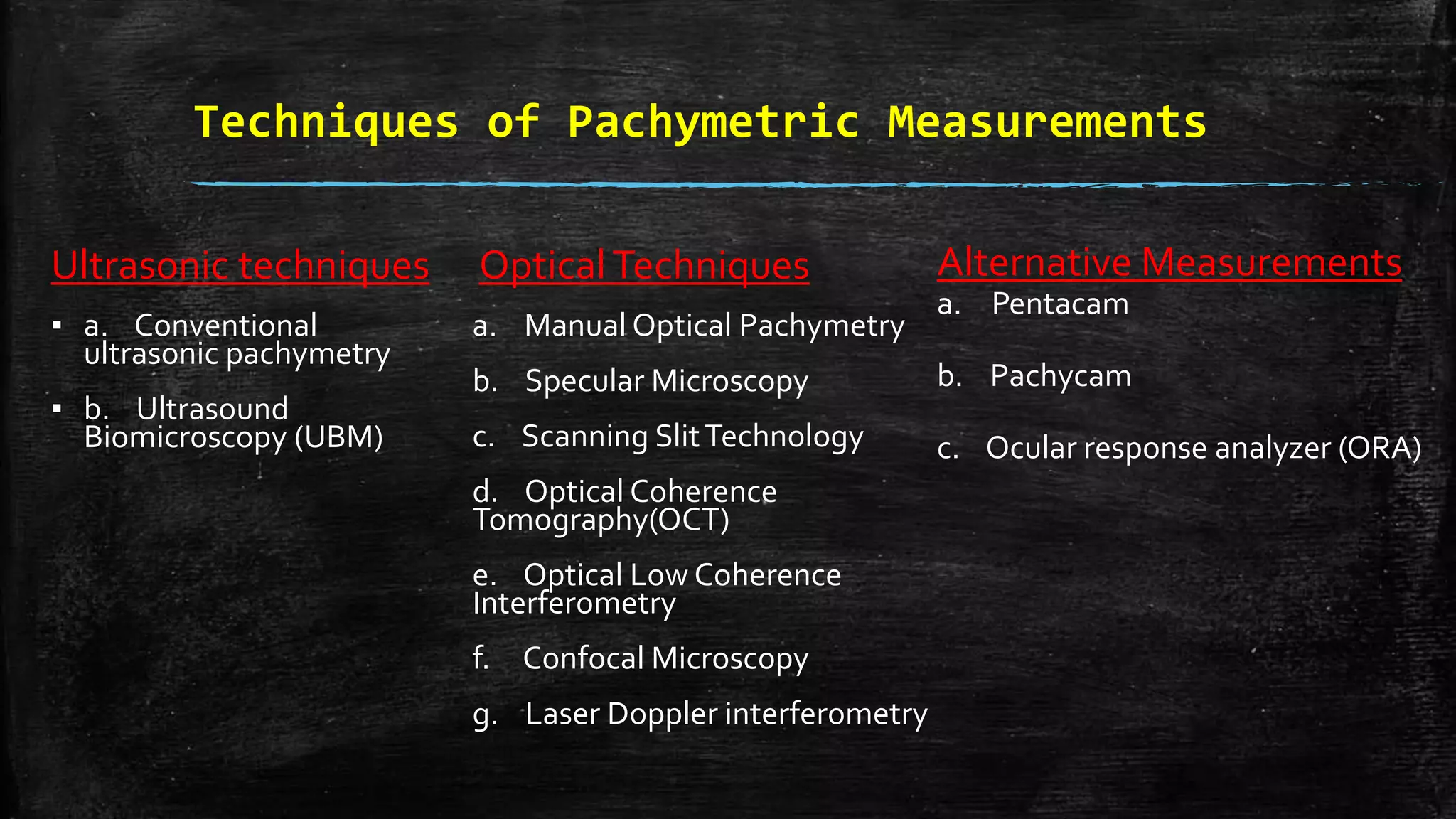
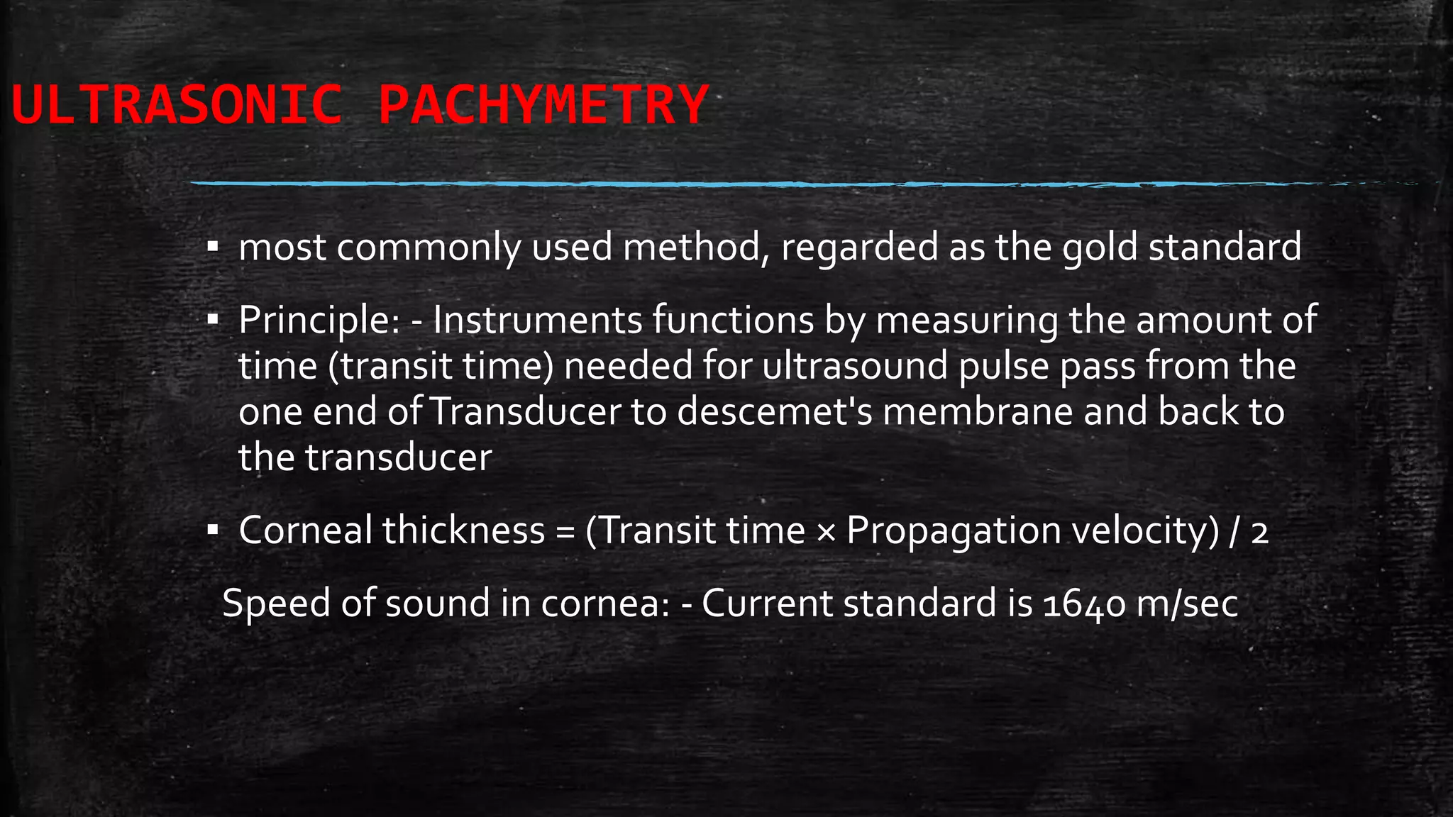
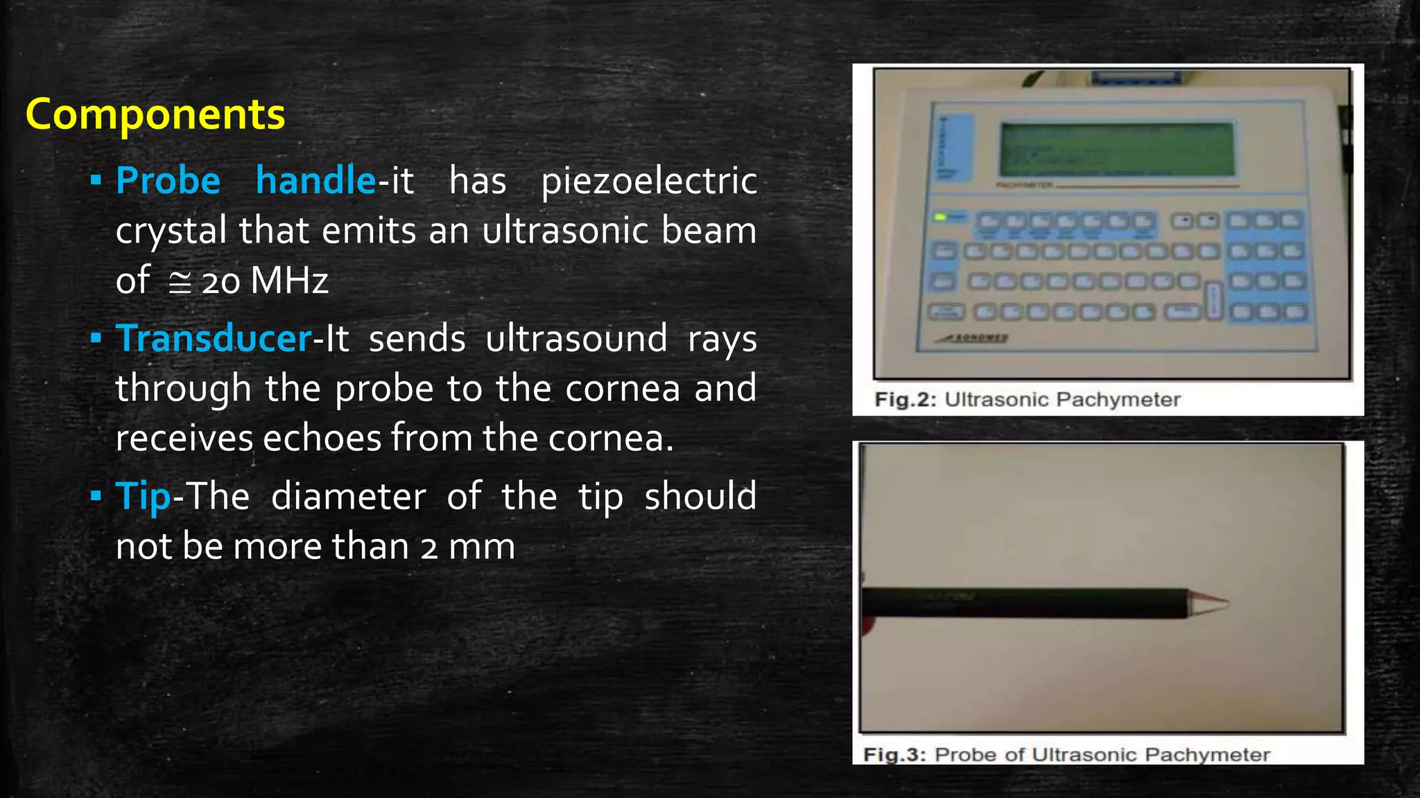
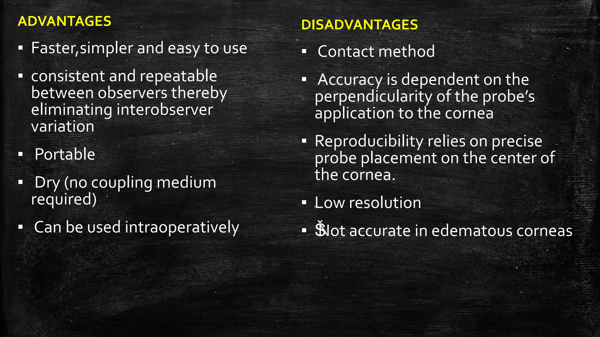
SlideShare a Scribd company logo
PACHYMETRY
SIVATEJA CHALLA
SSSIHMS
Introduction
▪ Pachymetry (Greek words: Pachos = thick + metry = to measure) is term
used for the measurement of corneal thickness.
▪ It is an important indicator of health status of the cornea especially of
corneal endothelial pump function
▪ The thickness of the cornea was first reported in ancient textbooks on
physiological optics (Helmholtz and gulstrand)
corneal thickness in normal eyes
▪ It ranges from 0.7 to 0.9 mm at the limbus and varies between 0.49 mm
and 0.56 mm at the centre
▪ The Central corneal thickness (CCT) reading of 0.7 mm or more is
indicative of endothelial decompensation.
▪ Peripheral corneal thickness is asymmetric so that temporal cornea is
thinnest followed by the inferior cornea.
IN NEWBORNS AND CHILDREN…
 Corneal configuration in newborns is similar to that of the adult cornea
 It has been found that cornea on day one is significantly thicker and
decreases in thickness as the child grows older.
 The average corneal thickness in infants is 585 ± 52 microns.
 The superior peripheral cornea is thinnest in newborn
Factors affecting central corneal thickness
▪ higher in younger patients, male patients and diabetic patients.
▪ The mean CCT of black children is thinner than that of white children
▪ The PITX2/Pitx2 mutation seen in Axenfeld-Rieger malformations results in
reduced corneal thickness .
▪ Central corneal thickness does not correlate with refraction or systemic
hypertension
Role in clinical practice
▪ Glaucoma: for applying correction factor in actual IOP determination
Congenital Glaucoma: to assess the amount of corneal edema.
▪ Refractive surgeries: preoperative screening and treatment plan of keratorefractive
procedures like LASIK, astigmatic keratotomy,even prior to radial keratotomy.
▪ Post operative follow up of keratoplasty patients to determine endothelial cell function and its
recovery and to become alert to early graft decompensation.
▪ Contact lens: To assess corneal edema
▪ Assessing cornea thinness as in corneal disorders like Terrien ’s and Pellucid marginal
degenerations, keratoconus, keratoglobus, post LASIK ectasia.
▪ Other cases of corneal decompensation as in herpetic endothelitis.
Role in Glaucoma
▪ Applanation tonometry is based on Imbert Fick’s law, which assumes that
cornea is a perfect flexible, dry, sphere which is infinitely thin.
▪ Therefore increase in the tissue in thicker cornea makes it less compliant and
subsequently leading to overestimation of IOP andViceversa
▪ Ocular HypertensionTreatment Study (OHTS) group published that central
corneal thickness (CCT) was an important independent risk factor for
progression from ocular hypertension to early glaucoma.
correction factor:
▪ It is recommended that in chronic eye diseases like glaucoma and
glaucoma suspects for every increase in central corneal thickness of 50
microns, the correction done is to decrease the recorded IOP by 2.5mm
Hg.
▪ For acute onset diseases it was recommended to correct by 10 mm Hg for
every 50 microns.
facts about cct in glaucoma
Bechmann in 2000 found following association of CCT with different forms of glaucoma.
1) Increased CCT measurements are found in patients with ocular hypertension, which
can lead to falsely elevated IOP readings,
2) Decreased CCT is found in patients with low tension glaucoma, resulting in falsely
reduced IOP measurements.
3) CCT was found to be lower in patients with Pseudoexfoliation syndrome (PXS) and in
Primary open angle Glaucoma (POAG).
4) There is no difference in corneal thickness in individuals with Pigmentary Glaucoma
(PG) and Primary angle Closure Glaucoma (PACG).
Evidence Based Guideline for Corneal
Pachymetry in Glaucoma
▪ Patients who have risk factors for developing Primary Open Angle
Glaucoma
▪ Elevated intraocular pressure repeatedly measured >24 mm Hg
▪ ŠAfrican descent
▪ ŠAdvancing age (>65 years old)
▪ ŠFamily history of glaucoma
▪ Diabetes mellitus (though it is controversial)
Pachymetry in refractive surgery
▪ preoperative assessment of candidates LASIK. It is recommended
that before undertaking residual stromal thickness of 250-300 μm
must be ensured.
▪ Patient with adequate thickness to their corneas may be candidates
for LASIK while those with thinner corneas may be safer considering
PRK / LASEK as their treatment option
▪ It is important to evaluate the outcome of laser refractive surgical
procedures, especially in candidates for enhancement surgery.
Pachymetry in contact lens use
▪ Moreover assessment of corneal thickness before contact lens
prescription is another important thing to analyze
▪ A study showed that corneal edema in contact lens wearers resolved
in 2-15 days after the use of soft contact lenses was stopped.
Therefore, it is recommended that the time period for the
pachymetric readings to stabilize is around 15 days. So, one must
discontinue contact lens atleast 15 days before surgery to get the
accurate pachymetric readings.
Techniques of Pachymetric Measurements
Spot measurements: traditional optical pachymetry, specular and confocal microscopy,
ultrasound pachymetry, and optical low-coherence reflectometry.
Wide area mapping: slit scanning optical pachymetry and very high–frequency ultrasound
imaging.
Advantages of mappping over spot measurement
1. reveal abnormal patterns-keratoconus and pellucid marginal degeneration
2. preoperative planning for surgeries like astigmatic keratotomy,ICRS implantation,PTK,
DALK.
Techniques of Pachymetric Measurements
Ultrasonic techniques
▪ a. Conventional
ultrasonic pachymetry
▪ b. Ultrasound
Biomicroscopy (UBM)
OpticalTechniques
a. Manual Optical Pachymetry
b. Specular Microscopy
c. Scanning SlitTechnology
d. Optical Coherence
Tomography(OCT)
e. Optical Low Coherence
Interferometry
f. Confocal Microscopy
g. Laser Doppler interferometry
Alternative Measurements
a. Pentacam
b. Pachycam
c. Ocular response analyzer (ORA)
ULTRASONIC PACHYMETRY
▪ most commonly used method, regarded as the gold standard
▪ Principle: - Instruments functions by measuring the amount of
time (transit time) needed for ultrasound pulse pass from the
one end ofTransducer to descemet's membrane and back to
the transducer
▪ Corneal thickness = (Transit time × Propagation velocity) / 2
Speed of sound in cornea: - Current standard is 1640 m/sec
Components
▪ Probe handle-it has piezoelectric
crystal that emits an ultrasonic beam
of  20 MHz
▪ Transducer-It sends ultrasound rays
through the probe to the cornea and
receives echoes from the cornea.
▪ Tip-The diameter of the tip should
not be more than 2 mm
ADVANTAGES
▪ Faster,simpler and easy to use
▪ consistent and repeatable
between observers thereby
eliminating interobserver
variation
▪ Portable
▪ Dry (no coupling medium
required)
▪ Can be used intraoperatively
DISADVANTAGES
▪ Contact method
▪ Accuracy is dependent on the
perpendicularity of the probe’s
application to the cornea
▪ Reproducibility relies on precise
probe placement on the center of
the cornea.
▪ Low resolution
▪ ŠNot accurate in edematous corneas
ULTRASOUND BIOMICROSCOPY (UBM)
▪ high resolution ultrasound machine which images AS of eye.
▪ 12.5 - 50MHz probe,the depth of penetration is lesser (4 mm) than
conventional ultrasound.
Advantages
1.AS+corneal thickness.
Š2.Useful in cases where cornea is opaque.
Š3.Various layers of cornea can be identified.
Disadvantages
1.immersing the eye in acoupling fluid.
Š2.Contact method.
Š3. patient to lie supine
Š4.cannot be used intraoperatively.
Š5.Difficult to standardize
MANUAL OPTICAL PACHYMETRY
▪ This was the original method to measure corneal
thickness.
▪ The instrument contains two plano glass plates
that splits the image of the corneal parallelepiped
▪ There are two methods to measure corneal
thickness
1.just touch method
2.overlap method
▪ The corneal thickness is then directly read from the
scale
▪ Range is 0 to 1.2 mm, with a least gradation of 0.02
mm.
ADVANTAGES-non contact method
DISADVANTAGES-
1) Lack of accuracy in measurements; the usual range of
error with an optical pachymeter is ± 2%.
2) Lack of repeatability
3) Requires slit lamp and therefore has poor portability
and cannot be used in operating room
SPECULAR PACHYMETRY
▪ This is the oldest method to measure corneal thickness.
▪ Principle-This measures the distance between the
anterior and the posterior surfaces of cornea and depends
on the focusing of light rays through front back cornea
unlike sound waves in ultrasound pachymeter.
▪ There are 2 types of specular microscope
1.Contact 2.non contact
Advantages
1.Operator independent
2.Non invasive
3.Simultaneous measurement of cell
count
Disadvantages
1.The exact point where the reading is taken is
not known.
2.Time consuming.
3.Less reproducible
4.Impractical for use in operation room
5.Clinical use is limited to corneas free of
edema, scarring, deposits or opacities that may
distort light transmission.
SLIT-SCANNING PACHYMETRY
▪ elevation based system which
uses scanning slit technology
▪ capable of assessment of
multiple functions thickness
profile, anterior and posterior
topography, elevation, and
anterior chamber depth
▪ PrincipleIt measures anterior
and posterior corneal elevations
by comparing it to a best fit
sphere.
Advantages
1.It gives wide field pachymetry measures
across the entire cornea.
2.It also identifies the thinnest point in the
cornea
3. Corneal alignment is not required.
Š4.Can be used to calculate optical zones in
corneal refractive surgeries.
Disadvantages
▪ It overestimates corneal thickness by 5%.
▪ Tendency to underestimate corneal
thickness in Keratoconic, post-PRK, and
post-LASIK eyes
▪ the measurements are adjusted for
normal prolate shape of cornea. Change
of shape may interfere with the
reconstruction algorithms
▪ decreased accuracy in measuring corneal
thickness when clinically significant haze
is present.
ANTERIOR SEGMENT OPTICAL COHERENCE TOMOGRAPHY
▪ high- resolution, non- contact OCT customized for the anterior segment
▪ gives color coded map of the corneal thickness
Advantages
▪ Noncontact
▪ Rapid acquisition during the pachymetry scan ensures an accurate and
repeatable pachymetry map.
▪ ŠHigh Resolution
▪ ŠIt measures and documents both corneal flap thickness and residual stromal
thickness immediately following LASIK surgery.
▪ Measures through corneal opacity
OPTICAL LOW COHERENCE REFLECTOMETRY
▪ The instrument is attached to a slit lamp
▪ measure corneal thickness to a precision
of one micron.
▪ PRINCIPLEBased on Michelson
interferometer. It uses diode laser beam.
Advantages
▪ Precise 1 micron measurement
▪ Automatic alignment
▪ Non-contact
▪ Real-time data acquisition and display
▪ Convenient and easy
▪ Variability of measurements is
significantly lower
▪ Intraoperative measurements possible
Disadvantages
▪ Š Measures only central corneal
thickness
CONFOCAL MICROSCOPY
 This unique method offers the ability to examine
objects at high magnification ranging from 20X
to 500X.
 It also measure thickness of each layer by using
computerized scanning system providing the
total corneal thickness in studied area.
 Beside endothelium examination also measure
endothelial cell count (density) which is
comparable to specular microscopy.
 It offers the possibility to visualize structures
posterior to haze, scars or edema with in the
cornea.
Advantages
1.moderate to good repeatability,
particularly for measurements of thin
layers such as epithelial or Bowman’s layer
thickness.
2.As well as corneal thickness,epithelial
thickness,Bowman’s layer thickness, and
following LASIK surgery, flap thickness can
also be obtained.
3. The z-scan curve can be used to assess
the level and location of corneal haze
associated with the various corneal
dystrophies.
Disadvantages
1. Poor agreement between CMTF and
ultrasound pachymetry, the latter
apparently ov erestimating corneal
thickness.
2. The precision of measurements with
this technique will vary with contact
lens hydration, post-lens tear film
thickness and observation angle.
3. Slower data acquisition
4. Poor penetration of corneal opacity
Pentacam
▪ It is 3-Dimensional (3D) rotating scheimpflug camera.
▪ It can perform five functions in 2 sec.
1. Scheimpflug image of anterior segment
2. 3-D anterior chamber analyser
3. Pachymetry
4. Corneal topography
5. Cataract analyser
Pachymetry by pentacam is displayed as a color image over its entire
area from limbus to limbus.
Advantages
▪ Noninvasive, non contact
▪ Even minute eye movements are
captured and corrected simultaneously.
▪ gives precise representation and
repeatability.
Disadvantages
▪ It underestimates the corneal thickness in
comparison to ultrasonic pachymetry.
Applications
1.Preoperative planning for corneal refractive surgery
2.Glaucoma screening
3.IOP modification with regard to corneal thickness
4.Keratoconus detection & quantification.
PACHYCAM
▪ compact and portable non- contact pachymeter
with built-in keratometer. It can be mounted on
slit lamp. It automatically corrects the IOP
▪ Image acquisition is done with the help of a 3D
alignment screen.
Advantages
1. Noncontact
2. Compact,portable,light weight
OCULAR RESPONSE ANALYZER
▪ Newer modality for measuring bio mechanical
properties of cornea
▪ Utilises a rapid air impulse and maeasures delays
in inward and outward applanation of cornea
resulting in two different pressure values.the
difference in two pressure values gives corneal
thickness
THANK YOU

More Related Content

What's hot

Specular microscopy
Specular microscopySpecular microscopy
Specular microscopyRuchi sood
 
Slit lamp biomicroscopy
Slit lamp biomicroscopySlit lamp biomicroscopy
Slit lamp biomicroscopyHira Dahal
 
Humphrey visual field analyser (HVFA)
Humphrey visual field analyser (HVFA)Humphrey visual field analyser (HVFA)
Humphrey visual field analyser (HVFA)Azizul Islam
 
Optics of contact lens
Optics of contact lensOptics of contact lens
Optics of contact lensAayush Chandan
 
Measuring interpupillary distance
Measuring interpupillary distanceMeasuring interpupillary distance
Measuring interpupillary distanceIndra Prasad Sharma
 
Contact lens fitting in keratoconus copy
Contact lens fitting in keratoconus   copyContact lens fitting in keratoconus   copy
Contact lens fitting in keratoconus copykamal thakur
 
Anterior segment OCT & UBM
Anterior segment OCT & UBMAnterior segment OCT & UBM
Anterior segment OCT & UBMDinesh Madduri
 
Amblyopia Management
Amblyopia ManagementAmblyopia Management
Amblyopia Managementsiraj safi
 
Goldman applanation tonometry
 Goldman applanation tonometry Goldman applanation tonometry
Goldman applanation tonometryHossein Mirzaie
 
Ocular diagnostic dyes.pdf
Ocular diagnostic dyes.pdfOcular diagnostic dyes.pdf
Ocular diagnostic dyes.pdfMohan
 
Cycloplegic retinoscopy
Cycloplegic retinoscopy Cycloplegic retinoscopy
Cycloplegic retinoscopy R J
 
Gonioscopy presentation
Gonioscopy presentationGonioscopy presentation
Gonioscopy presentationHira Dahal
 

What's hot (20)

Specular microscopy
Specular microscopySpecular microscopy
Specular microscopy
 
Photostress recovery time
Photostress recovery timePhotostress recovery time
Photostress recovery time
 
Slit lamp biomicroscopy
Slit lamp biomicroscopySlit lamp biomicroscopy
Slit lamp biomicroscopy
 
Ophthalmic prisms
Ophthalmic prismsOphthalmic prisms
Ophthalmic prisms
 
Humphrey visual field analyser (HVFA)
Humphrey visual field analyser (HVFA)Humphrey visual field analyser (HVFA)
Humphrey visual field analyser (HVFA)
 
Optical aberrations
Optical aberrationsOptical aberrations
Optical aberrations
 
Optics of contact lens
Optics of contact lensOptics of contact lens
Optics of contact lens
 
Contrast sensitivity
Contrast sensitivityContrast sensitivity
Contrast sensitivity
 
Log mar chart
Log mar chartLog mar chart
Log mar chart
 
Measuring interpupillary distance
Measuring interpupillary distanceMeasuring interpupillary distance
Measuring interpupillary distance
 
binocular single vision
binocular single visionbinocular single vision
binocular single vision
 
Contact lens fitting in keratoconus copy
Contact lens fitting in keratoconus   copyContact lens fitting in keratoconus   copy
Contact lens fitting in keratoconus copy
 
Anterior segment OCT & UBM
Anterior segment OCT & UBMAnterior segment OCT & UBM
Anterior segment OCT & UBM
 
Amblyopia Management
Amblyopia ManagementAmblyopia Management
Amblyopia Management
 
Retinoscopy
RetinoscopyRetinoscopy
Retinoscopy
 
Goldman applanation tonometry
 Goldman applanation tonometry Goldman applanation tonometry
Goldman applanation tonometry
 
Ocular diagnostic dyes.pdf
Ocular diagnostic dyes.pdfOcular diagnostic dyes.pdf
Ocular diagnostic dyes.pdf
 
Cycloplegic retinoscopy
Cycloplegic retinoscopy Cycloplegic retinoscopy
Cycloplegic retinoscopy
 
Retinoscopy
RetinoscopyRetinoscopy
Retinoscopy
 
Gonioscopy presentation
Gonioscopy presentationGonioscopy presentation
Gonioscopy presentation
 

Similar to Pachymetry

Corneal pachymetry.ppt
Corneal pachymetry.pptCorneal pachymetry.ppt
Corneal pachymetry.pptAhmedAli191572
 
KERATOPROSTHESIS
KERATOPROSTHESISKERATOPROSTHESIS
KERATOPROSTHESISSSSIHMS-PG
 
Pre op corneal laser
Pre op corneal laserPre op corneal laser
Pre op corneal laserSumegha Tomar
 
Penetrating Keratoplasty
Penetrating Keratoplasty Penetrating Keratoplasty
Penetrating Keratoplasty Jigyasa Sahu
 
AS OCT & UBM - Dr Shylesh B Dabke
AS OCT & UBM - Dr Shylesh B DabkeAS OCT & UBM - Dr Shylesh B Dabke
AS OCT & UBM - Dr Shylesh B DabkeShylesh Dabke
 
Biometry instruments & equipment
Biometry instruments & equipmentBiometry instruments & equipment
Biometry instruments & equipmentDevdutta Nayak
 
Laser BASED PROCEDURES
Laser BASED PROCEDURES Laser BASED PROCEDURES
Laser BASED PROCEDURES Mahrukh Khan
 
501 UNIT 2CONTACT LENS FITTING.pptx
501 UNIT 2CONTACT LENS FITTING.pptx501 UNIT 2CONTACT LENS FITTING.pptx
501 UNIT 2CONTACT LENS FITTING.pptxRuchikaMaurya4
 
Keratoconus mangment
Keratoconus mangmentKeratoconus mangment
Keratoconus mangmentAmr Mounir
 
Ortho - k lenses by Ashith Tripathi
Ortho -  k lenses by Ashith Tripathi Ortho -  k lenses by Ashith Tripathi
Ortho - k lenses by Ashith Tripathi Ashith Tripathi
 
Corneal Topography Corneal Cross Linking Pediatric and Presbyopic Contact Lens
Corneal Topography Corneal Cross Linking Pediatric and Presbyopic Contact LensCorneal Topography Corneal Cross Linking Pediatric and Presbyopic Contact Lens
Corneal Topography Corneal Cross Linking Pediatric and Presbyopic Contact LensTahseen Jawaid
 
pachymetry confocal microscopy cornea ophthalmology diagnostics
pachymetry confocal microscopy cornea ophthalmology diagnosticspachymetry confocal microscopy cornea ophthalmology diagnostics
pachymetry confocal microscopy cornea ophthalmology diagnosticsPaavan Kalra
 
Excimer Laser Phototheraputic Keratectomy ( PTK )
 Excimer Laser Phototheraputic Keratectomy ( PTK ) Excimer Laser Phototheraputic Keratectomy ( PTK )
Excimer Laser Phototheraputic Keratectomy ( PTK )DiyarAlzubaidy
 
Corneal laser surgery
Corneal laser surgeryCorneal laser surgery
Corneal laser surgeryBAYHALQARNI
 

Similar to Pachymetry (20)

Pachymetry sivateja
Pachymetry sivatejaPachymetry sivateja
Pachymetry sivateja
 
Corneal pachymetry.ppt
Corneal pachymetry.pptCorneal pachymetry.ppt
Corneal pachymetry.ppt
 
pachy.pptx
pachy.pptxpachy.pptx
pachy.pptx
 
LASIK.pptx
LASIK.pptxLASIK.pptx
LASIK.pptx
 
keratoprosthesis
keratoprosthesiskeratoprosthesis
keratoprosthesis
 
KERATOPROSTHESIS
KERATOPROSTHESISKERATOPROSTHESIS
KERATOPROSTHESIS
 
Pre op corneal laser
Pre op corneal laserPre op corneal laser
Pre op corneal laser
 
Keratoplasty
KeratoplastyKeratoplasty
Keratoplasty
 
Penetrating Keratoplasty
Penetrating Keratoplasty Penetrating Keratoplasty
Penetrating Keratoplasty
 
AS OCT & UBM - Dr Shylesh B Dabke
AS OCT & UBM - Dr Shylesh B DabkeAS OCT & UBM - Dr Shylesh B Dabke
AS OCT & UBM - Dr Shylesh B Dabke
 
Biometry instruments & equipment
Biometry instruments & equipmentBiometry instruments & equipment
Biometry instruments & equipment
 
Laser BASED PROCEDURES
Laser BASED PROCEDURES Laser BASED PROCEDURES
Laser BASED PROCEDURES
 
501 UNIT 2CONTACT LENS FITTING.pptx
501 UNIT 2CONTACT LENS FITTING.pptx501 UNIT 2CONTACT LENS FITTING.pptx
501 UNIT 2CONTACT LENS FITTING.pptx
 
Keratoconus mangment
Keratoconus mangmentKeratoconus mangment
Keratoconus mangment
 
Ortho - k lenses by Ashith Tripathi
Ortho -  k lenses by Ashith Tripathi Ortho -  k lenses by Ashith Tripathi
Ortho - k lenses by Ashith Tripathi
 
Corneal Topography Corneal Cross Linking Pediatric and Presbyopic Contact Lens
Corneal Topography Corneal Cross Linking Pediatric and Presbyopic Contact LensCorneal Topography Corneal Cross Linking Pediatric and Presbyopic Contact Lens
Corneal Topography Corneal Cross Linking Pediatric and Presbyopic Contact Lens
 
pachymetry confocal microscopy cornea ophthalmology diagnostics
pachymetry confocal microscopy cornea ophthalmology diagnosticspachymetry confocal microscopy cornea ophthalmology diagnostics
pachymetry confocal microscopy cornea ophthalmology diagnostics
 
Excimer Laser Phototheraputic Keratectomy ( PTK )
 Excimer Laser Phototheraputic Keratectomy ( PTK ) Excimer Laser Phototheraputic Keratectomy ( PTK )
Excimer Laser Phototheraputic Keratectomy ( PTK )
 
Corneal laser surgery
Corneal laser surgeryCorneal laser surgery
Corneal laser surgery
 
Rosek2 part1
Rosek2 part1Rosek2 part1
Rosek2 part1
 

More from SSSIHMS-PG

Duanes retraction syndrome ..
Duanes retraction syndrome ..Duanes retraction syndrome ..
Duanes retraction syndrome ..SSSIHMS-PG
 
Visual evoked potential
Visual evoked potentialVisual evoked potential
Visual evoked potentialSSSIHMS-PG
 
Iridocorneal endothelial syndrome
Iridocorneal endothelial syndromeIridocorneal endothelial syndrome
Iridocorneal endothelial syndromeSSSIHMS-PG
 
Optic neuritis
Optic neuritisOptic neuritis
Optic neuritisSSSIHMS-PG
 
Supranuclear disorders of ocular motility
Supranuclear disorders of ocular motilitySupranuclear disorders of ocular motility
Supranuclear disorders of ocular motilitySSSIHMS-PG
 
Anatomy and physiology of cornea
Anatomy and physiology of corneaAnatomy and physiology of cornea
Anatomy and physiology of corneaSSSIHMS-PG
 
Congenital glaucomas
Congenital glaucomasCongenital glaucomas
Congenital glaucomasSSSIHMS-PG
 
neovascular glaucoma
neovascular glaucomaneovascular glaucoma
neovascular glaucomaSSSIHMS-PG
 
Optics of ametropia
Optics of ametropiaOptics of ametropia
Optics of ametropiaSSSIHMS-PG
 
Miotics and mydriatics
Miotics and mydriaticsMiotics and mydriatics
Miotics and mydriaticsSSSIHMS-PG
 
Refraction using a phoropter
Refraction using a phoropterRefraction using a phoropter
Refraction using a phoropterSSSIHMS-PG
 
Slit lamp biomicroscopy
Slit lamp biomicroscopySlit lamp biomicroscopy
Slit lamp biomicroscopySSSIHMS-PG
 
Uveitic glaucoma
Uveitic glaucomaUveitic glaucoma
Uveitic glaucomaSSSIHMS-PG
 
Complications of squint sx
Complications of squint sxComplications of squint sx
Complications of squint sxSSSIHMS-PG
 
HRT and GDx VCC
HRT and GDx VCCHRT and GDx VCC
HRT and GDx VCCSSSIHMS-PG
 
Vitreomacular traction
Vitreomacular tractionVitreomacular traction
Vitreomacular tractionSSSIHMS-PG
 
Angle recession glaucoma
Angle recession glaucomaAngle recession glaucoma
Angle recession glaucomaSSSIHMS-PG
 
Hvf progession
Hvf progessionHvf progession
Hvf progessionSSSIHMS-PG
 
Pigment dispersion syndrome
Pigment dispersion syndromePigment dispersion syndrome
Pigment dispersion syndromeSSSIHMS-PG
 

More from SSSIHMS-PG (20)

Duanes retraction syndrome ..
Duanes retraction syndrome ..Duanes retraction syndrome ..
Duanes retraction syndrome ..
 
Visual evoked potential
Visual evoked potentialVisual evoked potential
Visual evoked potential
 
Iridocorneal endothelial syndrome
Iridocorneal endothelial syndromeIridocorneal endothelial syndrome
Iridocorneal endothelial syndrome
 
Optic neuritis
Optic neuritisOptic neuritis
Optic neuritis
 
Supranuclear disorders of ocular motility
Supranuclear disorders of ocular motilitySupranuclear disorders of ocular motility
Supranuclear disorders of ocular motility
 
Anatomy and physiology of cornea
Anatomy and physiology of corneaAnatomy and physiology of cornea
Anatomy and physiology of cornea
 
Congenital glaucomas
Congenital glaucomasCongenital glaucomas
Congenital glaucomas
 
neovascular glaucoma
neovascular glaucomaneovascular glaucoma
neovascular glaucoma
 
Optics of ametropia
Optics of ametropiaOptics of ametropia
Optics of ametropia
 
Miotics and mydriatics
Miotics and mydriaticsMiotics and mydriatics
Miotics and mydriatics
 
Refraction using a phoropter
Refraction using a phoropterRefraction using a phoropter
Refraction using a phoropter
 
Slit lamp biomicroscopy
Slit lamp biomicroscopySlit lamp biomicroscopy
Slit lamp biomicroscopy
 
Uveitic glaucoma
Uveitic glaucomaUveitic glaucoma
Uveitic glaucoma
 
Complications of squint sx
Complications of squint sxComplications of squint sx
Complications of squint sx
 
HRT and GDx VCC
HRT and GDx VCCHRT and GDx VCC
HRT and GDx VCC
 
Vitreomacular traction
Vitreomacular tractionVitreomacular traction
Vitreomacular traction
 
Angle recession glaucoma
Angle recession glaucomaAngle recession glaucoma
Angle recession glaucoma
 
Trachoma
TrachomaTrachoma
Trachoma
 
Hvf progession
Hvf progessionHvf progession
Hvf progession
 
Pigment dispersion syndrome
Pigment dispersion syndromePigment dispersion syndrome
Pigment dispersion syndrome
 

Recently uploaded

Scientificity and feasibility study of non-invasive central arterial pressure...
Scientificity and feasibility study of non-invasive central arterial pressure...Scientificity and feasibility study of non-invasive central arterial pressure...
Scientificity and feasibility study of non-invasive central arterial pressure...Catherine Liao
 
"Central Hypertension"‚ in China: Towards the nation-wide use of SphygmoCor t...
"Central Hypertension"‚ in China: Towards the nation-wide use of SphygmoCor t..."Central Hypertension"‚ in China: Towards the nation-wide use of SphygmoCor t...
"Central Hypertension"‚ in China: Towards the nation-wide use of SphygmoCor t...Catherine Liao
 
Mastering Wealth: A Path to Financial Freedom
Mastering Wealth: A Path to Financial FreedomMastering Wealth: A Path to Financial Freedom
Mastering Wealth: A Path to Financial FreedomFatimaMary4
 
Gauri Gawande(9) Constipation Final.pptx
Gauri Gawande(9) Constipation Final.pptxGauri Gawande(9) Constipation Final.pptx
Gauri Gawande(9) Constipation Final.pptxgauripg8
 
Multiple sclerosis diet.230524.ppt3.pptx
Multiple sclerosis diet.230524.ppt3.pptxMultiple sclerosis diet.230524.ppt3.pptx
Multiple sclerosis diet.230524.ppt3.pptxMeenakshiGursamy
 
Book Trailer: PGMEE in a Nutshell (CEE MD/MS PG Entrance Examination)
Book Trailer: PGMEE in a Nutshell (CEE MD/MS PG Entrance Examination)Book Trailer: PGMEE in a Nutshell (CEE MD/MS PG Entrance Examination)
Book Trailer: PGMEE in a Nutshell (CEE MD/MS PG Entrance Examination)Dr. Aryan (Anish Dhakal)
 
TEST BANK For Williams' Essentials of Nutrition and Diet Therapy, 13th Editio...
TEST BANK For Williams' Essentials of Nutrition and Diet Therapy, 13th Editio...TEST BANK For Williams' Essentials of Nutrition and Diet Therapy, 13th Editio...
TEST BANK For Williams' Essentials of Nutrition and Diet Therapy, 13th Editio...kevinkariuki227
 
Non-Invasive assessment of arterial stiffness in advanced heart failure patie...
Non-Invasive assessment of arterial stiffness in advanced heart failure patie...Non-Invasive assessment of arterial stiffness in advanced heart failure patie...
Non-Invasive assessment of arterial stiffness in advanced heart failure patie...Catherine Liao
 
Alcohol_Dr. Jeenal Mistry MD Pharmacology.pdf
Alcohol_Dr. Jeenal Mistry MD Pharmacology.pdfAlcohol_Dr. Jeenal Mistry MD Pharmacology.pdf
Alcohol_Dr. Jeenal Mistry MD Pharmacology.pdfDr Jeenal Mistry
 
180-hour Power Capsules For Men In Ghana
180-hour Power Capsules For Men In Ghana180-hour Power Capsules For Men In Ghana
180-hour Power Capsules For Men In Ghanahealthwatchghana
 
Final CAPNOCYTOPHAGA INFECTION by Gauri Gawande.pptx
Final CAPNOCYTOPHAGA INFECTION by Gauri Gawande.pptxFinal CAPNOCYTOPHAGA INFECTION by Gauri Gawande.pptx
Final CAPNOCYTOPHAGA INFECTION by Gauri Gawande.pptxgauripg8
 
Prix Galien International 2024 Forum Program
Prix Galien International 2024 Forum ProgramPrix Galien International 2024 Forum Program
Prix Galien International 2024 Forum ProgramLevi Shapiro
 
THORACOTOMY . SURGICAL PERSPECTIVES VOL 1
THORACOTOMY . SURGICAL PERSPECTIVES VOL 1THORACOTOMY . SURGICAL PERSPECTIVES VOL 1
THORACOTOMY . SURGICAL PERSPECTIVES VOL 1DR SETH JOTHAM
 
1130525--家醫計畫2.0糖尿病照護研討會-社團法人高雄市醫師公會.pdf
1130525--家醫計畫2.0糖尿病照護研討會-社團法人高雄市醫師公會.pdf1130525--家醫計畫2.0糖尿病照護研討會-社團法人高雄市醫師公會.pdf
1130525--家醫計畫2.0糖尿病照護研討會-社團法人高雄市醫師公會.pdfKs doctor
 
Why invest into infodemic management in health emergencies
Why invest into infodemic management in health emergenciesWhy invest into infodemic management in health emergencies
Why invest into infodemic management in health emergenciesTina Purnat
 
5cl adbb 5cladba cheap and fine Telegram: +85297504341
5cl adbb 5cladba cheap and fine Telegram: +852975043415cl adbb 5cladba cheap and fine Telegram: +85297504341
5cl adbb 5cladba cheap and fine Telegram: +85297504341Sherrylee83
 
DECIPHERING COMMON ECG FINDINGS IN ED.pptx
DECIPHERING COMMON ECG FINDINGS IN ED.pptxDECIPHERING COMMON ECG FINDINGS IN ED.pptx
DECIPHERING COMMON ECG FINDINGS IN ED.pptxdrwaque
 
Is preeclampsia and spontaneous preterm delivery associate with vascular and ...
Is preeclampsia and spontaneous preterm delivery associate with vascular and ...Is preeclampsia and spontaneous preterm delivery associate with vascular and ...
Is preeclampsia and spontaneous preterm delivery associate with vascular and ...Catherine Liao
 
Aptopadesha Pramana / Pariksha: The Verbal Testimony
Aptopadesha Pramana / Pariksha: The Verbal TestimonyAptopadesha Pramana / Pariksha: The Verbal Testimony
Aptopadesha Pramana / Pariksha: The Verbal TestimonyDr KHALID B.M
 

Recently uploaded (20)

Scientificity and feasibility study of non-invasive central arterial pressure...
Scientificity and feasibility study of non-invasive central arterial pressure...Scientificity and feasibility study of non-invasive central arterial pressure...
Scientificity and feasibility study of non-invasive central arterial pressure...
 
"Central Hypertension"‚ in China: Towards the nation-wide use of SphygmoCor t...
"Central Hypertension"‚ in China: Towards the nation-wide use of SphygmoCor t..."Central Hypertension"‚ in China: Towards the nation-wide use of SphygmoCor t...
"Central Hypertension"‚ in China: Towards the nation-wide use of SphygmoCor t...
 
Mastering Wealth: A Path to Financial Freedom
Mastering Wealth: A Path to Financial FreedomMastering Wealth: A Path to Financial Freedom
Mastering Wealth: A Path to Financial Freedom
 
Gauri Gawande(9) Constipation Final.pptx
Gauri Gawande(9) Constipation Final.pptxGauri Gawande(9) Constipation Final.pptx
Gauri Gawande(9) Constipation Final.pptx
 
Multiple sclerosis diet.230524.ppt3.pptx
Multiple sclerosis diet.230524.ppt3.pptxMultiple sclerosis diet.230524.ppt3.pptx
Multiple sclerosis diet.230524.ppt3.pptx
 
Book Trailer: PGMEE in a Nutshell (CEE MD/MS PG Entrance Examination)
Book Trailer: PGMEE in a Nutshell (CEE MD/MS PG Entrance Examination)Book Trailer: PGMEE in a Nutshell (CEE MD/MS PG Entrance Examination)
Book Trailer: PGMEE in a Nutshell (CEE MD/MS PG Entrance Examination)
 
Contact dermaititis (irritant and allergic).pdf
Contact dermaititis (irritant and allergic).pdfContact dermaititis (irritant and allergic).pdf
Contact dermaititis (irritant and allergic).pdf
 
TEST BANK For Williams' Essentials of Nutrition and Diet Therapy, 13th Editio...
TEST BANK For Williams' Essentials of Nutrition and Diet Therapy, 13th Editio...TEST BANK For Williams' Essentials of Nutrition and Diet Therapy, 13th Editio...
TEST BANK For Williams' Essentials of Nutrition and Diet Therapy, 13th Editio...
 
Non-Invasive assessment of arterial stiffness in advanced heart failure patie...
Non-Invasive assessment of arterial stiffness in advanced heart failure patie...Non-Invasive assessment of arterial stiffness in advanced heart failure patie...
Non-Invasive assessment of arterial stiffness in advanced heart failure patie...
 
Alcohol_Dr. Jeenal Mistry MD Pharmacology.pdf
Alcohol_Dr. Jeenal Mistry MD Pharmacology.pdfAlcohol_Dr. Jeenal Mistry MD Pharmacology.pdf
Alcohol_Dr. Jeenal Mistry MD Pharmacology.pdf
 
180-hour Power Capsules For Men In Ghana
180-hour Power Capsules For Men In Ghana180-hour Power Capsules For Men In Ghana
180-hour Power Capsules For Men In Ghana
 
Final CAPNOCYTOPHAGA INFECTION by Gauri Gawande.pptx
Final CAPNOCYTOPHAGA INFECTION by Gauri Gawande.pptxFinal CAPNOCYTOPHAGA INFECTION by Gauri Gawande.pptx
Final CAPNOCYTOPHAGA INFECTION by Gauri Gawande.pptx
 
Prix Galien International 2024 Forum Program
Prix Galien International 2024 Forum ProgramPrix Galien International 2024 Forum Program
Prix Galien International 2024 Forum Program
 
THORACOTOMY . SURGICAL PERSPECTIVES VOL 1
THORACOTOMY . SURGICAL PERSPECTIVES VOL 1THORACOTOMY . SURGICAL PERSPECTIVES VOL 1
THORACOTOMY . SURGICAL PERSPECTIVES VOL 1
 
1130525--家醫計畫2.0糖尿病照護研討會-社團法人高雄市醫師公會.pdf
1130525--家醫計畫2.0糖尿病照護研討會-社團法人高雄市醫師公會.pdf1130525--家醫計畫2.0糖尿病照護研討會-社團法人高雄市醫師公會.pdf
1130525--家醫計畫2.0糖尿病照護研討會-社團法人高雄市醫師公會.pdf
 
Why invest into infodemic management in health emergencies
Why invest into infodemic management in health emergenciesWhy invest into infodemic management in health emergencies
Why invest into infodemic management in health emergencies
 
5cl adbb 5cladba cheap and fine Telegram: +85297504341
5cl adbb 5cladba cheap and fine Telegram: +852975043415cl adbb 5cladba cheap and fine Telegram: +85297504341
5cl adbb 5cladba cheap and fine Telegram: +85297504341
 
DECIPHERING COMMON ECG FINDINGS IN ED.pptx
DECIPHERING COMMON ECG FINDINGS IN ED.pptxDECIPHERING COMMON ECG FINDINGS IN ED.pptx
DECIPHERING COMMON ECG FINDINGS IN ED.pptx
 
Is preeclampsia and spontaneous preterm delivery associate with vascular and ...
Is preeclampsia and spontaneous preterm delivery associate with vascular and ...Is preeclampsia and spontaneous preterm delivery associate with vascular and ...
Is preeclampsia and spontaneous preterm delivery associate with vascular and ...
 
Aptopadesha Pramana / Pariksha: The Verbal Testimony
Aptopadesha Pramana / Pariksha: The Verbal TestimonyAptopadesha Pramana / Pariksha: The Verbal Testimony
Aptopadesha Pramana / Pariksha: The Verbal Testimony
 

Pachymetry

  • 2. Introduction ▪ Pachymetry (Greek words: Pachos = thick + metry = to measure) is term used for the measurement of corneal thickness. ▪ It is an important indicator of health status of the cornea especially of corneal endothelial pump function ▪ The thickness of the cornea was first reported in ancient textbooks on physiological optics (Helmholtz and gulstrand)
  • 3. corneal thickness in normal eyes ▪ It ranges from 0.7 to 0.9 mm at the limbus and varies between 0.49 mm and 0.56 mm at the centre ▪ The Central corneal thickness (CCT) reading of 0.7 mm or more is indicative of endothelial decompensation. ▪ Peripheral corneal thickness is asymmetric so that temporal cornea is thinnest followed by the inferior cornea.
  • 4. IN NEWBORNS AND CHILDREN…  Corneal configuration in newborns is similar to that of the adult cornea  It has been found that cornea on day one is significantly thicker and decreases in thickness as the child grows older.  The average corneal thickness in infants is 585 ± 52 microns.  The superior peripheral cornea is thinnest in newborn
  • 5. Factors affecting central corneal thickness ▪ higher in younger patients, male patients and diabetic patients. ▪ The mean CCT of black children is thinner than that of white children ▪ The PITX2/Pitx2 mutation seen in Axenfeld-Rieger malformations results in reduced corneal thickness . ▪ Central corneal thickness does not correlate with refraction or systemic hypertension
  • 6. Role in clinical practice ▪ Glaucoma: for applying correction factor in actual IOP determination Congenital Glaucoma: to assess the amount of corneal edema. ▪ Refractive surgeries: preoperative screening and treatment plan of keratorefractive procedures like LASIK, astigmatic keratotomy,even prior to radial keratotomy. ▪ Post operative follow up of keratoplasty patients to determine endothelial cell function and its recovery and to become alert to early graft decompensation. ▪ Contact lens: To assess corneal edema ▪ Assessing cornea thinness as in corneal disorders like Terrien ’s and Pellucid marginal degenerations, keratoconus, keratoglobus, post LASIK ectasia. ▪ Other cases of corneal decompensation as in herpetic endothelitis.
  • 7. Role in Glaucoma ▪ Applanation tonometry is based on Imbert Fick’s law, which assumes that cornea is a perfect flexible, dry, sphere which is infinitely thin. ▪ Therefore increase in the tissue in thicker cornea makes it less compliant and subsequently leading to overestimation of IOP andViceversa ▪ Ocular HypertensionTreatment Study (OHTS) group published that central corneal thickness (CCT) was an important independent risk factor for progression from ocular hypertension to early glaucoma.
  • 8. correction factor: ▪ It is recommended that in chronic eye diseases like glaucoma and glaucoma suspects for every increase in central corneal thickness of 50 microns, the correction done is to decrease the recorded IOP by 2.5mm Hg. ▪ For acute onset diseases it was recommended to correct by 10 mm Hg for every 50 microns.
  • 9. facts about cct in glaucoma Bechmann in 2000 found following association of CCT with different forms of glaucoma. 1) Increased CCT measurements are found in patients with ocular hypertension, which can lead to falsely elevated IOP readings, 2) Decreased CCT is found in patients with low tension glaucoma, resulting in falsely reduced IOP measurements. 3) CCT was found to be lower in patients with Pseudoexfoliation syndrome (PXS) and in Primary open angle Glaucoma (POAG). 4) There is no difference in corneal thickness in individuals with Pigmentary Glaucoma (PG) and Primary angle Closure Glaucoma (PACG).
  • 10. Evidence Based Guideline for Corneal Pachymetry in Glaucoma ▪ Patients who have risk factors for developing Primary Open Angle Glaucoma ▪ Elevated intraocular pressure repeatedly measured >24 mm Hg ▪ ŠAfrican descent ▪ ŠAdvancing age (>65 years old) ▪ ŠFamily history of glaucoma ▪ Diabetes mellitus (though it is controversial)
  • 11. Pachymetry in refractive surgery ▪ preoperative assessment of candidates LASIK. It is recommended that before undertaking residual stromal thickness of 250-300 μm must be ensured. ▪ Patient with adequate thickness to their corneas may be candidates for LASIK while those with thinner corneas may be safer considering PRK / LASEK as their treatment option ▪ It is important to evaluate the outcome of laser refractive surgical procedures, especially in candidates for enhancement surgery.
  • 12. Pachymetry in contact lens use ▪ Moreover assessment of corneal thickness before contact lens prescription is another important thing to analyze ▪ A study showed that corneal edema in contact lens wearers resolved in 2-15 days after the use of soft contact lenses was stopped. Therefore, it is recommended that the time period for the pachymetric readings to stabilize is around 15 days. So, one must discontinue contact lens atleast 15 days before surgery to get the accurate pachymetric readings.
  • 13. Techniques of Pachymetric Measurements Spot measurements: traditional optical pachymetry, specular and confocal microscopy, ultrasound pachymetry, and optical low-coherence reflectometry. Wide area mapping: slit scanning optical pachymetry and very high–frequency ultrasound imaging. Advantages of mappping over spot measurement 1. reveal abnormal patterns-keratoconus and pellucid marginal degeneration 2. preoperative planning for surgeries like astigmatic keratotomy,ICRS implantation,PTK, DALK.
  • 14. Techniques of Pachymetric Measurements Ultrasonic techniques ▪ a. Conventional ultrasonic pachymetry ▪ b. Ultrasound Biomicroscopy (UBM) OpticalTechniques a. Manual Optical Pachymetry b. Specular Microscopy c. Scanning SlitTechnology d. Optical Coherence Tomography(OCT) e. Optical Low Coherence Interferometry f. Confocal Microscopy g. Laser Doppler interferometry Alternative Measurements a. Pentacam b. Pachycam c. Ocular response analyzer (ORA)
  • 15. ULTRASONIC PACHYMETRY ▪ most commonly used method, regarded as the gold standard ▪ Principle: - Instruments functions by measuring the amount of time (transit time) needed for ultrasound pulse pass from the one end ofTransducer to descemet's membrane and back to the transducer ▪ Corneal thickness = (Transit time × Propagation velocity) / 2 Speed of sound in cornea: - Current standard is 1640 m/sec
  • 16. Components ▪ Probe handle-it has piezoelectric crystal that emits an ultrasonic beam of  20 MHz ▪ Transducer-It sends ultrasound rays through the probe to the cornea and receives echoes from the cornea. ▪ Tip-The diameter of the tip should not be more than 2 mm
  • 17. ADVANTAGES ▪ Faster,simpler and easy to use ▪ consistent and repeatable between observers thereby eliminating interobserver variation ▪ Portable ▪ Dry (no coupling medium required) ▪ Can be used intraoperatively DISADVANTAGES ▪ Contact method ▪ Accuracy is dependent on the perpendicularity of the probe’s application to the cornea ▪ Reproducibility relies on precise probe placement on the center of the cornea. ▪ Low resolution ▪ ŠNot accurate in edematous corneas
  • 18. ULTRASOUND BIOMICROSCOPY (UBM) ▪ high resolution ultrasound machine which images AS of eye. ▪ 12.5 - 50MHz probe,the depth of penetration is lesser (4 mm) than conventional ultrasound. Advantages 1.AS+corneal thickness. Š2.Useful in cases where cornea is opaque. Š3.Various layers of cornea can be identified. Disadvantages 1.immersing the eye in acoupling fluid. Š2.Contact method. Š3. patient to lie supine Š4.cannot be used intraoperatively. Š5.Difficult to standardize
  • 19.
  • 20. MANUAL OPTICAL PACHYMETRY ▪ This was the original method to measure corneal thickness. ▪ The instrument contains two plano glass plates that splits the image of the corneal parallelepiped ▪ There are two methods to measure corneal thickness 1.just touch method 2.overlap method
  • 21. ▪ The corneal thickness is then directly read from the scale ▪ Range is 0 to 1.2 mm, with a least gradation of 0.02 mm. ADVANTAGES-non contact method DISADVANTAGES- 1) Lack of accuracy in measurements; the usual range of error with an optical pachymeter is ± 2%. 2) Lack of repeatability 3) Requires slit lamp and therefore has poor portability and cannot be used in operating room
  • 22. SPECULAR PACHYMETRY ▪ This is the oldest method to measure corneal thickness. ▪ Principle-This measures the distance between the anterior and the posterior surfaces of cornea and depends on the focusing of light rays through front back cornea unlike sound waves in ultrasound pachymeter. ▪ There are 2 types of specular microscope 1.Contact 2.non contact
  • 23. Advantages 1.Operator independent 2.Non invasive 3.Simultaneous measurement of cell count Disadvantages 1.The exact point where the reading is taken is not known. 2.Time consuming. 3.Less reproducible 4.Impractical for use in operation room 5.Clinical use is limited to corneas free of edema, scarring, deposits or opacities that may distort light transmission.
  • 24. SLIT-SCANNING PACHYMETRY ▪ elevation based system which uses scanning slit technology ▪ capable of assessment of multiple functions thickness profile, anterior and posterior topography, elevation, and anterior chamber depth ▪ PrincipleIt measures anterior and posterior corneal elevations by comparing it to a best fit sphere.
  • 25. Advantages 1.It gives wide field pachymetry measures across the entire cornea. 2.It also identifies the thinnest point in the cornea 3. Corneal alignment is not required. Š4.Can be used to calculate optical zones in corneal refractive surgeries. Disadvantages ▪ It overestimates corneal thickness by 5%. ▪ Tendency to underestimate corneal thickness in Keratoconic, post-PRK, and post-LASIK eyes ▪ the measurements are adjusted for normal prolate shape of cornea. Change of shape may interfere with the reconstruction algorithms ▪ decreased accuracy in measuring corneal thickness when clinically significant haze is present.
  • 26. ANTERIOR SEGMENT OPTICAL COHERENCE TOMOGRAPHY ▪ high- resolution, non- contact OCT customized for the anterior segment ▪ gives color coded map of the corneal thickness
  • 27. Advantages ▪ Noncontact ▪ Rapid acquisition during the pachymetry scan ensures an accurate and repeatable pachymetry map. ▪ ŠHigh Resolution ▪ ŠIt measures and documents both corneal flap thickness and residual stromal thickness immediately following LASIK surgery. ▪ Measures through corneal opacity
  • 28. OPTICAL LOW COHERENCE REFLECTOMETRY ▪ The instrument is attached to a slit lamp ▪ measure corneal thickness to a precision of one micron. ▪ PRINCIPLEBased on Michelson interferometer. It uses diode laser beam.
  • 29. Advantages ▪ Precise 1 micron measurement ▪ Automatic alignment ▪ Non-contact ▪ Real-time data acquisition and display ▪ Convenient and easy ▪ Variability of measurements is significantly lower ▪ Intraoperative measurements possible Disadvantages ▪ Š Measures only central corneal thickness
  • 30. CONFOCAL MICROSCOPY  This unique method offers the ability to examine objects at high magnification ranging from 20X to 500X.  It also measure thickness of each layer by using computerized scanning system providing the total corneal thickness in studied area.  Beside endothelium examination also measure endothelial cell count (density) which is comparable to specular microscopy.  It offers the possibility to visualize structures posterior to haze, scars or edema with in the cornea.
  • 31. Advantages 1.moderate to good repeatability, particularly for measurements of thin layers such as epithelial or Bowman’s layer thickness. 2.As well as corneal thickness,epithelial thickness,Bowman’s layer thickness, and following LASIK surgery, flap thickness can also be obtained. 3. The z-scan curve can be used to assess the level and location of corneal haze associated with the various corneal dystrophies. Disadvantages 1. Poor agreement between CMTF and ultrasound pachymetry, the latter apparently ov erestimating corneal thickness. 2. The precision of measurements with this technique will vary with contact lens hydration, post-lens tear film thickness and observation angle. 3. Slower data acquisition 4. Poor penetration of corneal opacity
  • 32. Pentacam ▪ It is 3-Dimensional (3D) rotating scheimpflug camera. ▪ It can perform five functions in 2 sec. 1. Scheimpflug image of anterior segment 2. 3-D anterior chamber analyser 3. Pachymetry 4. Corneal topography 5. Cataract analyser Pachymetry by pentacam is displayed as a color image over its entire area from limbus to limbus.
  • 33. Advantages ▪ Noninvasive, non contact ▪ Even minute eye movements are captured and corrected simultaneously. ▪ gives precise representation and repeatability. Disadvantages ▪ It underestimates the corneal thickness in comparison to ultrasonic pachymetry. Applications 1.Preoperative planning for corneal refractive surgery 2.Glaucoma screening 3.IOP modification with regard to corneal thickness 4.Keratoconus detection & quantification.
  • 34. PACHYCAM ▪ compact and portable non- contact pachymeter with built-in keratometer. It can be mounted on slit lamp. It automatically corrects the IOP ▪ Image acquisition is done with the help of a 3D alignment screen. Advantages 1. Noncontact 2. Compact,portable,light weight
  • 35. OCULAR RESPONSE ANALYZER ▪ Newer modality for measuring bio mechanical properties of cornea ▪ Utilises a rapid air impulse and maeasures delays in inward and outward applanation of cornea resulting in two different pressure values.the difference in two pressure values gives corneal thickness