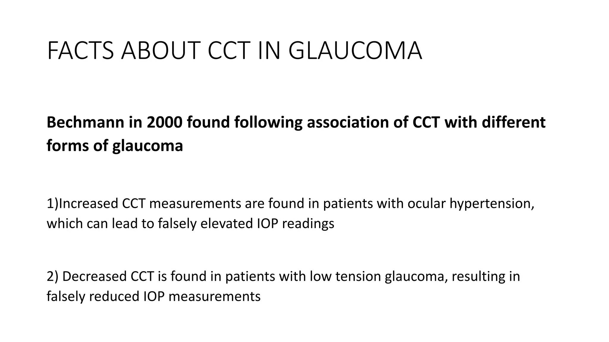
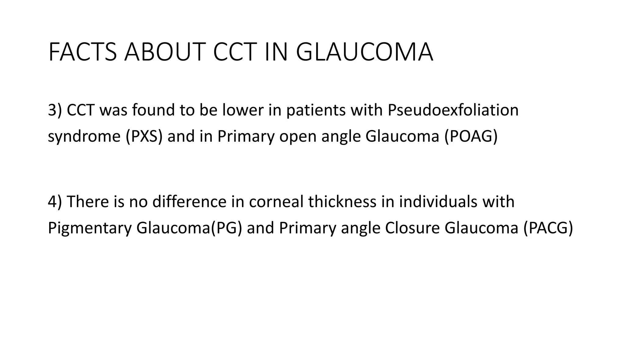
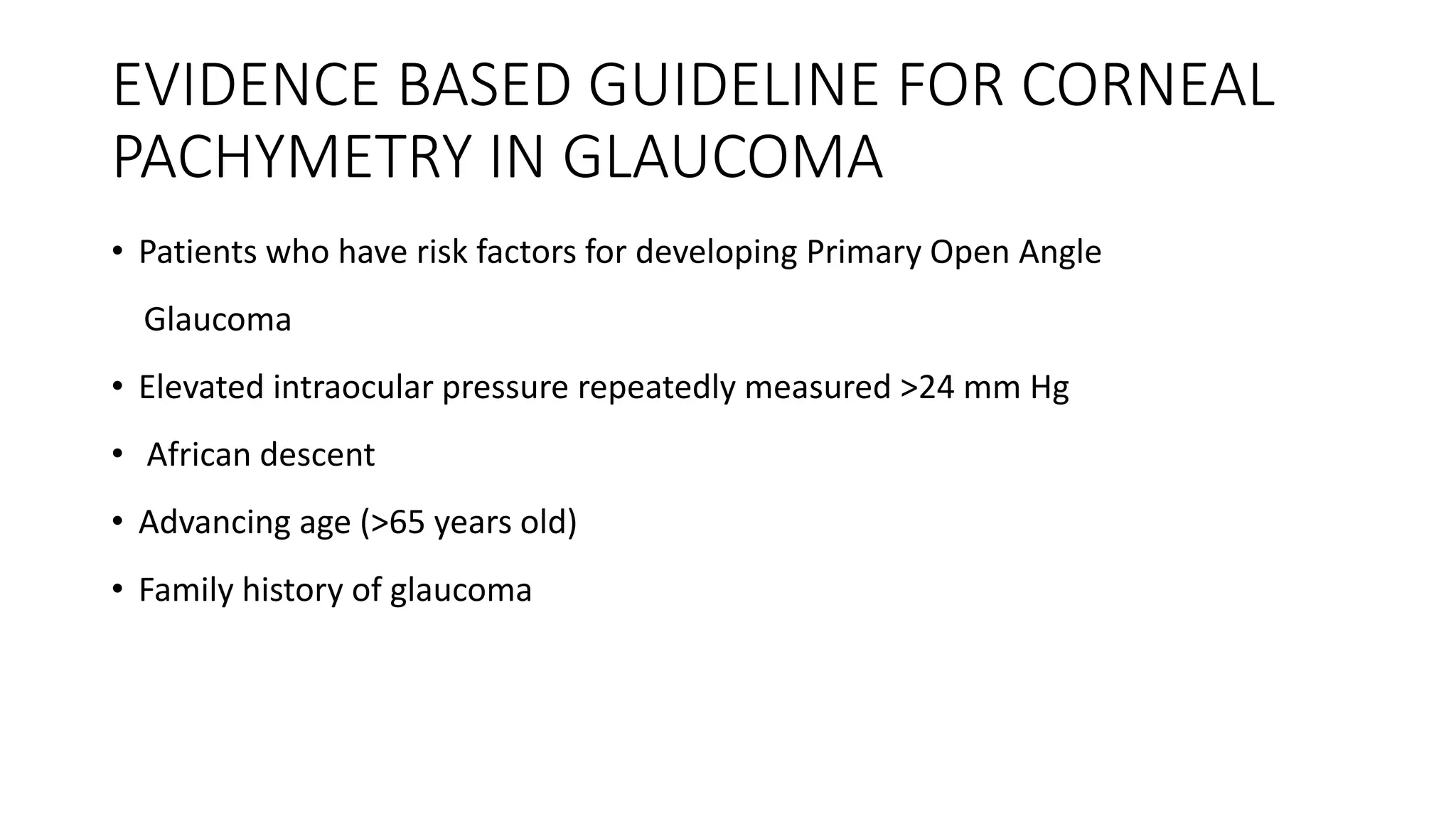
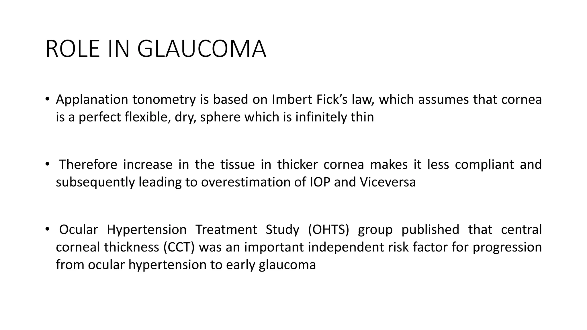
PACHYMETRY IN GLAUCOMA
EVALUATION
INTRODUCTION
• Pachymetry (Greek words: Pachos = thick + metry = to measure) is
term used for the measurement of corneal thickness
• It is an important indicator of health status of the cornea especially
of corneal endothelial pump function
• The thickness of the cornea was first reported in ancient textbooks
on physiological optics (Helmholtz and Gullstrand)
TYPES OF PACHYMETRY
Contact methods
• Ultrasound(mean CCT 544 μm)
• optical such as confocal
microscopy (CONFOSCAN)(mean
CCT 530 μm)
TYPES OF PACHYMETRY
Non-contact methods
• Optical biometry with a
single Scheimpflug camera
(SIRIUS or PENTACAM)
• Dual Scheimpflug camera
(GALILEI)
• Optical Coherence
Tomography (Visante)
• Optical Coherence
Pachymetry (ORBSCAN)
CORNEAL THICKNESS IN NORMAL EYE
• It ranges from 0.7 to 0.9 mm at the limbus and varies between 0.49
mm and 0.56 mm at the centre
• The Central corneal thickness (CCT) reading of 0.7 mm or more is
indicative of endothelial decompensation
• Peripheral corneal thickness is asymmetric so that temporal cornea is
thinnest followed by the inferior cornea
CORNEAL THICKNESS IN NEWBORN AND
CHILDREN
• Corneal configuration in newborns is similar to that of the adult
cornea
• It has been found that cornea on day one is significantly thicker and
decreases in thickness as the child grows older
• The average corneal thickness in infants is 585 ± 52 microns
• The superior peripheral cornea is thinnest in newborn
FACTORS AFFECTING CORNEAL THICKNESS
• Higher in younger patients, male patients and diabetic patients
• The mean CCT of black children is thinner than that of white children
• The PITX2/Pitx2 mutation seen in Axenfeld-Rieger malformations
results in reduced corneal thickness
• Central corneal thickness does not correlate with refraction or
systemic hypertension
ROLE IN CLINICAL PRACTICE
• Glaucoma: for applying correction factor in actual IOP determination
Congenital Glaucoma: to assess the amount of corneal edema
• Refractive surgeries: preoperative screening and treatment plan of
keratorefractive procedures like LASIK, astigmatic keratotomy,even prior to
radial keratotomy
 Post operative follow up of keratoplasty patients to determine endothelial
cell function and its recovery and to become alert to early graft
decompensation
ROLE IN CLINICAL PRACTICE
• Contact lens: To assess corneal edema
Assessing cornea thinness as in corneal disorders like Terrien ’s and
Pellucid marginal degenerations, keratoconus, keratoglobus, post
LASIK ectasia.
 Other cases of corneal decompensation as in herpetic endothelitis
FACTS ABOUT CCT IN GLAUCOMA
Bechmann in 2000 found following association of CCT with different
forms of glaucoma
1)Increased CCT measurements are found in patients with ocular hypertension,
which can lead to falsely elevated IOP readings
2) Decreased CCT is found in patients with low tension glaucoma, resulting in
falsely reduced IOP measurements
FACTS ABOUT CCT IN GLAUCOMA
3) CCT was found to be lower in patients with Pseudoexfoliation
syndrome (PXS) and in Primary open angle Glaucoma (POAG)
4) There is no difference in corneal thickness in individuals with
Pigmentary Glaucoma(PG) and Primary angle Closure Glaucoma (PACG)
EVIDENCE BASED GUIDELINE FOR CORNEAL
PACHYMETRY IN GLAUCOMA
• Patients who have risk factors for developing Primary Open Angle
Glaucoma
• Elevated intraocular pressure repeatedly measured >24 mm Hg
• African descent
• Advancing age (>65 years old)
• Family history of glaucoma
ROLE IN GLAUCOMA
• Applanation tonometry is based on Imbert Fick’s law, which assumes that cornea
is a perfect flexible, dry, sphere which is infinitely thin
• Therefore increase in the tissue in thicker cornea makes it less compliant and
subsequently leading to overestimation of IOP and Viceversa
• Ocular Hypertension Treatment Study (OHTS) group published that central
corneal thickness (CCT) was an important independent risk factor for progression
from ocular hypertension to early glaucoma
OCULAR HYPERTENSION TREATMENT STUDY
• The Ocular Hypertension Treatment Study (OHTS) highlighted the
prognostic significance of CCT in identifying patients with ocular
hypertension (OHT) who would progress to glaucoma
• A multivariate model which included the baseline characteristics of
1618 OHT individuals showed that thinner CCT was the most
important predictive factor for the development of POAG
OCULAR HYPERTENSION TREATMENT STUDY
• The routine measurement of CCT should be included in the initial
assessment of all glaucoma patients
• The prognostic value of CCT is well recognized in OHT patients, but its
predictive value in other glaucoma suspects and patients with
established glaucoma is less certain
Tonometry artefact
• IOP is the principal modifiable risk factor for the progression of established
glaucoma and for the development of glaucoma from OHT
• A 1mmHg decrease in IOP was associated with a 10% decreased risk of
both perimetric progression in the EMGTS and the development of
glaucoma in the OHTS
• It was found that found that the most accurate GAT reading was obtained
in eyes with CCT of approximately 520 μm
Tonometry artefact
• Every 100 μm deviation from this value would result in an error in
IOP measurement of 7 mmHg
• A positive correlation between CCT and IOP has been verified
Thick cornea would result in an artefactually increased IOP reading,
whereas a thin cornea would result in an artefactually reduced IOP
reading
CORRECTION FACTOR
• In chronic eye diseases like glaucoma and glaucoma suspects for
every increase in central corneal thickness of 50 microns, the
correction done is to decrease the recorded IOP by 2.5mmHg
• For acute onset diseases it was recommended to correct by 10 mmHg
for every 50 microns
BIOLOGICAL RISK FACTORS
• Extent of lamina movement was increased in individuals with thin corneas
compared with those with thick corneas(Lesk et al)
• After IOP reduction, the improvement in neuroretinal rim blood flow was also
smaller compared with patients with thicker corneas
• This supports the hypothesis that eyes with thinner CCT may have an increased
risk of developing glaucomatous damage due to laminas that are more
susceptible to being displaced in response to IOP changes
BIOLOGICAL RISK FACTOR
• Physical structures of the optic disc, including depth of cup and disc
area, have been reported by some studies to be correlated with CCT
• Another less likely hypothesis is that a thin cornea increases
trabecular exposure to oxidative damage
GENETIC ASSOCIATION
• CCT is one of the most heritable human traits and may explain some
aspects of the genetic predisposition to glaucoma
• Genome-wide association studies and candidate gene analyses have
uncovered associations between CCT and novel loci. These may
provide candidate genes for the interrogation of POAG
GENETIC ASSOCIATION
• Studies that confirmed a genetic link between CCT and open angle
glaucoma may enable us to determine the mechanisms underlying
this association
• Two missense mutations in a subtype of collagen-8, which if
inactivated results in thinning of the corneal stroma, were detected in
a group of White patients with very thin CCT and advanced POAG
GLAUCOMA TREATMENT
• Ocular hypotensive medications have been shown to alter corneal
thickness
• latanoprost and tafluprost and a significant decrease in CCT possibly
by modifying the corneal extracellular matrix
GLAUCOMA TREATMENT
• Topical b blockers have been associated with a reversible increase in CCT
• Topical carbonic anhydrase inhibitors can cause irreversible corneal
decompensation and increased corneal thickness in eyes with underlying
cornea diseases but they do not significantly alter CCT in healthy corneas
• Thicker CCT was associated with a less significant decrease in IOP after
starting ocular hypotensive medications in the OHTS
GLAUCOMA TREATMENT
• The percentage reduction in IOP after laser trabeculoplasty was
significantly greater in eyes with thinner corneas (CCT<555 μm )
VARIABILITY IN CENTRAL CORNEAL
THICKNESS
• CCT measurements vary with race, sex, age and other environmental
factors, which may confound the relationship between glaucoma and
corneal thickness
• Most epidemiological studies have found that men have thicker corneas
compared with women
• CCT also varies with age, with a small but significant inverse relationship
between CCT and age (range 2–10mm per decade) being reported in the
majority of cross-sectional studies
VARIABILITY IN CENTRAL CORNEAL
THICKNESS
• Hence, it may take up to 20 years for the change in CCT to be clinically
significant and to warrant remeasuring
• CCT is also influenced by environmental factors. Dry eyes and long-
term contact lens wear are associated with a decrease in CCT
• An indoor occupation and sleep are associated with an increase in
CCT
CORNEAL BIOMECHANICS
• CCT is regarded as a correlate of corneal rigidity, but this is only applicable to
structurally normal corneas
• An edematous cornea, though thicker, may result in lower GAT readings because
of reduced corneal rigidity .Conversely,scarred cornea, though thinner, may have
increased corneal rigidity leading to artefactually higher IOP readings
CORNEAL BIOMECHANICS
• Corneal cross-linking results in a significant increase in IOP 12 months after the
procedure despite CCT remaining unchanged. This is presumably because of
increased corneal rigidity
CONCLUSION
• The prognostic value of CCT is well recognized in OHT patients, but its
predictive value in other glaucoma suspects and patients with
established glaucoma is less certain
• Nevertheless, the routine measurement of CCT should be included in
the initial assessment of all glaucoma patients, as it aids in the
interpretation of IOP measurements, risk stratification and the setting
of a target IOP
CONCLUSION
• An adequately validated correction algorithm for GAT measurements
does not exist and better methods of tonometry which are less
influenced by corneal properties, should be developed and adopted
into clinical practice
• Most studies investigating the significance of CCT have included
patients with POAG, and the role of CCT in PACG and secondary
glaucoma is still poorly understood and warrants further investigation
THANK YOU

Pachymetry in glaucoma evaluation presentation

  • 1.
  • 2.
    INTRODUCTION • Pachymetry (Greekwords: Pachos = thick + metry = to measure) is term used for the measurement of corneal thickness • It is an important indicator of health status of the cornea especially of corneal endothelial pump function • The thickness of the cornea was first reported in ancient textbooks on physiological optics (Helmholtz and Gullstrand)
  • 3.
    TYPES OF PACHYMETRY Contactmethods • Ultrasound(mean CCT 544 μm) • optical such as confocal microscopy (CONFOSCAN)(mean CCT 530 μm)
  • 4.
    TYPES OF PACHYMETRY Non-contactmethods • Optical biometry with a single Scheimpflug camera (SIRIUS or PENTACAM) • Dual Scheimpflug camera (GALILEI) • Optical Coherence Tomography (Visante) • Optical Coherence Pachymetry (ORBSCAN)
  • 5.
    CORNEAL THICKNESS INNORMAL EYE • It ranges from 0.7 to 0.9 mm at the limbus and varies between 0.49 mm and 0.56 mm at the centre • The Central corneal thickness (CCT) reading of 0.7 mm or more is indicative of endothelial decompensation • Peripheral corneal thickness is asymmetric so that temporal cornea is thinnest followed by the inferior cornea
  • 6.
    CORNEAL THICKNESS INNEWBORN AND CHILDREN • Corneal configuration in newborns is similar to that of the adult cornea • It has been found that cornea on day one is significantly thicker and decreases in thickness as the child grows older • The average corneal thickness in infants is 585 ± 52 microns • The superior peripheral cornea is thinnest in newborn
  • 7.
    FACTORS AFFECTING CORNEALTHICKNESS • Higher in younger patients, male patients and diabetic patients • The mean CCT of black children is thinner than that of white children • The PITX2/Pitx2 mutation seen in Axenfeld-Rieger malformations results in reduced corneal thickness • Central corneal thickness does not correlate with refraction or systemic hypertension
  • 8.
    ROLE IN CLINICALPRACTICE • Glaucoma: for applying correction factor in actual IOP determination Congenital Glaucoma: to assess the amount of corneal edema • Refractive surgeries: preoperative screening and treatment plan of keratorefractive procedures like LASIK, astigmatic keratotomy,even prior to radial keratotomy  Post operative follow up of keratoplasty patients to determine endothelial cell function and its recovery and to become alert to early graft decompensation
  • 9.
    ROLE IN CLINICALPRACTICE • Contact lens: To assess corneal edema Assessing cornea thinness as in corneal disorders like Terrien ’s and Pellucid marginal degenerations, keratoconus, keratoglobus, post LASIK ectasia.  Other cases of corneal decompensation as in herpetic endothelitis
  • 10.
    FACTS ABOUT CCTIN GLAUCOMA Bechmann in 2000 found following association of CCT with different forms of glaucoma 1)Increased CCT measurements are found in patients with ocular hypertension, which can lead to falsely elevated IOP readings 2) Decreased CCT is found in patients with low tension glaucoma, resulting in falsely reduced IOP measurements
  • 11.
    FACTS ABOUT CCTIN GLAUCOMA 3) CCT was found to be lower in patients with Pseudoexfoliation syndrome (PXS) and in Primary open angle Glaucoma (POAG) 4) There is no difference in corneal thickness in individuals with Pigmentary Glaucoma(PG) and Primary angle Closure Glaucoma (PACG)
  • 12.
    EVIDENCE BASED GUIDELINEFOR CORNEAL PACHYMETRY IN GLAUCOMA • Patients who have risk factors for developing Primary Open Angle Glaucoma • Elevated intraocular pressure repeatedly measured >24 mm Hg • African descent • Advancing age (>65 years old) • Family history of glaucoma
  • 13.
    ROLE IN GLAUCOMA •Applanation tonometry is based on Imbert Fick’s law, which assumes that cornea is a perfect flexible, dry, sphere which is infinitely thin • Therefore increase in the tissue in thicker cornea makes it less compliant and subsequently leading to overestimation of IOP and Viceversa • Ocular Hypertension Treatment Study (OHTS) group published that central corneal thickness (CCT) was an important independent risk factor for progression from ocular hypertension to early glaucoma
  • 14.
    OCULAR HYPERTENSION TREATMENTSTUDY • The Ocular Hypertension Treatment Study (OHTS) highlighted the prognostic significance of CCT in identifying patients with ocular hypertension (OHT) who would progress to glaucoma • A multivariate model which included the baseline characteristics of 1618 OHT individuals showed that thinner CCT was the most important predictive factor for the development of POAG
  • 15.
    OCULAR HYPERTENSION TREATMENTSTUDY • The routine measurement of CCT should be included in the initial assessment of all glaucoma patients • The prognostic value of CCT is well recognized in OHT patients, but its predictive value in other glaucoma suspects and patients with established glaucoma is less certain
  • 16.
    Tonometry artefact • IOPis the principal modifiable risk factor for the progression of established glaucoma and for the development of glaucoma from OHT • A 1mmHg decrease in IOP was associated with a 10% decreased risk of both perimetric progression in the EMGTS and the development of glaucoma in the OHTS • It was found that found that the most accurate GAT reading was obtained in eyes with CCT of approximately 520 μm
  • 17.
    Tonometry artefact • Every100 μm deviation from this value would result in an error in IOP measurement of 7 mmHg • A positive correlation between CCT and IOP has been verified Thick cornea would result in an artefactually increased IOP reading, whereas a thin cornea would result in an artefactually reduced IOP reading
  • 18.
    CORRECTION FACTOR • Inchronic eye diseases like glaucoma and glaucoma suspects for every increase in central corneal thickness of 50 microns, the correction done is to decrease the recorded IOP by 2.5mmHg • For acute onset diseases it was recommended to correct by 10 mmHg for every 50 microns
  • 19.
    BIOLOGICAL RISK FACTORS •Extent of lamina movement was increased in individuals with thin corneas compared with those with thick corneas(Lesk et al) • After IOP reduction, the improvement in neuroretinal rim blood flow was also smaller compared with patients with thicker corneas • This supports the hypothesis that eyes with thinner CCT may have an increased risk of developing glaucomatous damage due to laminas that are more susceptible to being displaced in response to IOP changes
  • 20.
    BIOLOGICAL RISK FACTOR •Physical structures of the optic disc, including depth of cup and disc area, have been reported by some studies to be correlated with CCT • Another less likely hypothesis is that a thin cornea increases trabecular exposure to oxidative damage
  • 21.
    GENETIC ASSOCIATION • CCTis one of the most heritable human traits and may explain some aspects of the genetic predisposition to glaucoma • Genome-wide association studies and candidate gene analyses have uncovered associations between CCT and novel loci. These may provide candidate genes for the interrogation of POAG
  • 22.
    GENETIC ASSOCIATION • Studiesthat confirmed a genetic link between CCT and open angle glaucoma may enable us to determine the mechanisms underlying this association • Two missense mutations in a subtype of collagen-8, which if inactivated results in thinning of the corneal stroma, were detected in a group of White patients with very thin CCT and advanced POAG
  • 23.
    GLAUCOMA TREATMENT • Ocularhypotensive medications have been shown to alter corneal thickness • latanoprost and tafluprost and a significant decrease in CCT possibly by modifying the corneal extracellular matrix
  • 24.
    GLAUCOMA TREATMENT • Topicalb blockers have been associated with a reversible increase in CCT • Topical carbonic anhydrase inhibitors can cause irreversible corneal decompensation and increased corneal thickness in eyes with underlying cornea diseases but they do not significantly alter CCT in healthy corneas • Thicker CCT was associated with a less significant decrease in IOP after starting ocular hypotensive medications in the OHTS
  • 25.
    GLAUCOMA TREATMENT • Thepercentage reduction in IOP after laser trabeculoplasty was significantly greater in eyes with thinner corneas (CCT<555 μm )
  • 26.
    VARIABILITY IN CENTRALCORNEAL THICKNESS • CCT measurements vary with race, sex, age and other environmental factors, which may confound the relationship between glaucoma and corneal thickness • Most epidemiological studies have found that men have thicker corneas compared with women • CCT also varies with age, with a small but significant inverse relationship between CCT and age (range 2–10mm per decade) being reported in the majority of cross-sectional studies
  • 27.
    VARIABILITY IN CENTRALCORNEAL THICKNESS • Hence, it may take up to 20 years for the change in CCT to be clinically significant and to warrant remeasuring • CCT is also influenced by environmental factors. Dry eyes and long- term contact lens wear are associated with a decrease in CCT • An indoor occupation and sleep are associated with an increase in CCT
  • 28.
    CORNEAL BIOMECHANICS • CCTis regarded as a correlate of corneal rigidity, but this is only applicable to structurally normal corneas • An edematous cornea, though thicker, may result in lower GAT readings because of reduced corneal rigidity .Conversely,scarred cornea, though thinner, may have increased corneal rigidity leading to artefactually higher IOP readings
  • 29.
    CORNEAL BIOMECHANICS • Cornealcross-linking results in a significant increase in IOP 12 months after the procedure despite CCT remaining unchanged. This is presumably because of increased corneal rigidity
  • 30.
    CONCLUSION • The prognosticvalue of CCT is well recognized in OHT patients, but its predictive value in other glaucoma suspects and patients with established glaucoma is less certain • Nevertheless, the routine measurement of CCT should be included in the initial assessment of all glaucoma patients, as it aids in the interpretation of IOP measurements, risk stratification and the setting of a target IOP
  • 31.
    CONCLUSION • An adequatelyvalidated correction algorithm for GAT measurements does not exist and better methods of tonometry which are less influenced by corneal properties, should be developed and adopted into clinical practice • Most studies investigating the significance of CCT have included patients with POAG, and the role of CCT in PACG and secondary glaucoma is still poorly understood and warrants further investigation
  • 32.

Editor's Notes

  • #8 Paired-like homeodomain transcription factor 2 also known as pituitary homeobox 2 is a protein that in 
  • #13 Diabetes mellitus (though it is controversial)
  • #15 REVIEW ARTICLE : Central corneal thickness in glaucoma Sng et al ,March 2017. Volume 28 Number 2
  • #17 EarlyManifest Glaucoma Treatment Study (EMGTS)
  • #19 The target IOP is lower for a thin cornea and higher for a thick cornea.  Eyes with thick corneas have a true IOP that is lower than the measured IOP.  Thus, individuals with thicker corneas may be mis-classified as having ocular hypertension
  • #20 . imaged the lamina cribrosa with the confocal scanning laser ophthalmoscopy after lowering the IOP significantly and found tha It is still controversial whether there is an underlying biological association between corneal dimensions and the biomechanical properties of the lamina cribrosa or the peripapillary sclera
  • #21 with one report showing an inverse correlation between CCT and the partial pressure of oxygen in the anterior chamber angle (P¼0.048) in 124 patients undergoing ocular surgery
  • #22 , including genes encoding collagen-5 a-1, zinc finger 469 and fibrillin-1
  • #24 Conversely, in a large multicenter study which included 1181 White persons from 22 extended pedigrees, there was no genetic correlation between CCT and POAG, and CCT was deemed unlikely to be a useful surrogate endophenotype for POAG in this cohort
  • #25 Most studies have reported an association between the use of topical prostaglandin analogues, including Other studies reported that the use of topical prostaglandin analogues was not associated with a reduction in CCT [67] or even resulted in an increase in CCT [68], but some of these studies were limited by their cross-sectional and retrospective design
  • #27 In a retrospective study of 80 eyes of 47 consecutive patients who underwent selective laser trabeculoplasty as the primary treatment for POAG and OHT ,
  • #28 African (518–533mm) [7&&,74,75], Indian (511–514mm) [76,77], Mongolian (495 – 514mm) [78] and Japanese (517–532mm) [75,79,80] persons were found to have thinner corneas than White (542–558mm) [74,81,82], Hispanic (547mm) [83], Korean (554mm) [84] and Chinese (540–542mm) [85,86] persons in population-based studies Racial differences in CCT would need to be considered in the interpretation of IOP, andmayexplainthe higher rates of glaucoma in certain populations Wang et al. [87] foundthat the variationinCCTaccounted for up to 29.4% of the increased risk of glaucoma seen among African-American persons in a large multiethnic population in Northern California
  • #29 These factors The use of different devices to determine CCT has also resulted in variable measurements. In the context of glaucoma management, CCT measurement by ultrasound pachymetry is recommended, and this was used to determine CCT in the OHTS, the EMGTS and the EGPS should be considered in the interpretation of a glaucoma patient’s corneal thickness
  • #30 Corneal biomechanics may have a more significant impact on IOP measurement errors than CCT, which is merely a cornea dimension and not a biomechanical property
  • #31 Measurements of corneal biomechanical properties, such as corneal hysteresis, corneal resistance factor and corneal constant factor, can be obtained by the ORA and the Corvis ST Tonometer These parameters may be better correlated with the overall globe biomechanics than CCT, and may help to explain the susceptibility of some optic nerve heads to damage by IOP variations