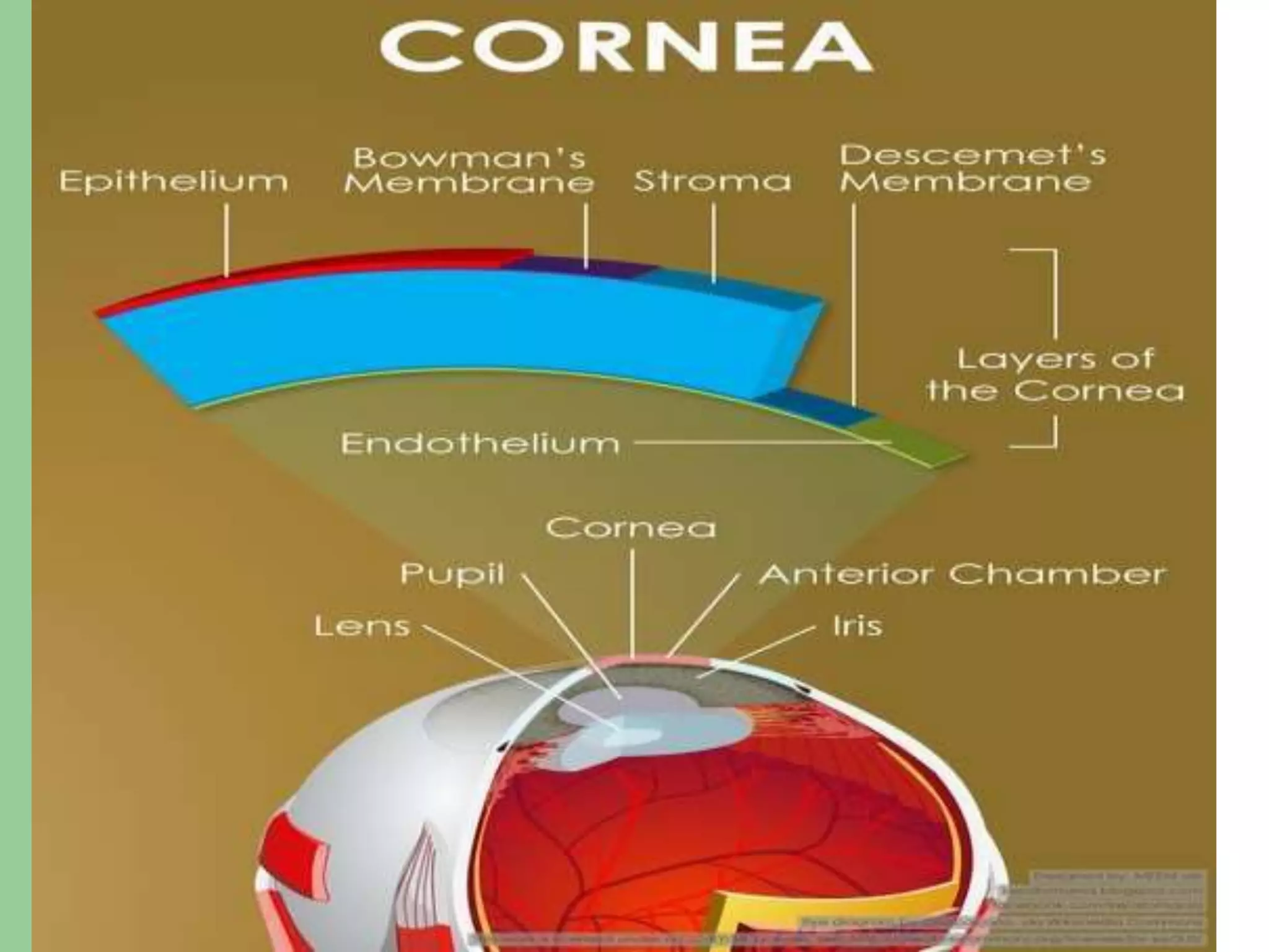
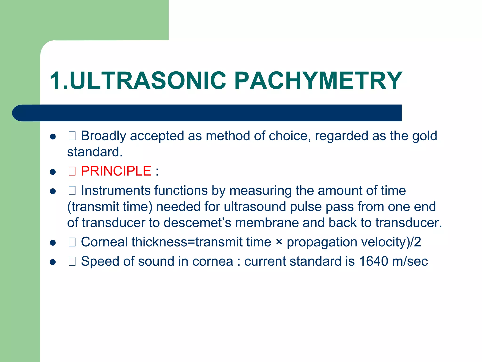
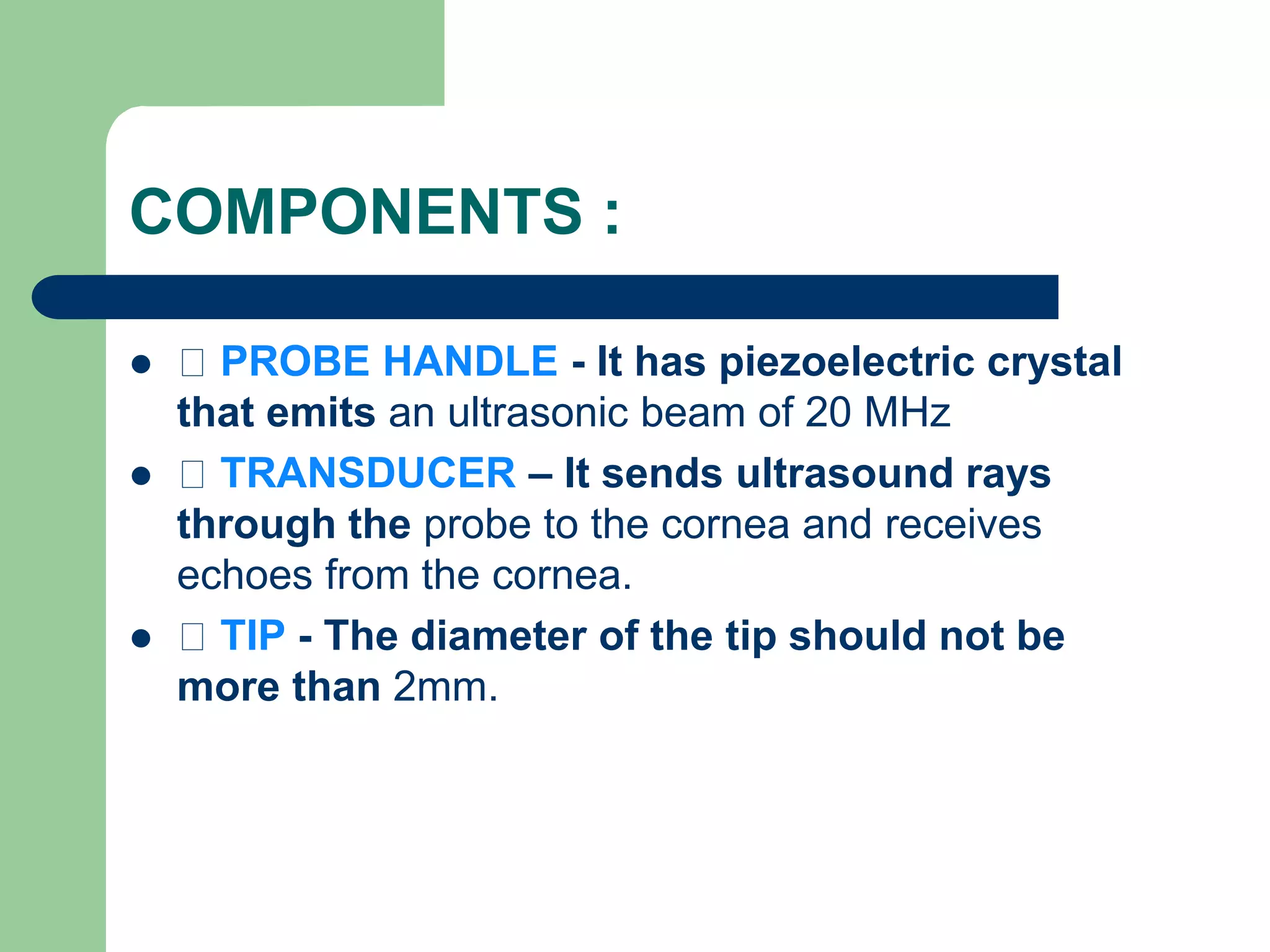
Corneal pachymetry
INTRODUCTION :
 Pachymetry (Greek words: Pachos= thick+metry)
is term used for measurement of corneal thickness.
 It is important indicator of health status of the
cornea especially of corneal endothelial function.
 The thickness of cornea was first reported in
ancient textbooks on physiological optics (Helmholtz
and Gullstrand).
CORNEAL THICKNESS IN
NORMAL EYES
 It ranges from 0.7-0.9 mm at the limbus and varies
between 0.49-0.56 mm at the center.
 The CCT reading of 0.7 mm or more is indicative
of endothelial decompensation.
 Peripheral corneal thickness is asymmetric so that
temporal cornea is thinnest followed by the inferior
cornea.
CORNEAL THICKNESS IN NEWBORNS AND
CHILDREN :
 Is similar to that of adult cornea.
 It has been found that cornea on day one
is significantly thicker and decreases in
thickness as child grows older.
 The average corneal thickness in infants is
585±52 microns.
 The superior peripheral cornea is thinnest
in newborn.
 Normal Values
 Normal corneal thickness – 500-575 microns.
 Abnormal results
 Abnormally thick or thin measurements may
indicate –
 corneal thinning.
 Corneal edema
 Lower or higher than suspected IOP.
FACTORS AFFECTING CCT
 The mean CCT in black children is thinner
than that of white children.
 CCT does not correlate with refraction or
systemic hypertension.
TECHNIQUES
ULTRASONIC OPTICAL ALTERNATIVE
MEASUREMENTS
• Conventional ultrasonic
pachymetry
Manual Pentacam
• Ultrasound
biomicroscpy (UBM).
Specular microscopy • Pachycam
Optical coherence
Tomography(OCT)
1.ULTRASONIC PACHYMETRY
 Broadly accepted as method of choice, regarded as the gold
standard.
 PRINCIPLE :
 Instruments functions by measuring the amount of time
(transmit time) needed for ultrasound pulse pass from one end
of transducer to descemet’s membrane and back to transducer.
 Corneal thickness=transmit time × propagation velocity)/2
 Speed of sound in cornea : current standard is 1640 m/sec
COMPONENTS :
 PROBE HANDLE - It has piezoelectric crystal
that emits an ultrasonic beam of 20 MHz
 TRANSDUCER – It sends ultrasound rays
through the probe to the cornea and receives
echoes from the cornea.
 TIP - The diameter of the tip should not be
more than 2mm.
ADVANTAGES DISADVANTAGES
• Faster ,simpler and easy to use. • Accuracy is dependent on the
perpendicularity of the probe’s
application to the cornea.
Portable Contact method.
• Dry(no coupling medium
required)
• Can be used intraoperatively. Not accurate in edematous cornea.
2. ULTRASOUND
BIOMICROSCOPY:
 High resolution ultrasound
machine which images anterior
segment of the eye.
 Using a 50-MHz imaging probe
 images of the cross-sectional
anterior chamber anatomy with
high resolution are achieved.
 12.5–50 MHz probe , the depth
of penetration is lesser (4mm)than
conventional ultrasound.
ADVANTAGES DISADVANTAGES
• Useful in cases where cornea is
opaque
The main limitation of UBM is that it
requires a water-bath coupling
media
and a very experienced examiner.
• Various layers of cornea can be
identified.
• Contact method and patient to lie
supine
• Cannot be used intraoperatively.
3.MANUAL OPTICAL PACHYMETER:
 This was the original method to measure
corneal thickness.
 The instrument contains two Plano glass
plates that splits the image of the corneal
parallelepiped.
 There are two methods to measure
corneal thickness.
 Just touch method.
 Overlap method.
 The corneal thickness is then directly read
from the scale.
 Range is 0-1.2mm, with a least gradation
of 0.02 mm.
ADVANTAGE DISADVANTAGE
Non contact method. • Lack of accuracy in
measurements, the usual range of
error with an optical pachymeter is
±2%
• Lack of repeatability.
• Requires slit lamp and therefore
has poor portability and cannot be
used in operating room.
4. SPECULAR PACHYMETRY :
 This is the oldest method to measure corneal
thickness.
 PRINCIPLE : This measures the distance
between the anterior and the posterior surfaces of
cornea and depends on the focusing of light rays
through front back cornea unlike sound waves in
ultrasound pachymeter.
 2 types 1.contact 2.Non contact
ADVANTAGES DISADVANTAGES
Operator independent. The exact point where reading is
taken is
not known.
Non invasive. • Time consuming.
Simultaneous measurement of
cell count.
• Impractical to use in operation
room.
6. ANTERIOR SEGMENT OPTICAL
COHERANCE TOMOGRAPHY :
 High resolution, non-contact OCT
customized anterior segment.
 Gives color coded map of corneal
thickness.
ADVANTAGES
 Non contact
 Rapid acquisition during the pachymetry scan ensures an
accurate and repeatable pachymetry map.
 High resolution
 It measures and documents both corneal flap thickness and
residual stromal thickness immediately following LASIK
surgery.
 Measures through corneal opacity.
10. PENTACAM
It is3-dimensional (3D) rotating
scheimpflug.
It can perform five functions in 2
sec.
1. Scheimpflug image of anterior
segment.
2. Pachymetry
3. 3-D anterior chamber
analyzer
4. Corneal topography.
5. Cataract analyzer
 Pachymetry by pentacam is displayed as a
color image over its entire area from limbus
to limbus..
ADVANTAGE DISADVANTAGE
Non invasive, non contact
gives precise representation and
repeatability.
Even minute eye movements are
captured and corrected simultaneously.
▪ It underestimates the corneal thickness in
comparison to ultrasonic pachymetry.
PACHYCAM
 compact and portable
non- contact pachymeter
with built-in keratometer.
It can be mounted on slit
lamp.
 Image acquisition is
done with the help of a
3D alignment screen.
 Advantages
1. Noncontact
2. Compact,portable,light weight
Corneal pachymetry.ppt

Corneal pachymetry.ppt

  • 1.
  • 2.
    INTRODUCTION :  Pachymetry(Greek words: Pachos= thick+metry) is term used for measurement of corneal thickness.  It is important indicator of health status of the cornea especially of corneal endothelial function.  The thickness of cornea was first reported in ancient textbooks on physiological optics (Helmholtz and Gullstrand).
  • 4.
    CORNEAL THICKNESS IN NORMALEYES  It ranges from 0.7-0.9 mm at the limbus and varies between 0.49-0.56 mm at the center.  The CCT reading of 0.7 mm or more is indicative of endothelial decompensation.  Peripheral corneal thickness is asymmetric so that temporal cornea is thinnest followed by the inferior cornea.
  • 5.
    CORNEAL THICKNESS INNEWBORNS AND CHILDREN :  Is similar to that of adult cornea.  It has been found that cornea on day one is significantly thicker and decreases in thickness as child grows older.  The average corneal thickness in infants is 585±52 microns.  The superior peripheral cornea is thinnest in newborn.
  • 6.
     Normal Values Normal corneal thickness – 500-575 microns.  Abnormal results  Abnormally thick or thin measurements may indicate –  corneal thinning.  Corneal edema  Lower or higher than suspected IOP.
  • 7.
    FACTORS AFFECTING CCT The mean CCT in black children is thinner than that of white children.  CCT does not correlate with refraction or systemic hypertension.
  • 8.
    TECHNIQUES ULTRASONIC OPTICAL ALTERNATIVE MEASUREMENTS •Conventional ultrasonic pachymetry Manual Pentacam • Ultrasound biomicroscpy (UBM). Specular microscopy • Pachycam Optical coherence Tomography(OCT)
  • 9.
    1.ULTRASONIC PACHYMETRY  Broadlyaccepted as method of choice, regarded as the gold standard.  PRINCIPLE :  Instruments functions by measuring the amount of time (transmit time) needed for ultrasound pulse pass from one end of transducer to descemet’s membrane and back to transducer.  Corneal thickness=transmit time × propagation velocity)/2  Speed of sound in cornea : current standard is 1640 m/sec
  • 10.
    COMPONENTS :  PROBEHANDLE - It has piezoelectric crystal that emits an ultrasonic beam of 20 MHz  TRANSDUCER – It sends ultrasound rays through the probe to the cornea and receives echoes from the cornea.  TIP - The diameter of the tip should not be more than 2mm.
  • 12.
    ADVANTAGES DISADVANTAGES • Faster,simpler and easy to use. • Accuracy is dependent on the perpendicularity of the probe’s application to the cornea. Portable Contact method. • Dry(no coupling medium required) • Can be used intraoperatively. Not accurate in edematous cornea.
  • 13.
    2. ULTRASOUND BIOMICROSCOPY:  Highresolution ultrasound machine which images anterior segment of the eye.  Using a 50-MHz imaging probe  images of the cross-sectional anterior chamber anatomy with high resolution are achieved.  12.5–50 MHz probe , the depth of penetration is lesser (4mm)than conventional ultrasound.
  • 14.
    ADVANTAGES DISADVANTAGES • Usefulin cases where cornea is opaque The main limitation of UBM is that it requires a water-bath coupling media and a very experienced examiner. • Various layers of cornea can be identified. • Contact method and patient to lie supine • Cannot be used intraoperatively.
  • 15.
    3.MANUAL OPTICAL PACHYMETER: This was the original method to measure corneal thickness.  The instrument contains two Plano glass plates that splits the image of the corneal parallelepiped.  There are two methods to measure corneal thickness.  Just touch method.  Overlap method.  The corneal thickness is then directly read from the scale.  Range is 0-1.2mm, with a least gradation of 0.02 mm.
  • 16.
    ADVANTAGE DISADVANTAGE Non contactmethod. • Lack of accuracy in measurements, the usual range of error with an optical pachymeter is ±2% • Lack of repeatability. • Requires slit lamp and therefore has poor portability and cannot be used in operating room.
  • 17.
    4. SPECULAR PACHYMETRY:  This is the oldest method to measure corneal thickness.  PRINCIPLE : This measures the distance between the anterior and the posterior surfaces of cornea and depends on the focusing of light rays through front back cornea unlike sound waves in ultrasound pachymeter.  2 types 1.contact 2.Non contact
  • 20.
    ADVANTAGES DISADVANTAGES Operator independent.The exact point where reading is taken is not known. Non invasive. • Time consuming. Simultaneous measurement of cell count. • Impractical to use in operation room.
  • 21.
    6. ANTERIOR SEGMENTOPTICAL COHERANCE TOMOGRAPHY :  High resolution, non-contact OCT customized anterior segment.  Gives color coded map of corneal thickness.
  • 23.
    ADVANTAGES  Non contact Rapid acquisition during the pachymetry scan ensures an accurate and repeatable pachymetry map.  High resolution  It measures and documents both corneal flap thickness and residual stromal thickness immediately following LASIK surgery.  Measures through corneal opacity.
  • 24.
    10. PENTACAM It is3-dimensional(3D) rotating scheimpflug. It can perform five functions in 2 sec. 1. Scheimpflug image of anterior segment. 2. Pachymetry 3. 3-D anterior chamber analyzer 4. Corneal topography. 5. Cataract analyzer  Pachymetry by pentacam is displayed as a color image over its entire area from limbus to limbus..
  • 25.
    ADVANTAGE DISADVANTAGE Non invasive,non contact gives precise representation and repeatability. Even minute eye movements are captured and corrected simultaneously. ▪ It underestimates the corneal thickness in comparison to ultrasonic pachymetry.
  • 26.
    PACHYCAM  compact andportable non- contact pachymeter with built-in keratometer. It can be mounted on slit lamp.  Image acquisition is done with the help of a 3D alignment screen.
  • 27.
     Advantages 1. Noncontact 2.Compact,portable,light weight