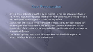
This document discusses otitis media, an inflammation of the middle ear. It begins with an introduction and case presentation of a 4 year old boy with ear pain, fever, and cough. It then covers the epidemiology, anatomy, classification, etiology, risk factors, pathology, clinical features, differential diagnosis, management, and complications of otitis media. The peak incidence is in children aged 1-4 years and is more common in boys. Acute otitis media, otitis media with effusion, chronic suppurative otitis media, and adhesive otitis media are subtypes discussed. Pneumoniae, influenzae, and Moraxella are common bacterial causes while viruses like R
2. ๏ Introduction
๏ Case Presentation
๏ Epidemiology
๏ Anatomy of the ear
๏ Classification
๏ Aetiology
๏ Risk factors
๏ Pathology
๏ Clinical Features
๏ Differential diagnoses
๏ Management
๏ Complications
๏ Prognosis
๏ Conclusion
๏ References
3. ๏ Otitis media can be defined as inflammation of the
cleft mucosal lining of the middle ear.
๏ May also involve inflammation of mastoid, petrous
apex, and peri-labyrinthine air cells.
๏ In most cases, it develops from an infection in the
upper respiratory tract (the nasopharynx) that then
extends into the middle ear via the Eustachian tube.
4. ๏ A.T is a 4 year old male brought in by his mother. He has had a low grade fever of
38.3 C for 3 days. He complained that his ears hurt with difficulty sleeping. He also
had a non productive cough that started the day before.
๏ Examination of both ears reveal significant redness and fluid in both middle ears
with no apparent involvement of the eardrum or tympanic membrane. Further
examination of the patientโs breathing and other manifestations indicate an upper
respiratory infection.
๏ The patientโs parents are chronic heavy smokers and the child is exposed to
second hand smoke in the home environment.
5. ๏ Acute otitis media is very common in childhood. It is one of the most common
condition for which medical care is provided for children in Nigeria.
๏ The prevalence is found to be 14.7%.
๏ The peak incidence is in children aged 1-4 years.
๏ The incidence is slightly higher in boys than girls.
๏ Some infants may experience their first attack shortly after birth and are
considered otitis-prone (i.e. at risk for recurrent otitis media).
6.
7. ๏ The human ear contains sense organs that serve two quite different functions: that
of hearing and that of postural equilibrium and coordination of head and eye
movements.
๏ Anatomically the ear has three distinguishable parts: the outer, middle, and inner
ear.
๏ The outer ear consists of the visible portion called the auricle or pinna, which
projects from the side of the head, and the short external auditory canal, the inner
end of which is closed by the tympanic membrane, commonly called the eardrum.
๏ The thin, semitransparent tympanic membrane, or eardrum, which forms the
boundary between the outer and middle ear, is stretched obliquely across the end
of the external canal.
๏ The uppermost small area of the membrane where the ring is open is slack and is
called the pars flaccida, but the far greater portion is tightly stretched and is called
the pars tensa.
8. ๏ The middle ear is a narrow, air-filled cavity in the temporal bone. It is spanned by a chain
of three tiny bonesโthe malleus (hammer), incus (anvil), and stapes (stirrup), collectively
called the auditory ossicles.
๏ The inner ear consists of two functional units: the vestibular apparatus, consisting of the
vestibule and semicircular canals, which contains the sensory organs of postural
equilibrium; and the snail-shell-like cochlea, which contains the sensory organ of hearing.
๏ These sensory organs are highly specialized endings of the eighth cranial nerve; the
vestibulocochlear nerve.
๏ The middle-ear space roughly resembles a rectangular room with four walls, a floor, and a
ceiling.
๏ The outer (lateral) wall of the middle-ear space is formed by the tympanic membrane.
๏ Its ceiling (superior wall) is a thin plate of bone that separates it from the cranial cavity
and brain above.
9. ๏ The floor (inferior wall) is also a thin bony plate separating the cavity from the
jugular vein and carotid artery below.
๏ The back (posterior) wall partly separates it from another cavity, the mastoid
antrum.
๏ In the front (anterior) wall is the opening of the eustachian or auditory tube,
which connects the middle ear with the nasopharynx.
๏ The inner (medial) wall, which separates the middle ear from the inner ear, or
labyrinth, is a part of the bony otic capsule of the inner ear. It has two small
openings, or fenestrae, one above the other.
๏ The upper one is the oval window (fenestrae vestibuli), which is closed by the
footplate of the stapes. The lower one is the round window (fenestrae rotundum),
which is covered by a thin membrane.
10. ๏ There are several subtypes of Otitis Media, as follows:
1. Acute otitis media (AOM)
2. Otitis media with effusion (OME)
3. Chronic suppurative otitis media (CSOM)
4. Adhesive otitis media
11. ๏ Acute Otitis Media (AOM)
โข AOM results from infection of fluid that has become trapped in the middle ear.
โข It can occur in suppurative, non-suppurative, and recurrent forms.
โข In non-suppurative AOM, inflammation of the middle ear cleft mucosa occurs
either without formation of an effusion or with a sterile effusion. This type of AOM
is often seen prior to, or in the resolution stage of, the acute suppurative otitis
media; however, resolution may occur before frank suppuration.
โข Recurrent AOM is defined as 3 episodes of acute suppurative otitis media in a 6-
month period, or 4 episodes in a 12-month period, with complete resolution of
symptoms and signs between the episodes.
โข Here, patients usually present with concurrent or recent symptoms of upper
respiratory infection (URI), such as cough, rhinorrhea or sinus congestion.
12. ๏ Otitis Media with Effusion (OME) -Serous OM/ Glue ear
โข This is characterized by non purulent effusion of the middle ear that may be serous or mucoid.
โข OME can occur within the resolution phase of AOM
โข Leading theory suggests that eustachian tube dysfunction is the main factor in OME, either by
anatomical blocks, allergic reaction (smoke) or upper respiratory tract infection.
โข This leads to a negative pressure within the middle ear that then causes transudate from the
mucosa.
โข OME often follows an episode of AOM. Symptoms that may be indicative of OME include the
following:
-Hearing loss: parents often describe the child as inattentive
-Tinnitus: occurs, though rarely
-Vertigo: may not be profound, but some unsteadiness or clumsiness may be reported
โข It should, however, be noted that although fluid is present in the middle ear in both conditions, the
fluid is not infected in OME as is seen in AOM patients.
13. ๏ Chronic Suppurative Otitis Media (CSOM)
โข Chronic suppurative otitis media is a persistent ear infection that results in tearing
or perforation of the eardrum.
โข Can occur with or without cholesteatoma (destructive tumor consisting of
keratinizing epithelial cells in the middle ear and/or mastoid process)
โข Surgery is the model modality of treatment with medical intervention used as an
adjunct.
โข It should be noted that CSOM is different from chronic otitis media in that the latter
is defined as middle ear effusion without perforation and persists for 1-3 months.
14. ๏ Adhesive Otitis Media
โข Adhesive otitis media occurs when a thin retracted ear drum becomes sucked into
the middle ear space and stuck.
โข It is an abacterial inflammation of the middle ear and the adjoining pneumatic
spaces.
โข Here, there is occlusion of the eustachian tube and adhesion of the tympanum.
15. The Aetiology of otitis media can be classified as follows;
๏ VIRAL PATHOGENS
โข Of the viral pathogens, the RSV (Respiratory Syncytial Virus) is the most common
cause of pneumonia and bronchiolitis in young persons and this can lead to upper
respiratory tract infection, not just in children, but in all age groups.
โข RSV was identified early as a pathogen that appeared to create long-term
pulmonary complications, primarily asthma, in as many as half of infants with
bronchiolitis.
โข Other viruses include: Rhino & Adeno Virus.
16. ๏ BACTERIAL PATHOGENS
โข Four bacteriaโnamely, Streptococcus pneumoniae, Heamophilus influenzae,
Moraxella catarrhalis, and Streptococcus pyogenes โare responsible for the
majority of episodes of otitis media in persons older than 6 weeks.
โข Other bacteria recovered and implicated include Staphylococcus aureus, Viridans
streptococci, and Pseudomonas aeruginosa.
โข It should be noted that pneumococcal infections are probably responsible for at
least 50% of otitis media episodes
17. ๏ Age - Children between 6 to 36 months are most likely to get ear infections.
๏ Attending daycare.
๏ Recent illness, such as a cold or sinus infection.
๏ History of allergies, like hay fever, also called allergic rhinitis, or sinusitis.
๏ Exposure to secondhand smoke.
๏ Having family members who are prone to ear infections; studies show a clear
genetic component for both acute and recurrent otitis media.
๏ Using a pacifier.
๏ Having a history of gastroesophageal reflux disease (GERD)
๏ Obesity.
18. 1. Catarrhal stage: Is characterized by occlusion of Eustachian tube and congestion
of middle ear.
2. Stage of exudation: Exudate collects in the middle ear and ear drum is pushed
laterally. Initially the exudate is mucoid, later it becomes purulent.
3. Stage of suppuration: Pus in the middle ear collects under tension, stretches the
drum & perforates it by pressure necrosis & the exudate starts escaping into
external auditory canal
4. Stage of healing: The infection starts resolving from any of the stages mentioned
& usually clears up completely without leaving any sequelae.
5. Stage of complications: Infection may spread to the mastoid antrum. Initially it
causes Catarrhal mastoiditis [congestion of the mastoid mucosa], stage of
coalescent mastoiditis & later empyema of the mastoid.
19. Patients usually present with following:
๏ Otalgia: Young children may exhibit signs of otalgia (ear pain) by pulling on the
affected ear or pulling on the hair. It apparently occurs more often when the child
is lying down (e.g., during the night, during nap time)
๏ Otorrhoea: Discharge may come from the middle ear through a recently
perforated tympanic membrane
๏ Headache
๏ Fever
๏ Loss of appetite
๏ Diarrhea
๏ Vomiting
21. ๏ History
1. History of previous Upper Respiratory Tract Infection
2. Otalgia, Otorrhoea
3. Fever, vomiting and diarrhea
4. Cough and nasal discharge
5. In older patients, hearing loss and stuffiness of the ear
6. History of exposure to risk factors e.g smoke, use of pacifiers in babies etc.
22. ๏ Examination
Pneumatic otoscopy remains the gold standard for OM diagnosis
1. Otoscopy shows signs of inflammation, beginning with reddening of the mucosa
and progressing to the formation of purulent middle ear effusion
2. In the posterior quadrants, the tympanic membrane may bulge, and the superficial
epithelial layer may exhibit a scalded appearance
3. Examination of the entire head and neck is also important because several
congenital anomalies are associated with OM e.g. Treacher Collins syndrome, cleft
palate, Downโs syndrome etc.
23. During examination of the tympanic membrane, the following parameters are to be
noted;
๏ Color: A normal membrane is pale grey. An opaque yellow or blue membrane
indicates Medial Ear Effusion, MEE. Dark red indicates recent trauma or blood
behind the membrane. Dark pink or light red is consistent with AOM.
๏ Position: The position of the tympanic membrane is key in differentiating AOM
from OME. Typically, in AOM, the membrane is bulging while in OME, the
membrane is retracted.
๏ Mobility: Abnormal movements can suggest various conditions or disorders.
Studies though, have shown that the most consistent physical finding in patients
with OME is reduced tympanic membrane motility during pneumatic otoscopy.
๏ Perforation: Single perforations are most common but the patient may have
multiple perforations also.
24. ๏ Investigations
In most cases of OM, no further investigations are necessary since the diagnosis is
clinical.
If symptoms are severe;
โข FBC: often reveals leukocytosis
โข Blood cultures may detect bacteremia during episodes of high fever.
โข A culture and sensitivity test of the ear discharge by tympanocentesis
(Tympanocentesis involves aspiration of the contents of the middle ear cleft by
piercing the tympanic membrane with a needle and collecting that material for
diagnostic examination) or ear swab is helpful in guiding antibiotic therapy in
patients in whom the first-line treatment is unsuccessful.
โข If recurrent AOM occurs along with recurrent infections in other systems, then an
underlying immune should be considered and appropriate investigations
requested.
25. โข Petromastoid Computed tomography (CT) scan: may be necessary to determine if
a complication has occurred.
โข Magnetic resonance imaging (MRI): might be more appropriate for diagnosing
suspected intracranial complications
โข Tympanometry may also be done to confirm Otitis media.
27. ๏ Control Infection
โข Arrest & reverse inflammation
โข Prevent suppuration and perforation
โข Relieve symptoms
โข Hasten resolution
โข Reduce risk of complications
AMPICILLIN
(50 mg/kg/day in 4 divided doses)
AMOXICILLIN
(40 mg/kg/day in 3 divided doses)
28. ๏ Local Therapy
Before Perforation:
โข Relieve earache
โข Control inflammation
โข Prevent perforation(spontaneous)
After Perforation:
โข Clear external acoustic canal toilet
โข Control infection
โข Repair TM
1. Decongestant nasal drops(ephedrine, oxymetazoline, xylometazoline)
2. Oral nasal decongestant (pseudoephedrine)
3. Analgesic & antipyretic(paracetamol)
29. ๏ Myringotomy
โข Tiny incision eardrum
โข Relieve pressure
โข Drain pus from the middle ear.
๏ A tympanostomy tube
โข Keep the middle ear aerated
โข Prevent re-accumulation of fluid.
๏ Indications for myringotomy
โข Symptoms are not relieved by antibiotics
โข TM bulges significantly
โข TM perforation is too small
โข Incomplete resolution
โข Persistent effusion beyond 12 weeks
30. Complications that may occur following the development of OM are as follows:
โข Acute infection of the mastoid air cells: resulting in a dangerous mastoid
abscess
โข Meningitis
โข Brain abscess
โข Epidural abscess: usually occurs through the temporal bone
โข Septicemia
โข Infection of the labyrinth
โข Facial nerve paralysis
31. ๏ At some point, surgery often is required to reconstruct the perforated eardrum
(tympanoplasty), clear the mastoid air cells of infection (mastoidectomy), and
remove the growth of excess tissue in the ear canal (removal of cholesteatoma).
Conductive hearing loss from acute otitis media typically resolves with time.
32. ๏ Otitis media is a common pediatric disease that can lead to various symptoms in
the neonate and infant. It is could be caused by both host factors and pathological
factors and, in the absence of complications, usually resolves spontaneously.
๏ Pneumatic otoscopy is the gold standard diagnostic test and a broad spectrum
antibiotic, usually Amoxicillin, is mostly employed.
33. ๏ Encyclopedia Britannica. Encyclopรฆdia Britannica Chicago: Encyclopรฆdia Britannica,
2013. Chicago: Encyclopedia Britannica, 2013.
๏ Moore KL, Dalley AF, Agur AMR. Clinically Oriented Anatomy, 6th edn. Williams & Wilkins.
2010.Basic Otorhinolaryngology by Rudolf Probst, Gerhard Grevers and Heinrich Iro
๏ American Academy of Pediatrics. Respiratory syncytial virus. Red Book: Report of the
Committee on Infectious Diseases. 27th ed. Elk Grove Village, Ill: 2006. 560-566.
๏ Medscape โOtitis media
๏ Lalwani AK. Current Diagnosis and Treatment in otolaryngology, 2nd edn. McGraw Hill.
2007.
๏ Probst R, Grevers G, Heinrich I. Basic otorhinolaryngology, 2nd edn. Georg Thieme Verlag,
Stuttgart. 2006.
๏ Wood DN, Nakas N, Gregory CW. International Journal of Pediatric Otorhinolaryngology.
Volume 76, Issue 9, September 2012
๏ https://www.ncbi.nlm.nih.gov/pubmed/16276700