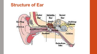
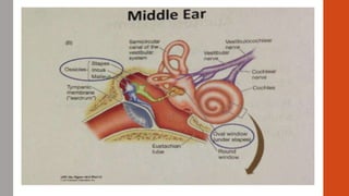
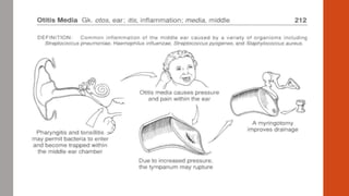
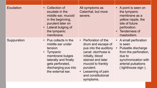
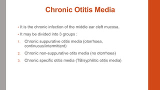
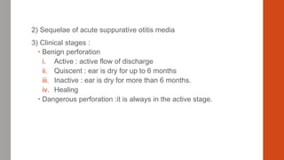
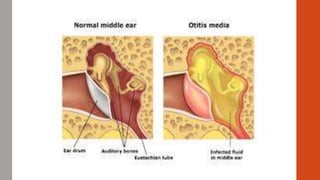
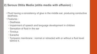
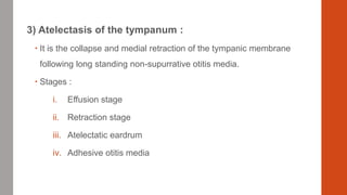
Otitis Media
By Dr. Rajal Sukhiyaji
Structure of Ear
Acute Suppurative Otitis Media
Definition :-
• It is an acute pyogenic infection of the mucosa of the middle ear
cleft.
Aetiology :-
• Age :- any, but more common in children
• Sex :- Male = Female
• Geographic variation :- None
• Reduced vitality
• Atmospheric pressure changes, as during flying and diving
• Upper respiratory tract infection
• Swimming
Pathophysiology
1) Eustachian tube :-
 Anatomical obstruction
 Forcible blowing of nose
 Source of infection :
i. Adenoiditis
ii. Tonsillitis
iii. Rhinitis
iv. Pharyngitis
v. Influenza
vi. Infections secondary to cleft palate.
 Feeding an infant with a bottle in supine position may permit
contaminated milk to enter the tube.
 Iatrogenic
2) External ear :- following traumatic rupture of the tympanic membrane.
3) Head injury :- fracture of temporal bone.
4) Haematogenous spread
 The causative organisms are as follows :-
• Streptococcus haemolyticus
• Staphylococcus aureus
• Haemophilus influenza
• Penumococcus
• Non-haemolytic Streptococcus.
Pathology :-
Stage Features Symptoms Signs
Catarrhal Occlusion of the
Eustachian tube.
Congestion in the
middle ear.
• Fullness and
heaviness in the
ear.
• Pain: severe,
worse at night in
recumbent position
• Deafness:
progressive
• Tinitus: bubbling
• Autophony: own
spoken words
echo in the ear.
• Constitutional
symptoms: fever,
malaise.
• Retraction of
tympanic
membrane:
early
• Congestion of
tympanic
membrane
Exudation • Collection of
exudate in the
middle ear, mucoid
in the beginning,
purulent later on
• Lateral bulging of
the tympanic
membrane.
All symptoms as
Catarrhal, but more
severe.
• A point is seen on
the tympanic
membrane as a
yellow nipple, the
site of future
perforation.
• Tenderness of
mastoidism.
Suppuration • Pus collects in the
middle ear under
tension
• Tympanic
membrane bulges
laterally and finally
gets perforated,
discharging pus into
the external ear.
• Perforation of the
drum and escape of
pus into the auditory
canal: otorrhoea is
initially, blood
stained and later
mucoid to frankly
purulent.
• Lessening of pain
and constitutional
symptoms.
• A small perforation
is seen
• Pulsatile discharge
from the perforation,
due to
synchronization with
arterial pulsations
( lighthouse sign ).
Healing • Resolution may begin
at any of the stages,
depending upon
patient’s resistance
and antibiotic therapy.
May begin at any stage.
Coalescent mastoiditis • Coalescent
mastoiditis:-Block of
the aditus to antrum
by granulations and
oedema, collection of
pus under tension
within the mastoid,
and coalescence of
mastoid air cells due
to break down of
septa between them.
• Catarrhal mastoiditis
• Empyema of mastoid.
Investigation
• Test of hearing
1. Tuning fork tests
2. Audiometry
• Microbiologic tests :on the ear discharge
• Radiography
Treatment
1) Systemic :-
• Broad spectrum antibiotics : Penicillin, Ampicillin, Amoxycillin
• Nasal decongestants
• Systemic decongestants
• NSAID
• Rest
2) Local :-
• Ear drops
• Aural toilet
3) Surgical : Myringotomy
Chronic Otitis Media
• It is the chronic infection of the middle ear cleft mucosa.
• It may be divided into 3 groups :
1. Chronic suppurative otitis media (otorrhoea,
continuous/intermittent)
2. Chronic non-suppurative otits media (no otorrhoea)
3. Chronic specific otitis media (TB/syphilitic otitis media)
1) Chronic Suppurative Otitis Media
• Definition :- CSOM is a chronic inflammatory process involving the
middle ear cleft producing irreversible pathological changes.
• Classification :
1)Benign or Tubotympanic type :
• Central perforation of the tympanic membrane.
• Limited to the tympanum and the Eustachian tube.
• No complication
2) Dangerous or atticoantral type :
• Marginal perforation
• Destructive cholesteatoma in the attic and the antrum
• Life threatening complications
Aetiology :
• Age : any
• Sex : Male = Female
• Low socioeconomic status
• Malnutrition
• Poor hygiene
• Persistence of acute suppurative media
Pathophysiology :
1) Microbiology : organisms are as follows.
 Benign tubotympanic perforation
i. Streptococci
ii. Staphylococci
iii. Pneumococci
 Atticoantral or dangerous perforation
i. Bacillus pyocyneu
ii. Bacillus proteus
iii. Bacillus coli
 Acute nacrotic otitis media
i. Haemolytic streptococci
2) Sequelae of acute suppurative otitis media
3) Clinical stages :
 Benign perforation
i. Active : active flow of discharge
ii. Quiscent : ear is dry for up to 6 months
iii. Inactive : ear is dry for more than 6 months.
iv. Healing
 Dangerous perforation :it is always in the active stage.
Pathology :
Reduced blood supply of ear drum
Necrosis start
Affects ossicles
Sclerosis of mastoid
Polyps may come
Tympanosclerosis due to hyaline degeneration of middle layer of mucosa
with calcification
Ossicular chain discontinuity
Adhesions of tympanic cavity
 Cholesteatoma :-
• It occurs in dangerous type otitis media. It is a sac lined by keratinising
stratified squamous epithelium in the middle ear cleft and contains
desquamated epithelium arranged like onion – skin layer.
• It has got consistency of a tooth paste with high destructive power.
• Hence, it is defined as a “ non malignant bone destroying disease of ear
“.
Symptoms :
1) Otorrhoea
2) Deafness ( mild to moderate degree )
3) Earache
4) Tinitus
5) Giddiness (due to labyrinthitis)
6) Bleeding or blood-stained discharge (due to
granulations or polyps)
7) Swelling in the mastoid region due to mastoid
abscess.
Signs :
1) Perforation
• Central in benign type
• Marginal in dangerous type
2) Tenderness over the mastoid : due to mastoiditis
Investigation :
1) Examination of nose/pharynx
2) Hearing tests
3) Microbiologic studies : on the ear discharge
4) Radiology of mastoid
5) Tests for patency of Eustachian tube :
 If the ear drops reaches the throat, the tube is patent.
6) Otomicroscopy
7) Fistula test
8) Examination of the CNS for intracranial complications.
Treatment :
1. Conservative treatment:
 Removal of septic foci like adenoids, tonsils, sinusitis.
 Aural toilet :
 Prevention of entry of water into the ear.
 Cleaning of the ear with a dry swab stick or suction.
 Ear drops : otorrhoea :
 antiseptic or antibiotic ear drops, as in acute suppurative
otitis media. The drops are changed if there is no
response in 6 or 7 days.
 Chemical cautery
 Good nutrition
 Improvement of general condition
2) Surgical treatment :
 Adenoidectomy, septoplasty
 Aural polypectomy : Removal of polyps and granulation followed by
chemical cauterization or by tympanoplasty
 Myringoplasty
 Tympanoplasty : reconstructive surgery of the damaged ossicles and the
membrane.
 Depending upon extent of disease and degree of deafness, various
surgical procedures are undertaken like,
oAtticotomy
oModified radical mastoidectomy
oRadical mastoidectomy
oMastoidectomy with tympanoplasty
oCombined approach tympanoplasty (CAT)
Comparison of Benign and Dangerous
Types of CSOM
Variable Benign type Dangerous type
Site Tubotympanic Atticoantral
Perforation Central Attic or marginal
Discharge • Intermittent
• Mucopurulent / purulent
• Without foul smell
• White/yellowish
• Copious
• Bleeding uncommon
• More with upper respiratory
tract infection.
• Continuous
• Purulent
• Foul smelling
• Yellowish/brownish/greenis
• Often scanty
• Bleeding possible
• No effect of upper respiratory
tract infection.
Deafness • Conductive, mild to
moderate
• Conductive/mixed, mild to
severe
Polyp Occasional Common
Cholesteatoma Very rare Almost always present
Complications Very rare Common
Radiography of mastoid Cellular/sclerotic Sclerotic with erosion
2)Chronic non-suppurative otitis
media
• Characterized by accumulation of a non-purulent effusion in
the middle ear cleft.
• The following diseases occur as a result.
1) Eustachian catarrh :-
 Due to the obstruction of the Eustachian tube, the air in the
middle ear gets absorbed and the eardrum becomes
retracted.
 Patient experiences conductive deafness
2) Serous Otitis Media (otitis media with effusion) :
 Fluid having a consistency of glue in the middle ear, producing conductive
deafness.
 Features :
 Deafness
 Impairment of speech and language development in children
 Sensation of fluid in the ear
 Tinnitus
 Earache
 Tympanic membrane : normal or retracted with or without a fluid level
behind it.
3) Atelectasis of the tympanum :
 It is the collapse and medial retraction of the tympanic membrane
following long standing non-supurrative otitis media.
 Stages :
i. Effusion stage
ii. Retraction stage
iii. Atelectatic eardrum
iv. Adhesive otitis media
4) Tympanosclerosis :
 It is a condition characterized by white plaques (chalk patches) occurring
in the tympanic membrane and surrounding ossicles.
 It results from healed chronic otitis media.
 There is hyaline degeneration of middle ear mucosa which undergoes
calcification and appears like the flakes of white snow.
Treatment :-
1) Treat the causative factors like diseases of nose, sinuses or pharynx
2) Eustachian catheterization
3) Myringopuncture :- Injection of air into the middle ear may give relief.
4) Myringotomy : Surgical incision into the eardrum, to relieve pressure or drain
fluid.
5) Grommet : In recurrent cases a temporary Teflon tube is inserted for
ventilation through the eardrum
6) Adenoidectomy : In obstructive cases
7) Tympanoplasty / ossiculoplasty
8) Hearing aid
9) Antibiotics, decongestants, steroid, mucolytics are used.
3) Tuberculous otitis media
• Not uncommon disease.
• Secondary to pulmonary TB
 Clinical features :
• Slow onset of disease
• Painless condition
• Discharge is thin, scanty and odourless
• Tympanic membrane is pale yellow to rosy pink in colour.
• Perforations in the membrane are usually multiple and may be associated
with pale granulations.
• Hearing loss is disproportionate to other symptoms.
 Treatment :-
• Antitubercular therapy
• Advanced cases may require surgical intervention.
Thank you

Otitis media

  • 1.
    Otitis Media By Dr.Rajal Sukhiyaji
  • 2.
  • 5.
    Acute Suppurative OtitisMedia Definition :- • It is an acute pyogenic infection of the mucosa of the middle ear cleft. Aetiology :- • Age :- any, but more common in children • Sex :- Male = Female • Geographic variation :- None • Reduced vitality • Atmospheric pressure changes, as during flying and diving • Upper respiratory tract infection • Swimming
  • 6.
    Pathophysiology 1) Eustachian tube:-  Anatomical obstruction  Forcible blowing of nose  Source of infection : i. Adenoiditis ii. Tonsillitis iii. Rhinitis iv. Pharyngitis v. Influenza vi. Infections secondary to cleft palate.  Feeding an infant with a bottle in supine position may permit contaminated milk to enter the tube.  Iatrogenic
  • 7.
    2) External ear:- following traumatic rupture of the tympanic membrane. 3) Head injury :- fracture of temporal bone. 4) Haematogenous spread  The causative organisms are as follows :- • Streptococcus haemolyticus • Staphylococcus aureus • Haemophilus influenza • Penumococcus • Non-haemolytic Streptococcus.
  • 9.
    Pathology :- Stage FeaturesSymptoms Signs Catarrhal Occlusion of the Eustachian tube. Congestion in the middle ear. • Fullness and heaviness in the ear. • Pain: severe, worse at night in recumbent position • Deafness: progressive • Tinitus: bubbling • Autophony: own spoken words echo in the ear. • Constitutional symptoms: fever, malaise. • Retraction of tympanic membrane: early • Congestion of tympanic membrane
  • 11.
    Exudation • Collectionof exudate in the middle ear, mucoid in the beginning, purulent later on • Lateral bulging of the tympanic membrane. All symptoms as Catarrhal, but more severe. • A point is seen on the tympanic membrane as a yellow nipple, the site of future perforation. • Tenderness of mastoidism. Suppuration • Pus collects in the middle ear under tension • Tympanic membrane bulges laterally and finally gets perforated, discharging pus into the external ear. • Perforation of the drum and escape of pus into the auditory canal: otorrhoea is initially, blood stained and later mucoid to frankly purulent. • Lessening of pain and constitutional symptoms. • A small perforation is seen • Pulsatile discharge from the perforation, due to synchronization with arterial pulsations ( lighthouse sign ).
  • 14.
    Healing • Resolutionmay begin at any of the stages, depending upon patient’s resistance and antibiotic therapy. May begin at any stage. Coalescent mastoiditis • Coalescent mastoiditis:-Block of the aditus to antrum by granulations and oedema, collection of pus under tension within the mastoid, and coalescence of mastoid air cells due to break down of septa between them. • Catarrhal mastoiditis • Empyema of mastoid.
  • 16.
    Investigation • Test ofhearing 1. Tuning fork tests 2. Audiometry • Microbiologic tests :on the ear discharge • Radiography
  • 17.
    Treatment 1) Systemic :- •Broad spectrum antibiotics : Penicillin, Ampicillin, Amoxycillin • Nasal decongestants • Systemic decongestants • NSAID • Rest 2) Local :- • Ear drops • Aural toilet 3) Surgical : Myringotomy
  • 18.
    Chronic Otitis Media •It is the chronic infection of the middle ear cleft mucosa. • It may be divided into 3 groups : 1. Chronic suppurative otitis media (otorrhoea, continuous/intermittent) 2. Chronic non-suppurative otits media (no otorrhoea) 3. Chronic specific otitis media (TB/syphilitic otitis media)
  • 19.
    1) Chronic SuppurativeOtitis Media • Definition :- CSOM is a chronic inflammatory process involving the middle ear cleft producing irreversible pathological changes. • Classification : 1)Benign or Tubotympanic type : • Central perforation of the tympanic membrane. • Limited to the tympanum and the Eustachian tube. • No complication 2) Dangerous or atticoantral type : • Marginal perforation • Destructive cholesteatoma in the attic and the antrum • Life threatening complications
  • 20.
    Aetiology : • Age: any • Sex : Male = Female • Low socioeconomic status • Malnutrition • Poor hygiene • Persistence of acute suppurative media
  • 21.
    Pathophysiology : 1) Microbiology: organisms are as follows.  Benign tubotympanic perforation i. Streptococci ii. Staphylococci iii. Pneumococci  Atticoantral or dangerous perforation i. Bacillus pyocyneu ii. Bacillus proteus iii. Bacillus coli  Acute nacrotic otitis media i. Haemolytic streptococci
  • 22.
    2) Sequelae ofacute suppurative otitis media 3) Clinical stages :  Benign perforation i. Active : active flow of discharge ii. Quiscent : ear is dry for up to 6 months iii. Inactive : ear is dry for more than 6 months. iv. Healing  Dangerous perforation :it is always in the active stage.
  • 23.
    Pathology : Reduced bloodsupply of ear drum Necrosis start Affects ossicles Sclerosis of mastoid Polyps may come Tympanosclerosis due to hyaline degeneration of middle layer of mucosa with calcification Ossicular chain discontinuity Adhesions of tympanic cavity
  • 24.
     Cholesteatoma :- •It occurs in dangerous type otitis media. It is a sac lined by keratinising stratified squamous epithelium in the middle ear cleft and contains desquamated epithelium arranged like onion – skin layer. • It has got consistency of a tooth paste with high destructive power. • Hence, it is defined as a “ non malignant bone destroying disease of ear “.
  • 25.
    Symptoms : 1) Otorrhoea 2)Deafness ( mild to moderate degree ) 3) Earache 4) Tinitus 5) Giddiness (due to labyrinthitis) 6) Bleeding or blood-stained discharge (due to granulations or polyps) 7) Swelling in the mastoid region due to mastoid abscess.
  • 27.
    Signs : 1) Perforation •Central in benign type • Marginal in dangerous type 2) Tenderness over the mastoid : due to mastoiditis
  • 29.
    Investigation : 1) Examinationof nose/pharynx 2) Hearing tests 3) Microbiologic studies : on the ear discharge 4) Radiology of mastoid 5) Tests for patency of Eustachian tube :  If the ear drops reaches the throat, the tube is patent. 6) Otomicroscopy 7) Fistula test 8) Examination of the CNS for intracranial complications.
  • 31.
    Treatment : 1. Conservativetreatment:  Removal of septic foci like adenoids, tonsils, sinusitis.  Aural toilet :  Prevention of entry of water into the ear.  Cleaning of the ear with a dry swab stick or suction.  Ear drops : otorrhoea :  antiseptic or antibiotic ear drops, as in acute suppurative otitis media. The drops are changed if there is no response in 6 or 7 days.  Chemical cautery  Good nutrition  Improvement of general condition
  • 32.
    2) Surgical treatment:  Adenoidectomy, septoplasty  Aural polypectomy : Removal of polyps and granulation followed by chemical cauterization or by tympanoplasty  Myringoplasty  Tympanoplasty : reconstructive surgery of the damaged ossicles and the membrane.  Depending upon extent of disease and degree of deafness, various surgical procedures are undertaken like, oAtticotomy oModified radical mastoidectomy oRadical mastoidectomy oMastoidectomy with tympanoplasty oCombined approach tympanoplasty (CAT)
  • 33.
    Comparison of Benignand Dangerous Types of CSOM Variable Benign type Dangerous type Site Tubotympanic Atticoantral Perforation Central Attic or marginal Discharge • Intermittent • Mucopurulent / purulent • Without foul smell • White/yellowish • Copious • Bleeding uncommon • More with upper respiratory tract infection. • Continuous • Purulent • Foul smelling • Yellowish/brownish/greenis • Often scanty • Bleeding possible • No effect of upper respiratory tract infection. Deafness • Conductive, mild to moderate • Conductive/mixed, mild to severe
  • 34.
    Polyp Occasional Common CholesteatomaVery rare Almost always present Complications Very rare Common Radiography of mastoid Cellular/sclerotic Sclerotic with erosion
  • 36.
    2)Chronic non-suppurative otitis media •Characterized by accumulation of a non-purulent effusion in the middle ear cleft. • The following diseases occur as a result. 1) Eustachian catarrh :-  Due to the obstruction of the Eustachian tube, the air in the middle ear gets absorbed and the eardrum becomes retracted.  Patient experiences conductive deafness
  • 37.
    2) Serous OtitisMedia (otitis media with effusion) :  Fluid having a consistency of glue in the middle ear, producing conductive deafness.  Features :  Deafness  Impairment of speech and language development in children  Sensation of fluid in the ear  Tinnitus  Earache  Tympanic membrane : normal or retracted with or without a fluid level behind it.
  • 38.
    3) Atelectasis ofthe tympanum :  It is the collapse and medial retraction of the tympanic membrane following long standing non-supurrative otitis media.  Stages : i. Effusion stage ii. Retraction stage iii. Atelectatic eardrum iv. Adhesive otitis media
  • 39.
    4) Tympanosclerosis : It is a condition characterized by white plaques (chalk patches) occurring in the tympanic membrane and surrounding ossicles.  It results from healed chronic otitis media.  There is hyaline degeneration of middle ear mucosa which undergoes calcification and appears like the flakes of white snow.
  • 40.
    Treatment :- 1) Treatthe causative factors like diseases of nose, sinuses or pharynx 2) Eustachian catheterization 3) Myringopuncture :- Injection of air into the middle ear may give relief. 4) Myringotomy : Surgical incision into the eardrum, to relieve pressure or drain fluid. 5) Grommet : In recurrent cases a temporary Teflon tube is inserted for ventilation through the eardrum 6) Adenoidectomy : In obstructive cases 7) Tympanoplasty / ossiculoplasty 8) Hearing aid 9) Antibiotics, decongestants, steroid, mucolytics are used.
  • 41.
    3) Tuberculous otitismedia • Not uncommon disease. • Secondary to pulmonary TB  Clinical features : • Slow onset of disease • Painless condition • Discharge is thin, scanty and odourless • Tympanic membrane is pale yellow to rosy pink in colour. • Perforations in the membrane are usually multiple and may be associated with pale granulations. • Hearing loss is disproportionate to other symptoms.
  • 42.
     Treatment :- •Antitubercular therapy • Advanced cases may require surgical intervention.
  • 43.