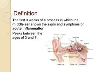
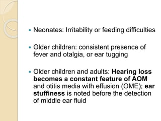
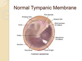
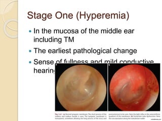
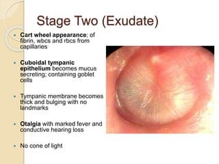
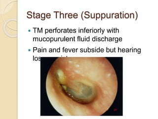
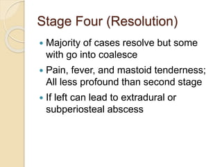
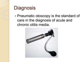
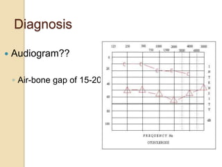
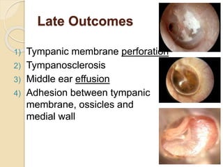
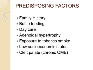
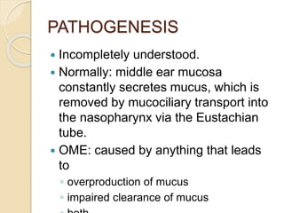
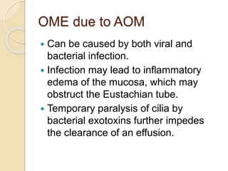
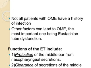
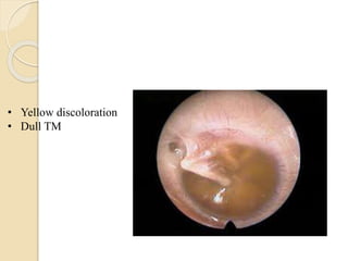
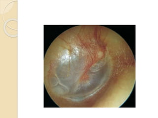
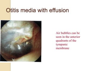
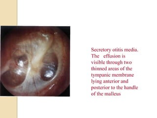
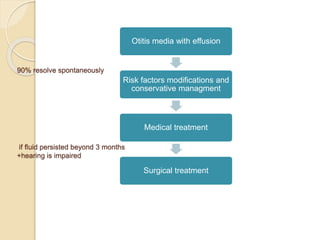
SlideShare a Scribd company logo
Acute Otitis Media
Definition
• The first 3 weeks of a process in which the
middle ear shows the signs and symptoms of
acute inflammation
• Peaks between the
ages of 3 and 7.
 Neonates: Irritability or feeding difficulties
 Older children: consistent presence of
fever and otalgia, or ear tugging
 Older children and adults: Hearing loss
becomes a constant feature of AOM
and otitis media with effusion (OME); ear
stuffiness is noted before the detection
of middle ear fluid
Normal Tympanic Membrane
Otitis Media Stages
Tympanic Membrane View with
Otoscope
Stage One (Hyperemia)
 In the mucosa of the middle ear
including TM
 The earliest pathological change
 Sense of fullness and mild conductive
hearing loss
Stage Two (Exudate)
 Cart wheel appearance; of
fibrin, wbcs and rbcs from
capillaries
 Cuboidal tympanic
epithelium becomes mucus
secreting; containing goblet
cells
 Tympanic membrane becomes
thick and bulging with no
landmarks
 Otalgia with marked fever and
conductive hearing loss
 No cone of light
Stage Three (Suppuration)
 TM perforates inferiorly with
mucopurulent fluid discharge
 Pain and fever subside but hearing
loss persists
Stage Four (Resolution)
 Majority of cases resolve but some
with go into coalesce
 Pain, fever, and mastoid tenderness;
All less profound than second stage
 If left can lead to extradural or
subperiosteal abscess
Clinical Course
 OM is a self limiting disease
 Healing with resolution of tissues toward
normal even without treatment
 Most common cause is S. Pneumonia
 Clinical course is shortened and
terminated earlier by adequate antibiotic
treatment
Diagnosis
 History and physical exam
 Head & neck exam
Diagnosis
 Pneumatic otoscopy is the standard of
care in the diagnosis of acute and
chronic otitis media.
Tympanogram
 Type B curve
Diagnosis
 Audiogram??
◦ Air-bone gap of 15-20dB
Treatment - AOM
 Best time to treat is before the beginning of the
3rd stage
 Antibiotics - consider drug resistance patterns 
amoxicillin
 All antibiotics must be given for 7-10 days.
 In the coalescence stage patient usually needs IV
antibiotics for 24 hours followed be cortical
mastoidectomy .
Antibiotics
 In children under the age of 5;
◦ H. Influenza
◦ Augmentin (Amoxacillin and clavulanic
acid)
 In adults and children above the age of 5;
 Streptococcus (Esp. S. Pneumonia)
◦ Ampicillin / Amoxacillin/ Erythromycin
Late Outcomes
1) Tympanic membrane perforation
2) Tympanosclerosis
3) Middle ear effusion
4) Adhesion between tympanic
membrane, ossicles and
medial wall
OTITIS MEDIA WITH EFFUSION
secretory otitis media
glue ear
 It's defined as the presence of fluid (non
purulent) in the middle ear.
 Fluid  mucoid I serious
 It's the leading cause of hearing loss in
childhood.
OTITIS MEDIA WITH
EFFUSION
(Secretory/nonsuppurative)
 “Glue ear”: describing the thick and glue like
fluid in the middle ear.
 persistence of fluid (for weeks to months)
within the middle ear resulting in conductive
deafness.
 the most common cause of hearing loss in
children in the developed world and has
peaks in incidence 2-5 years of age
 occurs after an episode of AOM, and children
with OME are far more likely to suffer from
recurrent AOM.
 may be responsible for developmental and
educational impairment, and if untreated may
result in permanent middle-ear changes
PREDISPOSING FACTORS
 Family History
 Bottle feeding
 Day care
 Adenoidal hypertrophy
 Exposure to tobacco smoke
 Low socioeconomic status
 Cleft palate (chronic OME)
PATHOGENESIS
 Incompletely understood.
 Normally: middle ear mucosa
constantly secretes mucus, which is
removed by mucociliary transport into
the nasopharynx via the Eustachian
tube.
 OME: caused by anything that leads
to
◦ overproduction of mucus
◦ impaired clearance of mucus
OME due to AOM
 Can be caused by both viral and
bacterial infection.
 Infection may lead to inflammatory
edema of the mucosa, which may
obstruct the Eustachian tube.
 Temporary paralysis of cilia by
bacterial exotoxins further impedes
the clearance of an effusion.
 Not all patients with OME have a history
of infection
 Other factors can lead to OME, the
most important one being Eustachian
tube dysfunction.
Functions of the ET include:
 1)Protection of the middle ear from
nasopharyngeal secretions.
 2)Clearance of secretions of the middle
THE EUSTACHIAN TUBE
 Is normally blocked and opens temporarily with
swallowing
 Air in the middle ear space is absorbed resulting
in sub-atmospheric pressure when the ET is
closed. When the tube opens temporarily with
each swallow, atmospheric air is drawn into the
middle ear to equalize the pressure.
 The result of any tubal obstruction is a decrease
in the intra-tympanic pressure due to oxygen
consumption from the middle ear.
 Because the walls of the middle ear cant
collapse, the negative pressure is maintained
and transduction of fluids from the vasculature of
the middle ear may result in OME.
OTHER CAUSES
 Recurrent URTI (sinusitis/tonsillitis/rhinitis)
culture from OME shows mainly
pneumococcus/H.influenza.
 Nasopharyngeal obstruction:
 In adults the MCC of unilateral middle ear
effusion is nasopharyngeal CA ( carcinoma or
lymphoma)
 Allergy ; allergic edema of the ET is the
principal cause of obstruction
 Barotrauma (diving)
 During descent the outside pressure and the
pressure in the nasopharynx is higher than
middle ear pressure causing locking of the
ET.
CLINICAL FINDINGS
 Can be symptomatic or Asymptomatic
 The most common symptom of OME is
hearing loss.
 In younger children, the only symptom
may be delayed speech development
or behavioral problems
 Another common symptom is a “blocked”
feeling in the ear.
 Ear ache is uncommon
 the Mother may notice that her child
tends to do traction to his auricle
• Yellow discoloration
• Dull TM
Otitis media with effusion
Air bubbles can be
seen in the anterior
quadrants of the
tympanic
membrane
Secretory otitis media.
The effusion is
visible through two
thinned areas of the
tympanic membrane
lying anterior and
posterior to the handle
of the malleus
Management
90% resolve spontaneously without any
medical intervention .
Initial management should identify the
underlying cause , in adults nasopharyngeal
tumors should be excluded .
90% resolve spontaneously
if fluid persisted beyond 3 months
+hearing is impaired
Otitis media with effusion
Risk factors modifications and
conservative managment
Medical treatment
Surgical treatment
The following modifications
may help:
• Avoiding secondhand smoke
• Breastfeeding whenever possible
• Avoiding feeding, either by breast or bottle, while
completely supine
• Avoiding exposure to a large number of children,
particularly in daycare centers
• Avoiding exposure to children who are known to be
affected
• Avoiding known allergens
When to refer to an ENT specialist ?
• An otolaryngologist should be consulted
whenever the primary care physician
(PCP) is concerned about persistent
conductive hearing loss in children,
especially those with signs of language
development delay or if the disease is
Medical treatment
• Pharmacologic management of otitis
media with effusion (OME) includes
administration of antimicrobial agents,
steroids, antihistamines and
decongestants, and mucolytics.
However, their use is not
recommended with concerns
regarding side effects and cost, as
well as a lack of evidence for long-
term therapeutic effectiveness.
1. Antimicrobial agents
• short-term benefit.
• When the otitis media with effusion
becomes chronic (3 mo), the effectiveness
of antimicrobials diminishes.
2. steriods
• In 3 placebo-controlled randomized
clinical trials, oral steroids alone did
not improve otitis media with effusion
clearance within 2 weeks of treatment.
When oral steroids are combined with
antibiotics, the rate of clearance of
middle ear effusion does not improve
compared with the rate with antibiotics
alone.
3. Antihistamines and
decongestants
• There is no difference in the clearance
rates of the effusion between use of
an antihistamine/decongestant or
placebo.
• However, nasal obstruction,
rhinorrhea, and sinusitis often
accompany otitis media, and
antihistamines and decongestants
may be considered for the relief of
these associated symptoms.
Indications for Surgical
Intervention
Surgery has become the most widely accepted therapeutic
intervention for persistent otitis media with effusion
(OME), and it is clearly effective. The interventions
include myringotomy ( procedure to create a hole in the
ear drum) with or without tube insertion, adenoidectomy,
or both.
Indications for Surgical
Intervention
• surgical intervention was supported if
fluid persisted beyond 3 months, but
observation is an option if hearing is not
impaired, therefore, continuous hearing
assessment is a must.
• For patients with hearing loss and otitis
media with effusion, a loss of 40 dB or
greater is an absolute indication for
pressure equalization tube insertion. A
loss in the range of 21-40 dB is a relative
indication with a very low threshold for
placement.
Myringotomy
• is a surgical procedure in which a tiny
incision is created in
the eardrum (tympanic membrane) to
relieve pressure caused by excessive
buildup of fluid, or to drain pus from
the middle ear.
• Myringotomy and aspiration is useful
to treat patients with moderate to
severe hearing loss as they recover
normal middle ear function.
Myringotomy with PET
(Grommet tube) insertion
• When myringotomy is performed alone
without the placement of pressure
equalization tubes, this procedure is worse
in long-term follow-up in children but when
its performed with placement of pressure
equalization tubes, there is improvement
in hearing, duration of middle ear effusion
(MEE), time to recurrence, and less need
for repeated procedures.
Myringotomy & Tympanostomy
Tube
Myringotomy Tympanostomy Tube
Mercado 2011 ©
Mercado 2011 ©
Types of Tubes
Shepard Grommet Soileau Tytan®
Titanium Ventilation Tubes Spoon Bobbins Goode T-Tubes®
Armstrong Beveled
Grommets, Modified
Paparella-Type Vent Tubes
TriuneTubes
A
All tubes are designed to permit ventilation of the middle ear and
mastoid system.
Typically, the tubes self-extrude 9-12 months after placement.
Complications
• Persistent otorrhea (most common)
• Tympanosclerosis (scarring of the ear
drum)
• Persistent perforation
These complications is also known to increase
with the placement of tympanostomy tubes (T-
tubes) that are designed to stay in the tympanic
membrane longer than the typical grommet tube.
Long-Term Monitoring
• No standard of care for the follow-up of
patients with otitis media with effusion (OME)
has been established.
• Some caregivers follow up with the patient 3
weeks after the placement of the tubes and
then every 6 months thereafter, until the
tubes extrude or are removed.
• Patients are instructed that if more than 2
episodes of otorrhea occur before the 6-
month follow-up is scheduled, they should
see their otolaryngologist (ENT) instead of or
in addition to their primary care physician
(PCP).

More Related Content

Similar to Otitis Media ppt .pptx

Chronic Otitis Media Effusion
Chronic Otitis Media EffusionChronic Otitis Media Effusion
Chronic Otitis Media Effusion
healthuseful
 
Otitis Media With Effusion.pptx
Otitis Media With Effusion.pptxOtitis Media With Effusion.pptx
Otitis Media With Effusion.pptx
crezynaz
 
Inflammatory condition of middle ear
Inflammatory condition of middle earInflammatory condition of middle ear
Inflammatory condition of middle ear
ankittiwari0822
 
MIDDLE EAR MSN
MIDDLE EAR MSNMIDDLE EAR MSN
MIDDLE EAR MSN
SareenaBuwaji
 
Otitis Media
Otitis Media Otitis Media
Otitis Media
Boluwatife Afolabi
 
otitis media.pdf
otitis media.pdfotitis media.pdf
otitis media.pdf
Chanchal Riya sharma
 
Otitis media with effusion
Otitis media with effusionOtitis media with effusion
Otitis media with effusion
shriyashsinha
 
SECRETORY_OTITIS_MEDIA.pptx
SECRETORY_OTITIS_MEDIA.pptxSECRETORY_OTITIS_MEDIA.pptx
SECRETORY_OTITIS_MEDIA.pptx
SurabhiKumari55
 
DISORDERS OF EUSTACHIAN TUBE.pptx
DISORDERS OF EUSTACHIAN TUBE.pptxDISORDERS OF EUSTACHIAN TUBE.pptx
DISORDERS OF EUSTACHIAN TUBE.pptx
KarishmaMishra13
 
Otitis Media.pptx
Otitis Media.pptxOtitis Media.pptx
Otitis Media.pptx
Tamanna Dahiya
 
ACUTE OTITIS MEDIA infection of the middle ear
ACUTE OTITIS MEDIA infection of the middle earACUTE OTITIS MEDIA infection of the middle ear
ACUTE OTITIS MEDIA infection of the middle ear
paultembo7
 
Otitis Media 3.pptx
Otitis Media 3.pptxOtitis Media 3.pptx
Otitis Media 3.pptx
paultembo7
 
3RD SEMINAR VSN,,.pptx
3RD SEMINAR VSN,,.pptx3RD SEMINAR VSN,,.pptx
3RD SEMINAR VSN,,.pptx
VchinnariBai
 
otittismedia-200825060054-230321082330-b4b9dde2.pdf
otittismedia-200825060054-230321082330-b4b9dde2.pdfotittismedia-200825060054-230321082330-b4b9dde2.pdf
otittismedia-200825060054-230321082330-b4b9dde2.pdf
ssuser8f10bd
 
Asom
AsomAsom
Dis of mid ear,dr.s.s.bakshi,27.03.17
Dis of mid ear,dr.s.s.bakshi,27.03.17Dis of mid ear,dr.s.s.bakshi,27.03.17
Dis of mid ear,dr.s.s.bakshi,27.03.17
ophthalmgmcri
 
Disease of middle ear,dr.s.s.bakshi,27.03.17
Disease of middle ear,dr.s.s.bakshi,27.03.17Disease of middle ear,dr.s.s.bakshi,27.03.17
Disease of middle ear,dr.s.s.bakshi,27.03.17
ophthalmgmcri
 
Csom mucosal
Csom mucosalCsom mucosal
Csom mucosal
amit jha
 
ACUTE AND CHRONIC OTITIS MEDIA notes.pptx
ACUTE AND CHRONIC OTITIS MEDIA notes.pptxACUTE AND CHRONIC OTITIS MEDIA notes.pptx
ACUTE AND CHRONIC OTITIS MEDIA notes.pptx
ruth444mukami
 

Similar to Otitis Media ppt .pptx (20)

Chronic Otitis Media Effusion
Chronic Otitis Media EffusionChronic Otitis Media Effusion
Chronic Otitis Media Effusion
 
Otitis Media With Effusion.pptx
Otitis Media With Effusion.pptxOtitis Media With Effusion.pptx
Otitis Media With Effusion.pptx
 
Inflammatory condition of middle ear
Inflammatory condition of middle earInflammatory condition of middle ear
Inflammatory condition of middle ear
 
MIDDLE EAR MSN
MIDDLE EAR MSNMIDDLE EAR MSN
MIDDLE EAR MSN
 
Otitis Media
Otitis Media Otitis Media
Otitis Media
 
otitis media.pdf
otitis media.pdfotitis media.pdf
otitis media.pdf
 
Otitis media with effusion
Otitis media with effusionOtitis media with effusion
Otitis media with effusion
 
SECRETORY_OTITIS_MEDIA.pptx
SECRETORY_OTITIS_MEDIA.pptxSECRETORY_OTITIS_MEDIA.pptx
SECRETORY_OTITIS_MEDIA.pptx
 
DISORDERS OF EUSTACHIAN TUBE.pptx
DISORDERS OF EUSTACHIAN TUBE.pptxDISORDERS OF EUSTACHIAN TUBE.pptx
DISORDERS OF EUSTACHIAN TUBE.pptx
 
Otitis Media.pptx
Otitis Media.pptxOtitis Media.pptx
Otitis Media.pptx
 
ACUTE OTITIS MEDIA infection of the middle ear
ACUTE OTITIS MEDIA infection of the middle earACUTE OTITIS MEDIA infection of the middle ear
ACUTE OTITIS MEDIA infection of the middle ear
 
Otitis Media 3.pptx
Otitis Media 3.pptxOtitis Media 3.pptx
Otitis Media 3.pptx
 
3RD SEMINAR VSN,,.pptx
3RD SEMINAR VSN,,.pptx3RD SEMINAR VSN,,.pptx
3RD SEMINAR VSN,,.pptx
 
otittismedia-200825060054-230321082330-b4b9dde2.pdf
otittismedia-200825060054-230321082330-b4b9dde2.pdfotittismedia-200825060054-230321082330-b4b9dde2.pdf
otittismedia-200825060054-230321082330-b4b9dde2.pdf
 
Otittis media
Otittis mediaOtittis media
Otittis media
 
Asom
AsomAsom
Asom
 
Dis of mid ear,dr.s.s.bakshi,27.03.17
Dis of mid ear,dr.s.s.bakshi,27.03.17Dis of mid ear,dr.s.s.bakshi,27.03.17
Dis of mid ear,dr.s.s.bakshi,27.03.17
 
Disease of middle ear,dr.s.s.bakshi,27.03.17
Disease of middle ear,dr.s.s.bakshi,27.03.17Disease of middle ear,dr.s.s.bakshi,27.03.17
Disease of middle ear,dr.s.s.bakshi,27.03.17
 
Csom mucosal
Csom mucosalCsom mucosal
Csom mucosal
 
ACUTE AND CHRONIC OTITIS MEDIA notes.pptx
ACUTE AND CHRONIC OTITIS MEDIA notes.pptxACUTE AND CHRONIC OTITIS MEDIA notes.pptx
ACUTE AND CHRONIC OTITIS MEDIA notes.pptx
 

More from BeerDilacshe1

pediatric meningitis.pptx
pediatric meningitis.pptxpediatric meningitis.pptx
pediatric meningitis.pptx
BeerDilacshe1
 
ENT13 - Sinusitis.ppt
ENT13 - Sinusitis.pptENT13 - Sinusitis.ppt
ENT13 - Sinusitis.ppt
BeerDilacshe1
 
dependency states.pptx
dependency states.pptxdependency states.pptx
dependency states.pptx
BeerDilacshe1
 
tastimonail..docx
tastimonail..docxtastimonail..docx
tastimonail..docx
BeerDilacshe1
 
Instructional Leadership Practices of Selected Principals in Maldives - A Cas...
Instructional Leadership Practices of Selected Principals in Maldives - A Cas...Instructional Leadership Practices of Selected Principals in Maldives - A Cas...
Instructional Leadership Practices of Selected Principals in Maldives - A Cas...
BeerDilacshe1
 
DEQO XASAN.docx
DEQO XASAN.docxDEQO XASAN.docx
DEQO XASAN.docx
BeerDilacshe1
 
cabdulkaadir.pdf
cabdulkaadir.pdfcabdulkaadir.pdf
cabdulkaadir.pdf
BeerDilacshe1
 
GENECOLOGY-1 (Autosaved).docx
GENECOLOGY-1 (Autosaved).docxGENECOLOGY-1 (Autosaved).docx
GENECOLOGY-1 (Autosaved).docx
BeerDilacshe1
 
Instructional Leadership Practices of Selected Principals in Maldives - A Cas...
Instructional Leadership Practices of Selected Principals in Maldives - A Cas...Instructional Leadership Practices of Selected Principals in Maldives - A Cas...
Instructional Leadership Practices of Selected Principals in Maldives - A Cas...
BeerDilacshe1
 
GENECOLOGY-1 (Autosaved).docx
GENECOLOGY-1 (Autosaved).docxGENECOLOGY-1 (Autosaved).docx
GENECOLOGY-1 (Autosaved).docx
BeerDilacshe1
 
cv.docx
cv.docxcv.docx
cv.docx
BeerDilacshe1
 
gastroenteritisppt-120817094816-phpapp01.pdf
gastroenteritisppt-120817094816-phpapp01.pdfgastroenteritisppt-120817094816-phpapp01.pdf
gastroenteritisppt-120817094816-phpapp01.pdf
BeerDilacshe1
 
tastimonail..docx
tastimonail..docxtastimonail..docx
tastimonail..docx
BeerDilacshe1
 
cabdulkaadir.pdf
cabdulkaadir.pdfcabdulkaadir.pdf
cabdulkaadir.pdf
BeerDilacshe1
 
tastimonail..docx
tastimonail..docxtastimonail..docx
tastimonail..docx
BeerDilacshe1
 
ASTHMA AND COPD .pptx
ASTHMA AND COPD .pptxASTHMA AND COPD .pptx
ASTHMA AND COPD .pptx
BeerDilacshe1
 

More from BeerDilacshe1 (16)

pediatric meningitis.pptx
pediatric meningitis.pptxpediatric meningitis.pptx
pediatric meningitis.pptx
 
ENT13 - Sinusitis.ppt
ENT13 - Sinusitis.pptENT13 - Sinusitis.ppt
ENT13 - Sinusitis.ppt
 
dependency states.pptx
dependency states.pptxdependency states.pptx
dependency states.pptx
 
tastimonail..docx
tastimonail..docxtastimonail..docx
tastimonail..docx
 
Instructional Leadership Practices of Selected Principals in Maldives - A Cas...
Instructional Leadership Practices of Selected Principals in Maldives - A Cas...Instructional Leadership Practices of Selected Principals in Maldives - A Cas...
Instructional Leadership Practices of Selected Principals in Maldives - A Cas...
 
DEQO XASAN.docx
DEQO XASAN.docxDEQO XASAN.docx
DEQO XASAN.docx
 
cabdulkaadir.pdf
cabdulkaadir.pdfcabdulkaadir.pdf
cabdulkaadir.pdf
 
GENECOLOGY-1 (Autosaved).docx
GENECOLOGY-1 (Autosaved).docxGENECOLOGY-1 (Autosaved).docx
GENECOLOGY-1 (Autosaved).docx
 
Instructional Leadership Practices of Selected Principals in Maldives - A Cas...
Instructional Leadership Practices of Selected Principals in Maldives - A Cas...Instructional Leadership Practices of Selected Principals in Maldives - A Cas...
Instructional Leadership Practices of Selected Principals in Maldives - A Cas...
 
GENECOLOGY-1 (Autosaved).docx
GENECOLOGY-1 (Autosaved).docxGENECOLOGY-1 (Autosaved).docx
GENECOLOGY-1 (Autosaved).docx
 
cv.docx
cv.docxcv.docx
cv.docx
 
gastroenteritisppt-120817094816-phpapp01.pdf
gastroenteritisppt-120817094816-phpapp01.pdfgastroenteritisppt-120817094816-phpapp01.pdf
gastroenteritisppt-120817094816-phpapp01.pdf
 
tastimonail..docx
tastimonail..docxtastimonail..docx
tastimonail..docx
 
cabdulkaadir.pdf
cabdulkaadir.pdfcabdulkaadir.pdf
cabdulkaadir.pdf
 
tastimonail..docx
tastimonail..docxtastimonail..docx
tastimonail..docx
 
ASTHMA AND COPD .pptx
ASTHMA AND COPD .pptxASTHMA AND COPD .pptx
ASTHMA AND COPD .pptx
 

Recently uploaded

MS Wine Day 2024 Arapitsas Advancements in Wine Metabolomics Research
MS Wine Day 2024 Arapitsas Advancements in Wine Metabolomics ResearchMS Wine Day 2024 Arapitsas Advancements in Wine Metabolomics Research
MS Wine Day 2024 Arapitsas Advancements in Wine Metabolomics Research
Panagiotis Arapitsas
 
Food Processing and Preservation Presentation.pptx
Food Processing and Preservation Presentation.pptxFood Processing and Preservation Presentation.pptx
Food Processing and Preservation Presentation.pptx
dengejnr13
 
在线办理(UCM毕业证书)加州大学美熹德分校毕业证文凭证书一模一样
在线办理(UCM毕业证书)加州大学美熹德分校毕业证文凭证书一模一样在线办理(UCM毕业证书)加州大学美熹德分校毕业证文凭证书一模一样
在线办理(UCM毕业证书)加州大学美熹德分校毕业证文凭证书一模一样
7sl9gbvd
 
Kitchen Audit at restaurant as per FSSAI act
Kitchen Audit at restaurant as per FSSAI actKitchen Audit at restaurant as per FSSAI act
Kitchen Audit at restaurant as per FSSAI act
MuthuMK13
 
Best hotel in keerthy hotel manage ment
Best hotel in keerthy hotel manage   mentBest hotel in keerthy hotel manage   ment
Best hotel in keerthy hotel manage ment
keerthyhotelmangemen
 
Vietnam Mushroom Market Growth, Demand and Challenges of the Key Industry Pla...
Vietnam Mushroom Market Growth, Demand and Challenges of the Key Industry Pla...Vietnam Mushroom Market Growth, Demand and Challenges of the Key Industry Pla...
Vietnam Mushroom Market Growth, Demand and Challenges of the Key Industry Pla...
IMARC Group
 

Recently uploaded (6)

MS Wine Day 2024 Arapitsas Advancements in Wine Metabolomics Research
MS Wine Day 2024 Arapitsas Advancements in Wine Metabolomics ResearchMS Wine Day 2024 Arapitsas Advancements in Wine Metabolomics Research
MS Wine Day 2024 Arapitsas Advancements in Wine Metabolomics Research
 
Food Processing and Preservation Presentation.pptx
Food Processing and Preservation Presentation.pptxFood Processing and Preservation Presentation.pptx
Food Processing and Preservation Presentation.pptx
 
在线办理(UCM毕业证书)加州大学美熹德分校毕业证文凭证书一模一样
在线办理(UCM毕业证书)加州大学美熹德分校毕业证文凭证书一模一样在线办理(UCM毕业证书)加州大学美熹德分校毕业证文凭证书一模一样
在线办理(UCM毕业证书)加州大学美熹德分校毕业证文凭证书一模一样
 
Kitchen Audit at restaurant as per FSSAI act
Kitchen Audit at restaurant as per FSSAI actKitchen Audit at restaurant as per FSSAI act
Kitchen Audit at restaurant as per FSSAI act
 
Best hotel in keerthy hotel manage ment
Best hotel in keerthy hotel manage   mentBest hotel in keerthy hotel manage   ment
Best hotel in keerthy hotel manage ment
 
Vietnam Mushroom Market Growth, Demand and Challenges of the Key Industry Pla...
Vietnam Mushroom Market Growth, Demand and Challenges of the Key Industry Pla...Vietnam Mushroom Market Growth, Demand and Challenges of the Key Industry Pla...
Vietnam Mushroom Market Growth, Demand and Challenges of the Key Industry Pla...
 

Otitis Media ppt .pptx

  • 2. Definition • The first 3 weeks of a process in which the middle ear shows the signs and symptoms of acute inflammation • Peaks between the ages of 3 and 7.
  • 3.  Neonates: Irritability or feeding difficulties  Older children: consistent presence of fever and otalgia, or ear tugging  Older children and adults: Hearing loss becomes a constant feature of AOM and otitis media with effusion (OME); ear stuffiness is noted before the detection of middle ear fluid
  • 5. Otitis Media Stages Tympanic Membrane View with Otoscope
  • 6. Stage One (Hyperemia)  In the mucosa of the middle ear including TM  The earliest pathological change  Sense of fullness and mild conductive hearing loss
  • 7. Stage Two (Exudate)  Cart wheel appearance; of fibrin, wbcs and rbcs from capillaries  Cuboidal tympanic epithelium becomes mucus secreting; containing goblet cells  Tympanic membrane becomes thick and bulging with no landmarks  Otalgia with marked fever and conductive hearing loss  No cone of light
  • 8. Stage Three (Suppuration)  TM perforates inferiorly with mucopurulent fluid discharge  Pain and fever subside but hearing loss persists
  • 9. Stage Four (Resolution)  Majority of cases resolve but some with go into coalesce  Pain, fever, and mastoid tenderness; All less profound than second stage  If left can lead to extradural or subperiosteal abscess
  • 10. Clinical Course  OM is a self limiting disease  Healing with resolution of tissues toward normal even without treatment  Most common cause is S. Pneumonia  Clinical course is shortened and terminated earlier by adequate antibiotic treatment
  • 11. Diagnosis  History and physical exam  Head & neck exam
  • 12. Diagnosis  Pneumatic otoscopy is the standard of care in the diagnosis of acute and chronic otitis media.
  • 15. Treatment - AOM  Best time to treat is before the beginning of the 3rd stage  Antibiotics - consider drug resistance patterns  amoxicillin  All antibiotics must be given for 7-10 days.  In the coalescence stage patient usually needs IV antibiotics for 24 hours followed be cortical mastoidectomy .
  • 16. Antibiotics  In children under the age of 5; ◦ H. Influenza ◦ Augmentin (Amoxacillin and clavulanic acid)  In adults and children above the age of 5;  Streptococcus (Esp. S. Pneumonia) ◦ Ampicillin / Amoxacillin/ Erythromycin
  • 17. Late Outcomes 1) Tympanic membrane perforation 2) Tympanosclerosis 3) Middle ear effusion 4) Adhesion between tympanic membrane, ossicles and medial wall
  • 18. OTITIS MEDIA WITH EFFUSION secretory otitis media glue ear  It's defined as the presence of fluid (non purulent) in the middle ear.  Fluid  mucoid I serious  It's the leading cause of hearing loss in childhood.
  • 19. OTITIS MEDIA WITH EFFUSION (Secretory/nonsuppurative)  “Glue ear”: describing the thick and glue like fluid in the middle ear.  persistence of fluid (for weeks to months) within the middle ear resulting in conductive deafness.  the most common cause of hearing loss in children in the developed world and has peaks in incidence 2-5 years of age  occurs after an episode of AOM, and children with OME are far more likely to suffer from recurrent AOM.  may be responsible for developmental and educational impairment, and if untreated may result in permanent middle-ear changes
  • 20. PREDISPOSING FACTORS  Family History  Bottle feeding  Day care  Adenoidal hypertrophy  Exposure to tobacco smoke  Low socioeconomic status  Cleft palate (chronic OME)
  • 21. PATHOGENESIS  Incompletely understood.  Normally: middle ear mucosa constantly secretes mucus, which is removed by mucociliary transport into the nasopharynx via the Eustachian tube.  OME: caused by anything that leads to ◦ overproduction of mucus ◦ impaired clearance of mucus
  • 22. OME due to AOM  Can be caused by both viral and bacterial infection.  Infection may lead to inflammatory edema of the mucosa, which may obstruct the Eustachian tube.  Temporary paralysis of cilia by bacterial exotoxins further impedes the clearance of an effusion.
  • 23.  Not all patients with OME have a history of infection  Other factors can lead to OME, the most important one being Eustachian tube dysfunction. Functions of the ET include:  1)Protection of the middle ear from nasopharyngeal secretions.  2)Clearance of secretions of the middle
  • 24. THE EUSTACHIAN TUBE  Is normally blocked and opens temporarily with swallowing  Air in the middle ear space is absorbed resulting in sub-atmospheric pressure when the ET is closed. When the tube opens temporarily with each swallow, atmospheric air is drawn into the middle ear to equalize the pressure.  The result of any tubal obstruction is a decrease in the intra-tympanic pressure due to oxygen consumption from the middle ear.  Because the walls of the middle ear cant collapse, the negative pressure is maintained and transduction of fluids from the vasculature of the middle ear may result in OME.
  • 25. OTHER CAUSES  Recurrent URTI (sinusitis/tonsillitis/rhinitis) culture from OME shows mainly pneumococcus/H.influenza.  Nasopharyngeal obstruction:  In adults the MCC of unilateral middle ear effusion is nasopharyngeal CA ( carcinoma or lymphoma)  Allergy ; allergic edema of the ET is the principal cause of obstruction  Barotrauma (diving)  During descent the outside pressure and the pressure in the nasopharynx is higher than middle ear pressure causing locking of the ET.
  • 26. CLINICAL FINDINGS  Can be symptomatic or Asymptomatic  The most common symptom of OME is hearing loss.  In younger children, the only symptom may be delayed speech development or behavioral problems  Another common symptom is a “blocked” feeling in the ear.  Ear ache is uncommon  the Mother may notice that her child tends to do traction to his auricle
  • 28.
  • 29. Otitis media with effusion Air bubbles can be seen in the anterior quadrants of the tympanic membrane
  • 30. Secretory otitis media. The effusion is visible through two thinned areas of the tympanic membrane lying anterior and posterior to the handle of the malleus
  • 31. Management 90% resolve spontaneously without any medical intervention . Initial management should identify the underlying cause , in adults nasopharyngeal tumors should be excluded .
  • 32. 90% resolve spontaneously if fluid persisted beyond 3 months +hearing is impaired Otitis media with effusion Risk factors modifications and conservative managment Medical treatment Surgical treatment
  • 33. The following modifications may help: • Avoiding secondhand smoke • Breastfeeding whenever possible • Avoiding feeding, either by breast or bottle, while completely supine • Avoiding exposure to a large number of children, particularly in daycare centers • Avoiding exposure to children who are known to be affected • Avoiding known allergens
  • 34. When to refer to an ENT specialist ? • An otolaryngologist should be consulted whenever the primary care physician (PCP) is concerned about persistent conductive hearing loss in children, especially those with signs of language development delay or if the disease is
  • 35. Medical treatment • Pharmacologic management of otitis media with effusion (OME) includes administration of antimicrobial agents, steroids, antihistamines and decongestants, and mucolytics. However, their use is not recommended with concerns regarding side effects and cost, as well as a lack of evidence for long- term therapeutic effectiveness.
  • 36. 1. Antimicrobial agents • short-term benefit. • When the otitis media with effusion becomes chronic (3 mo), the effectiveness of antimicrobials diminishes.
  • 37. 2. steriods • In 3 placebo-controlled randomized clinical trials, oral steroids alone did not improve otitis media with effusion clearance within 2 weeks of treatment. When oral steroids are combined with antibiotics, the rate of clearance of middle ear effusion does not improve compared with the rate with antibiotics alone.
  • 38. 3. Antihistamines and decongestants • There is no difference in the clearance rates of the effusion between use of an antihistamine/decongestant or placebo. • However, nasal obstruction, rhinorrhea, and sinusitis often accompany otitis media, and antihistamines and decongestants may be considered for the relief of these associated symptoms.
  • 39. Indications for Surgical Intervention Surgery has become the most widely accepted therapeutic intervention for persistent otitis media with effusion (OME), and it is clearly effective. The interventions include myringotomy ( procedure to create a hole in the ear drum) with or without tube insertion, adenoidectomy, or both.
  • 40. Indications for Surgical Intervention • surgical intervention was supported if fluid persisted beyond 3 months, but observation is an option if hearing is not impaired, therefore, continuous hearing assessment is a must. • For patients with hearing loss and otitis media with effusion, a loss of 40 dB or greater is an absolute indication for pressure equalization tube insertion. A loss in the range of 21-40 dB is a relative indication with a very low threshold for placement.
  • 41. Myringotomy • is a surgical procedure in which a tiny incision is created in the eardrum (tympanic membrane) to relieve pressure caused by excessive buildup of fluid, or to drain pus from the middle ear. • Myringotomy and aspiration is useful to treat patients with moderate to severe hearing loss as they recover normal middle ear function.
  • 42. Myringotomy with PET (Grommet tube) insertion • When myringotomy is performed alone without the placement of pressure equalization tubes, this procedure is worse in long-term follow-up in children but when its performed with placement of pressure equalization tubes, there is improvement in hearing, duration of middle ear effusion (MEE), time to recurrence, and less need for repeated procedures.
  • 43. Myringotomy & Tympanostomy Tube Myringotomy Tympanostomy Tube Mercado 2011 © Mercado 2011 ©
  • 44. Types of Tubes Shepard Grommet Soileau Tytan® Titanium Ventilation Tubes Spoon Bobbins Goode T-Tubes® Armstrong Beveled Grommets, Modified Paparella-Type Vent Tubes TriuneTubes A All tubes are designed to permit ventilation of the middle ear and mastoid system. Typically, the tubes self-extrude 9-12 months after placement.
  • 45. Complications • Persistent otorrhea (most common) • Tympanosclerosis (scarring of the ear drum) • Persistent perforation These complications is also known to increase with the placement of tympanostomy tubes (T- tubes) that are designed to stay in the tympanic membrane longer than the typical grommet tube.
  • 46. Long-Term Monitoring • No standard of care for the follow-up of patients with otitis media with effusion (OME) has been established. • Some caregivers follow up with the patient 3 weeks after the placement of the tubes and then every 6 months thereafter, until the tubes extrude or are removed. • Patients are instructed that if more than 2 episodes of otorrhea occur before the 6- month follow-up is scheduled, they should see their otolaryngologist (ENT) instead of or in addition to their primary care physician (PCP).