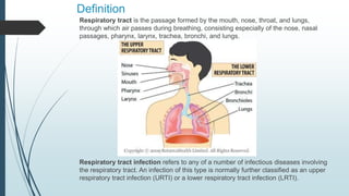
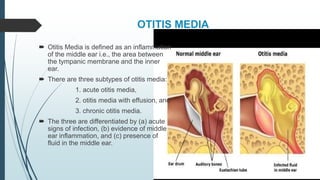
This document discusses respiratory tract infections, specifically focusing on infections of the upper respiratory tract. It defines otitis media as an inflammation of the middle ear and describes its three subtypes. Otitis media is common, especially in young children, and can be caused by bacteria like Streptococcus pneumoniae that enter the middle ear following a viral upper respiratory infection. Symptoms include ear pain, fever, and hearing loss. The document also discusses acute bacterial rhinosinusitis, noting that it is commonly caused by the same bacteria as otitis media and that differentiating between viral and bacterial infections is important to avoid overprescribing antibiotics.
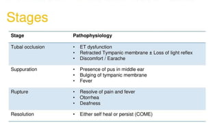
This document discusses various respiratory tract infections, including upper and lower respiratory tract infections. It covers topics such as otitis media (ear infection), pharyngitis (sore throat), sinusitis, bronchitis, bronchiolitis, and pneumonia. For each condition, it discusses etiology, clinical manifestations, diagnosis, treatment goals, and specific treatment options. Risk factors, pathogenesis, and monitoring of treatment response are also covered for some conditions. The document provides an overview of common respiratory infections seen in clinical practice.
This document discusses respiratory tract infections, which are infections that involve the respiratory tract. It describes upper respiratory tract infections such as sinusitis, pharyngitis, and otitis media, and lower respiratory tract infections such as bronchitis, bronchiolitis, and pneumonia. For each infection, it discusses the typical causative agents, affected age groups, characteristics, clinical features, and treatment approaches. It provides an overview of the pathophysiology of upper and lower respiratory tract infections.
Respiratory tract infections are classified as either upper respiratory tract infections (URTI) that involve the areas above the vocal cords such as the nose, sinuses and throat, or lower respiratory tract infections (LRTI) that involve the areas below the vocal cords such as the trachea and lungs. Common causes of URTI include viruses and bacteria while LRTI are usually caused by bacteria or viruses with pneumonia being the most common LRTI. Symptoms vary depending on the location of the infection but may include cough, sore throat, runny nose and fever. Treatment involves medications to reduce symptoms and antibiotics if caused by bacteria.
Gastroenteritis is an infection of the small and large intestines that causes diarrhea and vomiting. In 2015, nearly 2 billion cases of gastroenteritis were observed worldwide, resulting in approximately 1.3 million deaths, with 80% of deaths occurring in India. Gastroenteritis can be caused by bacteria like E. coli and Campylobacter, parasites like Cryptosporidium and Giardia, or viruses. Symptoms include diarrhea, vomiting, abdominal pain, and fever. Treatment involves oral rehydration, antibiotics in some bacterial cases, and managing symptoms. Prevention relies on proper hygiene and sanitation.
Bronchitis is an inflammation of the bronchial tubes caused by viral or bacterial infection or irritants like smoke. It is classified as acute (lasting days to weeks) or chronic (lasting months). Acute bronchitis is usually caused by cold/flu viruses while chronic bronchitis is often caused by long-term smoke inhalation. Symptoms include cough, wheezing, chest tightness and mucus production. Treatment focuses on reducing inflammation, opening airways, treating infection if present, and addressing underlying causes like smoking.
Upper respiratory tract infections are caused by viruses or bacteria that infect the nose, sinuses, pharynx, or larynx. They include conditions like the common cold, sinusitis, tonsillitis, laryngitis, and pharyngitis. A nurse will assess symptoms, check for signs of infection, monitor breathing, help clear secretions, manage pain, and educate patients on treatment plans and avoiding exacerbating factors. Proper nursing care is important for relieving symptoms and supporting healing from upper respiratory infections.
Urinary Tract Infection with Nursing ManagementSwatilekha Das
Urinary Tract Infection introduction, definition, common microorganisms, classification, predisposing factors, clinical manifestations, pathophysiology, diagnostic studies, medical management and nursing management along with assessment, nursing diagnosis, goal, nursing interventions and expected outcome after the intervention.
Laryngitis is inflammation of the larynx or voice box caused by infection, irritation or overuse. It causes hoarseness, coughing and difficulty speaking. Acute laryngitis lasts less than 3 weeks and is usually caused by a cold or flu virus. Chronic laryngitis persists over 3 weeks and can be caused by smoking, acid reflux, vocal misuse or inhaled irritants. Treatment focuses on voice rest, hydration, steam inhalation and medication if caused by infection. Prevention involves avoiding irritants, smoking and overusing the voice.
Pertusis or Whooping cough class presentation Abhilasha verma
Pertussis, also known as whooping cough, is a highly contagious respiratory disease caused by the bacterium Bordetella pertussis. It is characterized by severe coughing fits that can end in a "whooping" sound. It primarily affects children under 5 years old. The disease spreads through respiratory droplets when an infected person coughs or sneezes. It can be prevented through active immunization with the DPT vaccine, which is recommended in 5 doses for children up to age 6.
This document provides an overview of urinary tract infections (UTIs). It discusses the terminology, classification, epidemiology, etiology, pathogenesis, risk factors, clinical presentation, diagnosis, and treatment of UTIs. UTIs can affect different parts of the urinary tract and are classified as uncomplicated or complicated depending on underlying conditions. Escherichia coli is the most common cause. Diagnosis involves urinalysis, urine culture, and imaging tests. Treatment depends on the site and severity of infection, and commonly involves short courses of antibiotics like trimethoprim-sulfamethoxazole or fluoroquinolones.
Mumps is a viral infection caused by a paramyxovirus that typically causes swelling of the parotid glands. It has an incubation period of 12-25 days and is transmitted through respiratory droplets. While many cases are asymptomatic, common symptoms include fever, headache, sore throat, and swelling of the parotid glands. Diagnosis is usually made clinically through physical examination. Treatment focuses on relieving symptoms through rest, fluids, fever medication, and application of warm or cold compresses. Vaccination with the MMR or MMRV vaccines can help prevent mumps.
This document discusses upper respiratory tract infections (URTI). URTIs are commonly caused by viruses like rhinovirus, coronavirus, and influenza. They often involve the nose, sinuses, throat, and larynx, causing symptoms like nasal congestion, sore throat, cough. While most cases are viral, some bacterial infections like Streptococcus and Mycoplasma pneumoniae can also cause URTIs. Treatment involves rest, hydration, analgesics, and sometimes antibiotics for bacterial infections. Vaccines are recommended for influenza prevention.
Pharyngitis, commonly known as a sore throat, is inflammation of the pharynx usually caused by viral infections like the common cold. Symptoms include soreness, difficulty swallowing, and fever. While most cases are viral and self-limiting, bacterial infections like strep throat require antibiotics. Proper diagnosis involves examination of the throat and potential testing. Treatment focuses on relieving symptoms for viral cases and antibiotics for bacterial infections. Maintaining good hygiene can help prevent pharyngitis.
Pneumonia is an inflammation of the lung parenchyma caused by various microorganisms, including bacteria, mycobacteria, fungi, and viruses.
Pneumonitis is a more general term that describes the inflammatory process in the lung tissue that may predispose and Pneumonia is an inflammation of the lung parenchyma that is caused by a microbial agent.
place the patient at risk for microbial invasion.
Pneumonia is classified into four: community-acquired pneumonia (CAP) and hospital-acquired pneumonia (HAP), pneumonia in the immunocompromised host, and aspiration pneumonia.
1. Pneumonia, lung abscess, and pleural effusion are respiratory infections that can affect the lungs. Pneumonia is an inflammation of the lung tissues that is usually caused by microorganisms. Lung abscess occurs when an area of lung tissue becomes necrotic and fills with pus. Pleural effusion is an excess collection of fluid in the pleural space surrounding the lungs.
2. The document discusses the causes, pathophysiology, clinical manifestations, diagnosis, and treatment of each condition. It provides details on the different types of pneumonia and outlines strategies for educating clients to prevent respiratory infections.
This document defines pneumonia as an inflammation of the lungs caused by infection. It can affect 450 million people annually and is a major cause of death worldwide. Pneumonia has several classifications including bacterial, atypical, fungal, and viral. It also distinguishes between community acquired pneumonia and hospital acquired pneumonia. Risk factors include age, lung diseases like emphysema, and conditions that weaken the immune system. The document outlines the pathophysiology of pneumonia and discusses some common causes by age group like bacteria in adults and viruses in children.
Endocarditis is an inflammation of the inner layer of the heart. It is usually caused by bacteria, fungi, or viruses entering the bloodstream through invasive procedures like dental work or surgery. The microbes accumulate on the heart valves, forming vegetations that can damage the valves over time. Symptoms may include fever, fatigue, loss of appetite, and heart murmurs. Diagnosis involves blood cultures, echocardiography, and other tests. Treatment requires antibiotics and sometimes valve replacement surgery. Complications can include heart failure, embolism, and stroke if left untreated.
Tuberculosis is caused by the bacterium Mycobacterium tuberculosis. It most commonly affects the lungs. Risk factors include malnutrition, poverty, crowding, and immunocompromised states. Transmission occurs via airborne droplets from the lungs of active cases. Diagnosis involves microscopy, culture, molecular tests, chest imaging and the Mantoux skin test. Complications include cavitary lesions, caseous pneumonia, and disseminated disease. Treatment requires long-term antibiotic therapy.
Pulmonary TB is a bacterial infection of the lungs that can cause a range of symptoms, including chest pain, breathlessness, and severe coughing. Pulmonary TB can be life-threatening if a person does not receive treatment. People with active TB can spread the bacteria through the air.
Urinary tract infections (UTIs) occur when bacteria enter the urinary tract. UTIs are common in children and can affect any part of the urinary system, including the kidneys, ureters, bladder, and urethra. The most frequent cause is E. coli bacteria. Left untreated, UTIs can spread and potentially cause renal failure or sepsis. Treatment involves antibiotics and supportive care like fluids and pain medication. Proper hygiene and toilet training can help prevent recurrent UTIs.
This document discusses tonsillitis, an inflammation of the tonsils. It defines tonsillitis and lists its common causes as various bacteria like streptococcus, staphylococcus, and pneumococcus. Risk factors include poor oral hygiene, poor nutrition, and upper respiratory tract infections. The document outlines the clinical features of tonsillitis such as sore throat, dysphagia, fever, and enlarged tonsils. It also discusses the diagnosis, management with antibiotics and other treatments, and potential complications of tonsillitis like peritonsillar and parapharyngeal abscesses.
This document provides information about meningitis, including definitions, causes, types, symptoms, diagnosis, treatment and nursing management. It defines meningitis as inflammation of the meninges, or protective membranes covering the brain and spinal cord. It can be caused by viruses, bacteria, fungi, parasites, toxins or malignancies. The main types discussed are pyogenic (bacterial), viral, fungal/aseptic and tuberculous meningitis. Bacterial meningitis requires urgent treatment with antibiotics to prevent complications like brain damage or hydrocephalus. Nursing goals include pain management, temperature control and seizure prevention through interventions like medication administration and environmental modifications.
aids and hiv in children. it is the topic in child health nursing. it include definition, etiology, types, signs and symptoms, pathophysiology, clinical stages, diagnosis and management of pediatric hiv or aids.
Pneumonia is an inflammatory condition of the lungs caused by microbial agents like bacteria, viruses, and fungi. It affects millions of people worldwide annually and is a common cause of death, especially in young children and older adults. Symptoms include cough, fever, shortness of breath, and chest pain. Diagnosis involves physical exam, chest x-ray, and tests of respiratory samples. Treatment focuses on antibiotics targeting the causative organism as well as oxygen therapy, breathing exercises, and ensuring adequate nutrition and hydration. Complications can include lung abscesses, empyema, and respiratory failure. With treatment, most cases stabilize within a week but full recovery may take several weeks.
Otitis media is an inflammation of the middle ear that commonly affects children under 10 years old. It can be caused by bacteria like Streptococcus pneumoniae or viruses. Common symptoms include ear pain, fever, hearing loss, and discharge from the ear. Without treatment, complications can include mastoiditis, cholesteatoma, meningitis, and permanent hearing loss. Diagnosis is usually based on examination of the eardrum. Treatment involves antibiotics, analgesics, and occasionally surgery to drain the ear or place tubes. Nursing care focuses on pain management, communication strategies due to hearing loss, and ensuring complete treatment to prevent recurrence.
Upper respiratory tract infections are common illnesses that affect the nasal passages, sinuses, pharynx and larynx. The common cold is the most frequent viral illness, often caused by rhinoviruses. Other viral infections like influenza and RSV can cause pharyngitis. Bacterial sinusitis is usually preceded by a viral infection. Acute laryngitis is commonly caused by inhalation of irritants or viral infections. Croup is most often caused by parainfluenza viruses in young children. Nasopharyngeal carcinoma is associated with Epstein-Barr virus and more common in Chinese populations. Laryngeal tumors include non-cancerous lesions like nodules and papillomas as well as
Otitis media is an infection of the middle ear that is usually caused by bacteria or viruses. It is common in young children due to the short, horizontal shape of the eustachian tubes. Risk factors include exposure to cigarette smoke, overcrowding, bottle feeding, and allergies. Acute otitis media presents with severe ear pain over days to weeks and visible inflammation of the eardrum. Chronic otitis media is characterized by persistent fluid buildup behind an intact eardrum, while chronic suppurative otitis media involves long-term drainage through a perforated eardrum. Treatment involves antibiotics, antihistamines, tubes, or surgery depending on the type and severity of
This document discusses upper respiratory tract infections (URTIs) with a focus on acute otitis media (AOM). It defines AOM and outlines its typical symptoms, causes, risk factors, and diagnostic criteria. The document describes the pathophysiology of AOM, which usually develops following a viral upper respiratory infection that impairs the eustachian tube. Common bacterial pathogens that may cause secondary bacterial infection include Streptococcus pneumoniae, Haemophilus influenzae, and Moraxella catarrhalis. The goals of AOM treatment are outlined as well as considerations for prevention through vaccination.
This document provides an overview of acute otitis media (AOM). It begins with a brief history of AOM and then discusses the definition, clinical features, significance, and etiopathogenesis of the condition. Regarding etiology, it notes that AOM is often associated with upper respiratory tract infections and discusses various bacterial, viral, and fungal pathogens implicated in AOM. It also explores the roles of allergy, eustachian tube dysfunction, genetics, immunity, and other factors in AOM development. The document aims to comprehensively summarize our current understanding of AOM.
This document discusses otitis media, an inflammation of the middle ear. It begins with an introduction and case presentation of a 4 year old boy with ear pain, fever, and cough. It then covers the epidemiology, anatomy, classification, etiology, risk factors, pathology, clinical features, differential diagnosis, management, and complications of otitis media. The peak incidence is in children aged 1-4 years and is more common in boys. Acute otitis media, otitis media with effusion, chronic suppurative otitis media, and adhesive otitis media are subtypes discussed. Pneumoniae, influenzae, and Moraxella are common bacterial causes while viruses like R
Acute otitis media (AOM) refers to inflammation of the middle ear caused by bacterial infection, usually following a viral upper respiratory infection. Common bacteria include Streptococcus pneumoniae, Haemophilus influenzae, and Moraxella catarrhalis. Symptoms include ear pain, fever, and bulging of the eardrum. Treatment involves antibiotics or watchful waiting. Otitis media with effusion refers to persistent middle ear fluid for over 3 months causing hearing loss but no other symptoms. Risk factors are similar to AOM. Treatment options include observation, medical therapies, or ear tube surgery.
Upper respiratory tract infections like the common cold, sinusitis, and pharyngitis are caused by viruses and bacteria that infect the nose, sinuses, and throat.
The common cold is usually caused by rhinoviruses and presents with nasal congestion and discharge. Sinusitis occurs when the sinuses become infected, often following a viral upper respiratory infection, and can cause facial pain and tenderness. Pharyngitis, or a sore throat, is commonly caused by streptococcus bacteria or viruses like adenovirus. Accurate diagnosis involves examining symptoms, signs, and testing mucus samples. Treatment focuses on relieving symptoms and in some bacterial cases using antibiotics.
This document discusses different types of ear infections:
- Otitis externa is inflammation of the external ear canal and can be caused by bacteria, fungi, or other factors. Acute cases involve pain while chronic cases involve pruritus. Treatment involves cleaning and antibiotics.
- Acute otitis media is a common childhood infection involving the middle ear. It is usually caused by viruses but can become bacterial. Symptoms include ear pain, fever, and hearing loss. Treatment is usually with amoxicillin or other antibiotics.
- Otitis media with effusion involves fluid in the middle ear without infection. It often follows acute otitis media and can cause temporary hearing loss. Most cases
Acute respiratory infections (ARI) are responsible for 20% of childhood deaths under 5 years of age, with pneumonia accounting for 90% of ARI mortality. Children under 2 years old, malnourished children, children with HIV, and those with poor access to healthcare or poorly educated parents are most at risk. ARIs are a common cause of outpatient visits (20-60%) and admissions (12-45%) in children. Common respiratory infections affecting children include the common cold, acute epiglottitis, croup, ear infections, tonsillitis, sinusitis, and pneumonia. Bacteria and viruses can cause ARIs, with symptoms and severity depending on the specific infectious agent.
ACUTE OTITIS MEDIA infection of the middle earpaultembo7
Acute otitis media refers to the acute inflammation of the middle ear cavity. There are several types including acute suppurative otitis media, acute necrotizing otitis media, otitis media with effusions, and recurrent otitis media. Acute suppurative otitis media is usually caused by viral upper respiratory infections allowing bacterial infection of the middle ear via the eustachian tube. It involves five stages from tubal occlusion to resolution or complications. Treatment focuses on relieving symptoms, draining pus, and preventing recurrence through antibiotics, myringotomy, and addressing predisposing factors. Otitis media with effusions is characterized by non-purulent effusion
Acute otitis media refers to the acute inflammation of the middle ear cavity. There are several types including acute suppurative otitis media, which is caused by pyogenic bacteria invading the middle ear through the eustachian tube. It presents in stages from tubal occlusion to resolution or complications. Treatment involves relieving symptoms, draining pus, and preventing recurrence with antibiotics. Otitis media with effusions involves sterile fluid accumulation in the middle ear due to eustachian tube malfunction or increased mucus secretion. Recurrent otitis media describes repeated acute infections. Aero-otitis media results from pressure changes during air travel or diving that the eustachian tubes cannot equalize
This document provides information on otitis media with effusion (OME), including its definition, causes, characteristics, diagnosis, and epidemiology. Specifically:
- OME is the chronic accumulation of fluid in the middle ear for at least 12 weeks, usually presenting as hearing impairment. It is often preceded by acute otitis media or upper respiratory infection.
- The fluid results from inflammation of the Eustachian tube epithelium that prevents drainage of the middle ear. Histological examination shows replacement of normal epithelium with mucus-secreting cells.
- Diagnosis involves otoscopy, pneumatic otoscopy, and tympanometry which can classify effusions. Type B
Provides detailed in formation on otitis media.It is subdivided into:
Table of content
Literature review
Patient information
medical and surgical management
nursing careplan
and it is well referenced.
It provides more information on better management of ENT patient.
Can be used by anyone in the medical or nursing field.
This document provides information on upper and lower respiratory pathologies including sinusitis, otitis externa, otitis media, and allergic rhinitis. It defines the different types of sinusitis and describes the anatomy and development of the paranasal sinuses. It also discusses the etiology, signs and symptoms, diagnosis, and treatment of otitis externa, otitis media, and allergic rhinitis.
Acute Otitis Media (AOM) is an infection of the middle ear caused by bacteria or viruses. It is common in young children, especially between 6-18 months of age. Risk factors include daycare attendance, lack of breastfeeding, exposure to tobacco smoke, and underlying conditions like cleft palate. AOM is diagnosed based on signs of bulging of the eardrum, fluid, or pus behind the eardrum seen on pneumatic otoscopy. Common bacteria that cause AOM include Streptococcus pneumoniae, Haemophilus influenzae, and Moraxella catarrhalis. Recurrent AOM is defined as three or more episodes within 6 months or four episodes within
This document discusses acute suppurative otitis media (ASOM), an acute inflammation of the middle ear caused by pyogenic bacteria. It notes that ASOM is most common in young children, especially during winter months. Common bacteria that cause ASOM include Streptococcus pneumoniae, Haemophilus influenzae, and Moraxella catarrhalis. Treatment involves antibiotics, analgesics, ear drainage via myringotomy if needed, and prevention through childhood vaccinations. Complications can include mastoiditis, petrositis, and Gradenigo's syndrome involving pain and eye muscle palsies.
TEST BANK For Community Health Nursing A Canadian Perspective, 5th Edition by...Donc Test
TEST BANK For Community Health Nursing A Canadian Perspective, 5th Edition by Stamler, Verified Chapters 1 - 33, Complete Newest Version Community Health Nursing A Canadian Perspective, 5th Edition by Stamler, Verified Chapters 1 - 33, Complete Newest Version Community Health Nursing A Canadian Perspective, 5th Edition by Stamler Community Health Nursing A Canadian Perspective, 5th Edition TEST BANK by Stamler Test Bank For Community Health Nursing A Canadian Perspective, 5th Edition Pdf Chapters Download Test Bank For Community Health Nursing A Canadian Perspective, 5th Edition Pdf Download Stuvia Test Bank For Community Health Nursing A Canadian Perspective, 5th Edition Study Guide Test Bank For Community Health Nursing A Canadian Perspective, 5th Edition Ebook Download Stuvia Test Bank For Community Health Nursing A Canadian Perspective, 5th Edition Questions and Answers Quizlet Test Bank For Community Health Nursing A Canadian Perspective, 5th Edition Studocu Test Bank For Community Health Nursing A Canadian Perspective, 5th Edition Quizlet Test Bank For Community Health Nursing A Canadian Perspective, 5th Edition Stuvia Community Health Nursing A Canadian Perspective, 5th Edition Pdf Chapters Download Community Health Nursing A Canadian Perspective, 5th Edition Pdf Download Course Hero Community Health Nursing A Canadian Perspective, 5th Edition Answers Quizlet Community Health Nursing A Canadian Perspective, 5th Edition Ebook Download Course hero Community Health Nursing A Canadian Perspective, 5th Edition Questions and Answers Community Health Nursing A Canadian Perspective, 5th Edition Studocu Community Health Nursing A Canadian Perspective, 5th Edition Quizlet Community Health Nursing A Canadian Perspective, 5th Edition Stuvia Community Health Nursing A Canadian Perspective, 5th Edition Test Bank Pdf Chapters Download Community Health Nursing A Canadian Perspective, 5th Edition Test Bank Pdf Download Stuvia Community Health Nursing A Canadian Perspective, 5th Edition Test Bank Study Guide Questions and Answers Community Health Nursing A Canadian Perspective, 5th Edition Test Bank Ebook Download Stuvia Community Health Nursing A Canadian Perspective, 5th Edition Test Bank Questions Quizlet Community Health Nursing A Canadian Perspective, 5th Edition Test Bank Studocu Community Health Nursing A Canadian Perspective, 5th Edition Test Bank Quizlet Community Health Nursing A Canadian Perspective, 5th Edition Test Bank Stuvia
Promoting Wellbeing - Applied Social Psychology - Psychology SuperNotesPsychoTech Services
A proprietary approach developed by bringing together the best of learning theories from Psychology, design principles from the world of visualization, and pedagogical methods from over a decade of training experience, that enables you to: Learn better, faster!
Local Advanced Lung Cancer: Artificial Intelligence, Synergetics, Complex Sys...Oleg Kshivets
Overall life span (LS) was 1671.7±1721.6 days and cumulative 5YS reached 62.4%, 10 years – 50.4%, 20 years – 44.6%. 94 LCP lived more than 5 years without cancer (LS=2958.6±1723.6 days), 22 – more than 10 years (LS=5571±1841.8 days). 67 LCP died because of LC (LS=471.9±344 days). AT significantly improved 5YS (68% vs. 53.7%) (P=0.028 by log-rank test). Cox modeling displayed that 5YS of LCP significantly depended on: N0-N12, T3-4, blood cell circuit, cell ratio factors (ratio between cancer cells-CC and blood cells subpopulations), LC cell dynamics, recalcification time, heparin tolerance, prothrombin index, protein, AT, procedure type (P=0.000-0.031). Neural networks, genetic algorithm selection and bootstrap simulation revealed relationships between 5YS and N0-12 (rank=1), thrombocytes/CC (rank=2), segmented neutrophils/CC (3), eosinophils/CC (4), erythrocytes/CC (5), healthy cells/CC (6), lymphocytes/CC (7), stick neutrophils/CC (8), leucocytes/CC (9), monocytes/CC (10). Correct prediction of 5YS was 100% by neural networks computing (error=0.000; area under ROC curve=1.0).
Here is the updated list of Top Best Ayurvedic medicine for Gas and Indigestion and those are Gas-O-Go Syp for Dyspepsia | Lavizyme Syrup for Acidity | Yumzyme Hepatoprotective Capsules etc
Basavarajeeyam is a Sreshta Sangraha grantha (Compiled book ), written by Neelkanta kotturu Basavaraja Virachita. It contains 25 Prakaranas, First 24 Chapters related to Rogas& 25th to Rasadravyas.
Cell Therapy Expansion and Challenges in Autoimmune DiseaseHealth Advances
There is increasing confidence that cell therapies will soon play a role in the treatment of autoimmune disorders, but the extent of this impact remains to be seen. Early readouts on autologous CAR-Ts in lupus are encouraging, but manufacturing and cost limitations are likely to restrict access to highly refractory patients. Allogeneic CAR-Ts have the potential to broaden access to earlier lines of treatment due to their inherent cost benefits, however they will need to demonstrate comparable or improved efficacy to established modalities.
In addition to infrastructure and capacity constraints, CAR-Ts face a very different risk-benefit dynamic in autoimmune compared to oncology, highlighting the need for tolerable therapies with low adverse event risk. CAR-NK and Treg-based therapies are also being developed in certain autoimmune disorders and may demonstrate favorable safety profiles. Several novel non-cell therapies such as bispecific antibodies, nanobodies, and RNAi drugs, may also offer future alternative competitive solutions with variable value propositions.
Widespread adoption of cell therapies will not only require strong efficacy and safety data, but also adapted pricing and access strategies. At oncology-based price points, CAR-Ts are unlikely to achieve broad market access in autoimmune disorders, with eligible patient populations that are potentially orders of magnitude greater than the number of currently addressable cancer patients. Developers have made strides towards reducing cell therapy COGS while improving manufacturing efficiency, but payors will inevitably restrict access until more sustainable pricing is achieved.
Despite these headwinds, industry leaders and investors remain confident that cell therapies are poised to address significant unmet need in patients suffering from autoimmune disorders. However, the extent of this impact on the treatment landscape remains to be seen, as the industry rapidly approaches an inflection point.
TEST BANK For Basic and Clinical Pharmacology, 14th Edition by Bertram G. Kat...rightmanforbloodline
TEST BANK For Basic and Clinical Pharmacology, 14th Edition by Bertram G. Katzung, Verified Chapters 1 - 66, Complete Newest Version.
share - Lions, tigers, AI and health misinformation, oh my!.pptxTina Purnat
• Pitfalls and pivots needed to use AI effectively in public health
• Evidence-based strategies to address health misinformation effectively
• Building trust with communities online and offline
• Equipping health professionals to address questions, concerns and health misinformation
• Assessing risk and mitigating harm from adverse health narratives in communities, health workforce and health system
2. Definition
Respiratory tract is the passage formed by the mouth, nose, throat, and lungs,
through which air passes during breathing, consisting especially of the nose, nasal
passages, pharynx, larynx, trachea, bronchi, and lungs.
Respiratory tract infection refers to any of a number of infectious diseases involving
the respiratory tract. An infection of this type is normally further classified as an upper
respiratory tract infection (URTI) or a lower respiratory tract infection (LRTI).
4. OTITIS MEDIA
Otitis Media is defined as an inflammation
of the middle ear i.e., the area between
the tympanic membrane and the inner
ear.
There are three subtypes of otitis media:
1. acute otitis media,
2. otitis media with effusion, and
3. chronic otitis media.
The three are differentiated by (a) acute
signs of infection, (b) evidence of middle
ear inflammation, and (c) presence of
fluid in the middle ear.
5. 1. Acute otitis media involves the rapid onset of signs and symptoms of
inflammation in the middle ear that manifests clinically as one or more of the
following:
otalgia (denoted by pulling of the ear in some infants)
hearing loss
fever, or
irritability.
2. Otitis media with effusion (accumulation of liquid in the middle ear cavity) differs
from acute otitis media in that signs and symptoms of an acute infection are
absent.
6. EPIDEMIOLOGY
• There are more than 709 million cases of otitis media worldwide each year; half of these
cases occur in children under 5 years of age.
• Otitis media is a global problem and is found to be slightly more common in males than in
females.
• The specific number of cases per year is difficult to determine due to the lack of reporting
and varied incidence across many different geographical regions.
• The peak incidence of otitis media occurs between six and twelve months of life and
declines after age five.
• Approximately 80% of all children will experience a case of otitis media during their lifetime
and between 80% and 90% of all children will have otitis media with an effusion before
school age.
• Otitis media is less common in adults than in children, unless it occurs in
immunocompromised adults.
7. RISK FACTORS FOR OTITIS MEDIA
Winter season/outbreaks of respiratory syncytial or influenza virus
Attendance at day care centers
Lack of breast-feeding in infants
Early age of first diagnosis
Nasopharyngeal colonization with middle ear pathogens
Genetic predisposition
Siblings in the home
Lower socioeconomic status
Exposure to tobacco smoke
Use of a pacifier
Male gender
Immunodeficiency
Allergy
8. ETIOLOGY
S. pneumoniae
Haemophilus influenzae
Moraxella catarrhalis
PATHOPHYSIOLOGY
Acute bacterial otitis media usually follows a viral upper respiratory tract infection
that causes eustachian tube dysfunction and mucosal swelling in the middle ear.
Bacteria that colonize the nasopharynx thus enter the middle ear and are not
cleared properly by the mucociliary system.
Abnormal function of the eustachian tube can cause reflux transudation of liquid in
the middle ear and proliferation of bacteria, resulting in acute otitis media.
9. Due to etiological factor(URTI, Bacteria)
Exudates & edema in middle ear
Decrease retraction of tympanic membrane
Serous exudates in middle ear
Pus formation
Tympanic membrane rupture
ACUTE OTITIS MEDIA
10. Stages
1. Catarrhal stage: is characterized by occlusion of
Eustachian tube and congestion of middle ear.
2. Stage of exudation: Exudate collects in the middle ear
and ear drum is pushed laterally. Initially the exudate
is mucoid, later it becomes purulent.
3. Stage of suppuration: Pus in the middle ear collects
under tension, stretches the drum & perforates it by
pressure necrosis & the exudate starts escaping into
external auditory canal
4. Stage of healing: The infection starts resolving from
any of the stages mentioned & usually clears up
completely without leaving any sequelae.
5. Stage of complications: Infection may spread to the
mastoid antrum. Initially it causes Catarrhal mastoiditis
[congestion of the mastoid mucosa], stage of
Coalescent mastoiditis & later empyema of the
mastoid.
11.
12.
13. Clinical Presentation of Acute Otitis Media
The acute onset of signs and symptoms of middle ear
infection following cold symptoms of runny nose,
nasal congestion, or cough.
Signs and symptoms
o Pain that can be severe (more than 75% of patients)
o Children may be irritable, tug on the involved ear,
and have difficulty sleeping
o Fever is present in less than 25% of patients and,
when present, occurs more often in younger children
o Examination shows a discolored (gray), thickened,
bulging eardrum
o Pneumatic otoscopy or tympanometry demonstrates
an immobile eardrum; 50% of cases are bilateral.
o Draining middle ear fluid occurs (less than 3% of
patients) that usually reveals a bacterial etiology.
15. DIAGNOSTIC TESTS
Laboratory Studies – sepsis workup (gram staining, culture & sensitivity)
Imaging - study of choice is a contrast-enhanced CT scan of the temporal bones
MRI is more helpful in depicting fluid collections
Tympanometry may help with diagnosis in patients with OM with effusion
Diagnostic criteria for OM:
Bulging TM
Retracted TM
Impaired mobility of the TM
Loss of light reflex
Erythematous TM
Purulent otorrhea
Opacification of the TM
16. TREATMENT
First Line Second Line (10 day
course)
Third Line
Amoxicillin high dose
80-90 mg/kg/day
Divided twice daily.
If Penicillin Allergy, use
Macrolide (e.g.
Azithromycin)
1. Amoxicillin with
clavulanate
90 mg/kg/day divided twice
daily for 10 days
2. Cefuroxime
30 mg/kg/day divided twice
daily for 10 days
3. Cefprozil
30 mg/kg/day divided twice
daily for 10 days
4. Cefdinir
14 mg/kg/day divided one
to two times daily fo 10
days
5. Cefpodoxime
30 mg/kg once daily for 10
days
1. Strongly consider
Tympanocentesis for
bacterial culture.
2. Ceftriaxone
50 mg/kg IM daily for 3
days
3. Clindamycin
30-40 mg/kg/day divided
four times daily for 10
days.
17. Penicillin allergy
1. Consider Tympanocentesis
2. Clindamycin 30-40 mg/kg/day (max 1800 mg) divided four times daily for 10 days
3. Macrolide antibiotics (High bacterial resistance rate)
1. Clarithromycin 15 mg/kg/day divided twice daily for 10 days
2. Erythromicin 30-50mg/kg every 6-8 hours
3. Azithromycin
1. One dose of Azithromycin at 30 mg/kg (up to 1500 mg) or
2. Three days of Azithromycin at 20 mg/kg/day once daily (up to 500 mg/day) or
3. Azithromycin 10 mg/kg (max: 500 mg) day 1, then 5 mg/kg/day (max 250
mg) for 5 days
4. Fluoroquinolones (avoid under age 16 years)
1. Gatifloxacin
2. Levofloxacin
3. Moxifloxacin
19. SINUSES
They are hollow, airfilled cavities that are lined
by respiratory mucosa “pseudostratified
ciliated columnar epithelium”
There are four pairs of paranasal sinuses;
The frontal sinuses are located above the eyes,
in the frontal bone
The maxillary sinuses are located in the
cheekbones, under the eyes.
The ethmoid sinuses(6 – 10 per side), also
called ethmoid labyrinth are located between
the eyes and the nose.
The sphenoid sinuses(2) are located in the
body of sphenoid bone, behind the nose and
the eyes.
20. DEFINITION
Sinusitis is an inflammation and/or infection of the paranasal
sinuses, or membrane-lined air spaces, around the nose.
The term rhinosinusitis is now preferred because sinusitis
typically also involves the nasal mucosa.
Even though the majority of rhinosinusitis infections are viral
in origin, antibiotics are frequently prescribed.
It is thus important to differentiate between viral and bacterial
rhinosinusitis to avoid antibiotic overuse.
21. EPIDEMIOLOGY
Nearly 30 million cases of rhinosinusitis are diagnosed annually in USA.
Acute bacterial rhinosinusitis is overdiagnosed; thus, antibiotics are
overprescribed.
Most rhinosinusitis infections have a viral etiology, and yet, antibiotics are
frequently prescribed.
Adults with rhinosinusitis miss an average of 6 workdays/y with these
infections.
Patients with rhinosinusitis are significantly more likely to use the
emergency room, spend more than $500/y on medical care, and see a
medical specialist.
22. ETIOLOGY
Acute bacterial rhinosinusitis is caused, most often, by the same bacteria implicated in acute
otitis media: S. pneumoniae and H. influenzae.
These organisms are responsible for approximately 50% to 70% of bacterial causes of acute
bacterial rhinosinusitis in both adults and children.
M. catarrhalis is also sometimes implicated in adults and children (approximately 8%-16%).
Streptococcus pyogenes, Staphylococcus aureus, gram-negative bacilli, and anaerobes are
associated less frequently with acute bacterial rhinosinusitis.
Issues of bacterial resistance are similar to those found with acute otitis media.
23. PATHOPHYSIOLOGY
Mucosal edema resulting from a viral rhinosinusitis
obstruction of natural ostia
hypoxygenation
acidosis
vasodilation
increased secretion by goblet cells
ciliary dysfunction with poor mucous quality
retention of secretion and predisposition to bacterial infection.
24.
25. CLINICAL PRESENTATION
Acute bacterial sinusitis in adults most often
manifests with more than 7 days of nasal
congestion, purulent rhinorrhea, postnasal
drip, and facial pain and pressure, alone or
with associated referred pain to the ears and
teeth.
There may be a cough, often worsening at
night.
Children with acute sinusitis might not be able
to relay a history of postnasal drainage or
headaches, so cough and rhinorrhea are the
most commonly reported symptoms.
Other symptoms can include fever, nausea,
fatigue, impairments of smell and taste, and
halitosis.
26. Symptoms Associated with the Diagnosis of Chronic
Sinusitis
Facial pain or pressure
Facial congestion or fullness
Nasal obstruction or blockage
Nasal discharge, purulence, or postnasal drip
Hyposmia or anosmia
Headache
Fever
Halitosis
Fatigue
Dental pain
Cough
Ear pain, pressure, fullness
27. DIAGNOSIS
In a primary care setting, a good history and physical examination to detect the presence of
most or all of the commonly manifesting signs and symptoms can provide a reliable diagnosis
of acute sinusitis. The presence of purulent secretions has the highest positive predictive
value for diagnosing sinusitis clinically.
CT scan
28. Transillumination - A common practice before plain radiographs and CT scans
were widely available, transillumination is of limited use and has a high rate of
error.
Ultrasonography - Ultrasonography has not been proved accurate enough to
substitute for a radiographic evaluation. However, it may be considered to confirm
sinusitis in pregnant women, for whom radiographic studies could pose a risk.
Nasal Smear - By examining the cellular contents of the nasal secretions, one
might find polymorphonuclear cells and bacteria in sinusitis. In a viral infection,
these would not be found, and in allergic disease, one would expect to find
eosinophils.
Sinus Puncture
The most accurate way to determine the causative organism in sinusitis is a sinus
puncture. After anesthetization of the puncture site, the contents of the maxillary
sinus are aspirated under sterile technique, and bacterial cultures are performed to
identify the organism. Culture specimens obtained from nasal swabs correlate
poorly with sinus pathogens found by puncture because of contamination of the
swab with normal nasal flora. However, because sinus puncture is an invasive
procedure, it is not routinely performed.
29.
30. Treatment of Acute Sinusitis
Antibiotics, such as amoxicillin for 2 weeks, have been the recommended first-line
treatment of uncomplicated acute sinusitis. The antibiotic of choice must cover S.
pneumoniae, H. influenzae, and M. catarrhalis. Because rare intracranial and orbital
complications of acute bacterial sinusitis are caused by S. pneumoniae (most
commonly in the immunocompromised host), adequate coverage for this organism is
important. Amoxicillin-clavulanate (Augmentin) is also an appropriate first-line
treatment of uncomplicated acute sinusitis. The addition of clavulanate, a beta-
lactamase inhibitor, provides better coverage for H. influenzae and M. catarrhalis.
Because of S. pneumoniae resistance, higher doses of amoxicillin (90 mg/kg/day to a
maximum of 2 g/day) should be considered. These higher doses are effective
against S. pneumoniae because resistance is related to alteration in penicillin-binding
proteins, a mechanism distinct from the beta-lactamase enzymatic inactivation of H.
influenzae and M. catarrhalis.
31. Other options include cephalosporins such as cefpodoxime proxetil (Vantin) and
cefuroxime (Ceftin). In patients allergic to beta-lactams, trimethoprim-
sulfamethoxazole (Bactrim), clarithromycin (Biaxin), and azithromycin (Zithromax)
may be prescribed but might not be adequate coverage for H. influenzae or
resistant S. pneumoniae.
Penicillin, erythromycin (Suprax), and first-generation cephalosporins such as
cephalexin (Keflex, Keftab) are not recommended for treating acute sinusitis
because of inadequate antimicrobial coverage of the major organisms.
If treatment with one of these first-line agents has not shown a clinical response
within 72 hours of initial therapy, more broad-spectrum antibiotics should be
considered. These include the fluoroquinolones, gatifloxacin (Tequin), moxifloxacin
(Avelox), and levofloxacin (Levaquin), especially if amoxicillin-clavulanate,
cefpodoxime proxetil, and cefuroxime were previously prescribed.
32. Treatment of Chronic Sinusitis
Antibiotic therapy for chronic sinusitis is controversial and may be most appropriate
for acute exacerbation of chronic sinusitis. Medical therapy should include both a
broad-spectrum antibiotic and a topical intranasal steroid to address the strong
inflammatory component of this disease. Antibiotic therapy might need to be
continued for 4 to 6 weeks.
The antibiotics of choice include agents that cover organisms causing acute
sinusitis but also cover Staphylococcus species and anaerobes. These include
amoxicillin-clavulanate, cefpodoxime proxetil, cefuroxime, gatifloxacin,
moxifloxacin, and levofloxacin. Currently used topical intranasal steroids such as
fluticasone (Flonase), mometasone (Nasonex), budesonide (Rhinocort AQ), and
triamcinolone (Nasacort AQ) have a favorable safety profile and indications for the
pediatric age group. A short course of oral steroids may be used for extensive
mucosal thickening and congestion or nasal polyps.
33. Adjunctive Therapy
To temporarily alleviate the drainage and congestion associated with sinusitis,
decongestant nasal sprays oxymetazoline (Afrin) and phenylephrine hydrochloride
(Neo-Synephrine) may be used for 3 to 5 days. Long-term use of topical
decongestants can cause rhinitis medicamentosa, which is rebound congestion
caused by vasodilatation and inflammation. Oral decongestants
(pseudoephedrine) may be a reasonable alternative if the patient has no
contraindication such as hypertension. Mucolytic agents (guaifenesin) can help to
decrease the viscosity of the mucus for better clearance and are often found in
combination with decongestants. Some mucolytics are now available over the
counter. Saline spray or irrigation can help clear secretions. Topical corticosteroids
are not indicated for acute sinusitis but may be helpful for chronic sinusitis, nasal
polyps, and allergic and nonallergic rhinitis. Antihistamines are not indicated for
sinusitis but may be helpful for underlying allergic rhinitis.
38. PHARYNGITIS
Pharyngitis is inflammation of the pharynx, which is in the back of the
throat.
Pharyngitis is an acute infection of the oropharynx or nasopharynx.
It’s most often referred to simply as “sore throat.”
Pharyngitis can also cause scratchiness in the throat and difficulty
swallowing.
40. EPIDEMIOLOGY
In India, it is estimated that approximately 7 sore throat
episodes occur per child per year
There are as many as 20-30 million cases of streptococcal
pharyngitis which may occur annually in India.
41. PATHOPHYSIOLOGY
The mechanism by which group A Streptococcus causes pharyngitis is not
well defined.
Asymptomatic pharyngeal carriers of the organism may have an alteration
in host immunity (e.g., a breach in the pharyngeal mucosa) and the
bacteria of the oropharynx, allowing colonization to become infection.
Pathogenic factors associated with the organism itself also may play a
role.
These include pyrogenic toxins, hemolysins, streptokinase, and
proteinase.
42.
43. Clinical MANIFESTATIONS
General
A sore throat of sudden onset that is mostly self-limited.
Fever and constitutional symptoms resolving in about 3 to 5 days.
Clinical signs and symptoms are similar for viral causes as well as
nonstreptococcal bacterial causes.
Signs and symptoms
Sore throat
Pain on swallowing
Fever
Headache, nausea, vomiting and abdominal pain (especially children).
Erythema/inflammation of the tonsils and pharynx with or without patchy
exudates.
Enlarged, tender lymph nodes.
Red swollen uvula, petechiae on the soft palate, and a scarlatiniform rash.
Several symptoms that are not suggestive of group A streptococcus are cough,
conjunctivitis, coryza and diarrhea.
44.
45.
46. DIAGNOSIS
Physical exam
Throat culture
Blood tests
• Throat swab for C/S
• Rapid antigen testing against GABHS
• Kleb loeffler’s bacillus (KLB) (Corynebacterium diphtheriae)
• Leukocytosis
• Monospot test for EBV
47.
48. PHARMACOLOGICAL THERAPY
Antimicrobial therapy should be limited to those who have clinical and
epidemiologic features of group A streptococcal pharyngitis with a positive
laboratory test.
Pencillin V K- 250mg, TD
Pencillin benzathine- 1.2 million units IM OD
Pencillin G procaine- only in children 1.2 million units, OD
Amoxicillin- 500mg, TD
Erythromycin Estolate- 20-40mg/kg/day
Ethyl succinate- 40mg/kg/day
Cephalexin- 250-500mg, PO, QD
49. RECURRENT EPISODES OF PHARYNGITIS
DRUG DOSE MECHANISM OF ACTION A.D.R
Clindamycin 600mg It is a bacterial protein synthesis inhibitor by
inhibiting ribosomal translocation
Abdominal pain, cramp
and rash
Amoxicillin+
clavulonate
500mg, BD It inhibits cross-linkage between the linear
peptidoglycan polymer chains that make up
a major component of the cell walls of both
Gram-positive and Gram-negative bacteria
Insomnia, confusion and
anxiety
Pencillin
benzathine
1.2 million units
IM
β-Lactam antibiotics inhibit the formation of
peptidoglycan cross-links in the bacterial
cell wall, but have no direct effect on cell
wall degradation
Diarrhea,
hypersensitivity, nausea
and rash
Pencillin
benzathine+
rifampin
1.2million units
IM+
20M/kg/day
The β-lactam moiety of penicillin binds to
the enzyme that links the peptidoglycan
molecules in bacteria. The enzymes that
hydrolyze the peptidoglycan cross-links
continue to function, which weakens the cell
wall of the bacterium.
Neurotoxicity, urticaria
and rash.
50. NON PHARMACOLOGICAL THERAPY
(Pt. counselling points)
If a virus is causing your pharyngitis, home care can help relieve symptoms.
Home care includes:
drinking plenty of fluids to prevent dehydration
eating warm broth
gargling with warm salt water (1 teaspoon of salt per 8 ounces of water)
using a humidifier
resting until you feel better
For pain and fever relief, consider taking over-the-counter medication such
as acetaminophen or ibuprofen . Throat lozenges may also be helpful in
soothing a painful, scratchy throat.
51. LARYNGITIS AND LARYNGOTRACHEITIS
Laryngotracheobronchitis may affect people of any age, but it usually occurs
in children aged 6months to 6 years.
The peak incidence is in second year of life.
Thereafter, the enlarging caliber of the airway reduces the severity of the
manifestations of subglottic inflammation.
Vaccination has dramatically reduced rates of pertussis, including whooping
cough.
52. EPIGLOTTITIS
Epiglottitis occurs at a rate of 6-14 cases per 100,000 children,
according to estimates from other countries.
This condition typically occurs in children aged 2-7 years and has a
peak incidence in those aged 3 years.
Epiglottitis is estimated to occur at annual incidence of 9.7 cases per
million adults.
54. BRONCHITIS
•It is one of the top conditions for which patient seek medical care.
•Bronchitis is characterized by inflammation of the bronchial tubes, which are the
air passages that extend from the trachea into the small airway and alveoli.
•Triggers may be infectious agents, such as viruses or bacteria, or noninfectious
agents, such as smoking or inhalation of chemical pollutants or dust.
•Acute bronchitis is manifested by cough and occasionally sputum production
that last for no more than 3 weeks.
•Chronic bronchitis is defined clinically as cough with sputum expectoration for
at least 3 months during a period of 2 consecutive years.
55. ETIOLOGY
It is usually caused by infections, such as those caused by
Mycoplasma species
Chlamydia pneumoniae
Streptococcus pneumoniae
Moraxella catarrhalis
Haemophilus influenza
And viruses such as
Influenza
Parainfluenza
Adenovirus
Rhinovirus
Respiratory syncytial virus.
56. RISK FACTORS
Smoking
Exposed to second hand smoking
Immunocompromised
Elderly & Infants
GORD
Air pollution exposure
Infectious agents
57.
58.
59. CLINICAL PRESENTATION
Cough and sputum production (Cough is the most commonly
observed symptom).
Sore throat
Runny or stuffy nose
Muscle aches
Extreme fatigue
Fever
Nausea, vomiting and diarrhea.
Dyspnea and cyanosis.
60.
61. PHARMACOLOGICAL TREATMENT
Antibiotics
Preferred drugs
Usual Adult
Dose (mg)
MECHANISM OF ACTION A.D.R
Ampicillin 250-500mg It inhibits the third and final stage of
bacterial cell wall synthesis in binary
fission, which ultimately leads to cell
lysis.
Upset stomach, diarrhea,
vomiting
Amoxicillin 500-875mg This drug acts by inhibiting the
synthesis of bacterial cell walls
Nausea, vomiting, rashes
Amoxicillin-
Clavulanate
500-875mg It inhibits cross-linkage between the
linear peptidoglycan polymer chains
that make up a major component of the
cell walls of both Gram-positive and
Gram-negative bacteria
Insomnia, confusion and
anxiety
Ciprofloxacin 500-750mg It acts by inhibiting DNA gyrase, a
type II topoisomerase, and
topoisomerase IV, enzymes necessary
to separate bacterial DNA, thereby
inhibiting cell division.
Peripheral neuropathy,
Stevens-Johnson syndrome
62. Levofloxacin 500-750mg It acts by inhibiting DNA gyrase, a
type II topoisomerase, and
topoisomerase IV, enzymes
necessary to separate bacterial
DNA, thereby inhibiting cell
division.
Peripheral neuropathy,
hypersensitivity.
Moxifloxacin 400mg It acts by inhibiting DNA gyrase, a
type II topoisomerase, and
topoisomerase IV, enzymes
necessary to separate bacterial
DNA, thereby inhibiting cell
division.
Hepatitis, Stevens-Johnson
syndrome
Doxycycline 100mg They inhibit protein synthesis by
blocking the attachment of charged
aminoacyl-tRNA
Vomiting, diarrhea and
nausea.
Minocycline 100mg They inhibit protein synthesis by
blocking the attachment of charged
aminoacyl-tRNA
Upset stomach, diarrhea,
dizziness
Tetracycline HCL 500mg They inhibit protein synthesis by
blocking the attachment of charged
Nausea, vomiting
63. Supplemental Drugs
Azithromycin 250-500mg Azithromycin prevents bacteria from
growing by interfering with their protein
synthesis
Diarrhea, abdominal pain and
nausea
Erythromycin 500mg Erythromycin interferes with aminoacyl
translocation, preventing the transfer of
the tRNA bound at the A site of the rRNA
complex to the P site of the rRNA
complex
Diarrhea, nausea, abdominal
pain, and vomiting
Clarithromycin 250-500mg Clarithromycin prevents bacteria from
growing by interfering with their protein
synthesis
Extreme irritability, abdominal
pain and vomiting
64.
65. Pneumonia is a serious infection of the small bronchioles and alveoli that can involve
the pleura.
It occurs in a variety of situations and treatment must vary according to the situation.
It is classified as either community or hospital acquired depending on where the
patient contracted the infection.
It is very life-threatening in the elderly or people with illnesses that affect the immune
system.
Community-acquired pneumonia (CAP) is one of the most common infectious diseases
addressed by clinicians. CAP is an important cause of mortality and morbidity
worldwide.
69. PATHOPHYSIOLOGY (CAP)
CAP is usually acquired via inhalation or aspiration of pulmonary pathogenic
organisms into a lung segment or lobe.
Less commonly, CAP results from secondary bacteremia from a distant source,
such as Escherichia coli urinary tract infection and/or bacteremia.
CAP due to aspiration of oropharyngeal contents is the only form of CAP involving
multiple pathogens.
73. TREATMENT
Scenario Drugs Dose
Mild to moderate Amoxicillin 0.5gm, Q8h
Pneumonia Erythromycin 1-2gm, Q6h
Severe pneumonia Cefuroxime 7.50mg, Q8h
Pneumococcal disease Penicillin 500mg-2g/day
H.infleunza Amoxicillin 0.5gm, Q8h
Legionella species Erythromycin 250-500mg, Q6h
Eradication of the offending organism through selection of the appropriate antibiotic and complete
clinical cure are the goals of therapy for bacterial pneumonia.
S. pneumoniae- 7 to 10 days
Mycoplasma pneumoniae- 10 to 14
days
Chlamydia pneumoniae- 10 to 14
days
Duration of Therapy
74. REFERENCES
Text book of pharmacotherapy by JOSEPH T. DIPIRO, 1761- 1787.
Harrisons Manual of Medicine, 248- 254.
www.emedicine.medscape.com
THANK YOU!
Editor's Notes
1. The ethmoid and maxillary
sinuses are present at birth.
2. The frontal sinus develops about
the seven year of age .
3. The sphenoid about the fifth
year.