Downloaded 2,669 times


![Pathology
5. Stage of complications: Infection may
spread to the mastoid antrum. Initially it
causes Catarrhal mastoiditis [congestion of
the mastoid mucosa], stage of Coalescent
mastoiditis & later empyeme of the mastoid](https://image.slidesharecdn.com/otitismedia-131223002354-phpapp02/85/Otitis-media-8-320.jpg)

![3. Stage of suppuration
Perforation of Ear drum
Otorrhoea with mucoid purulent
discharge
Pulsatile discharge (ear discharge
with each arterial dilation)
[Lighthouse sign]](https://image.slidesharecdn.com/otitismedia-131223002354-phpapp02/85/Otitis-media-12-320.jpg)

![Diagnosis
Examination of nose and pharynx to find any
septic focus or an obstruction around the
Eustachian tube
Hearing test [voice test, tuning fork test,
audiometry]: Conductive deafness up to 60
db hearing loss
Radiology of the mastoid](https://image.slidesharecdn.com/otitismedia-131223002354-phpapp02/85/Otitis-media-27-320.jpg)


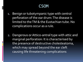
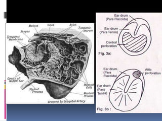
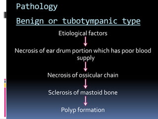
1. Otitis media is inflammation of the middle ear that can involve surrounding areas. It is commonly seen in children and caused by infections that spread from nearby areas like the throat. 2. The condition progresses through stages from initial congestion to collection of fluid/pus in the middle ear. Without treatment, the ear drum may rupture, allowing drainage of pus from the ear. 3. Treatment involves antibiotics and drainage procedures. Chronic or recurring cases may require surgery to repair damaged tissues and prevent complications like infection of the mastoid bone behind the ear.