Downloaded 1,319 times


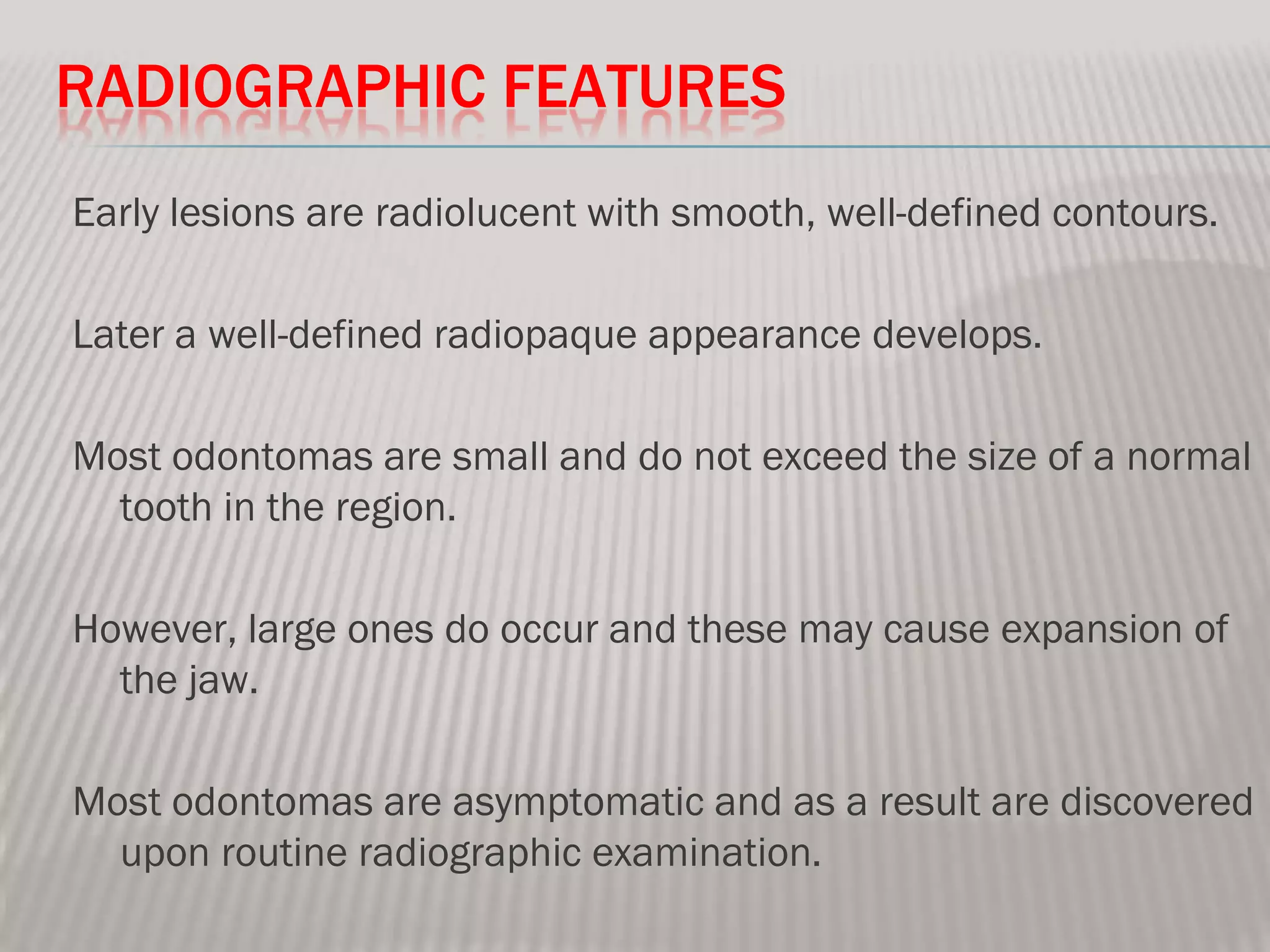


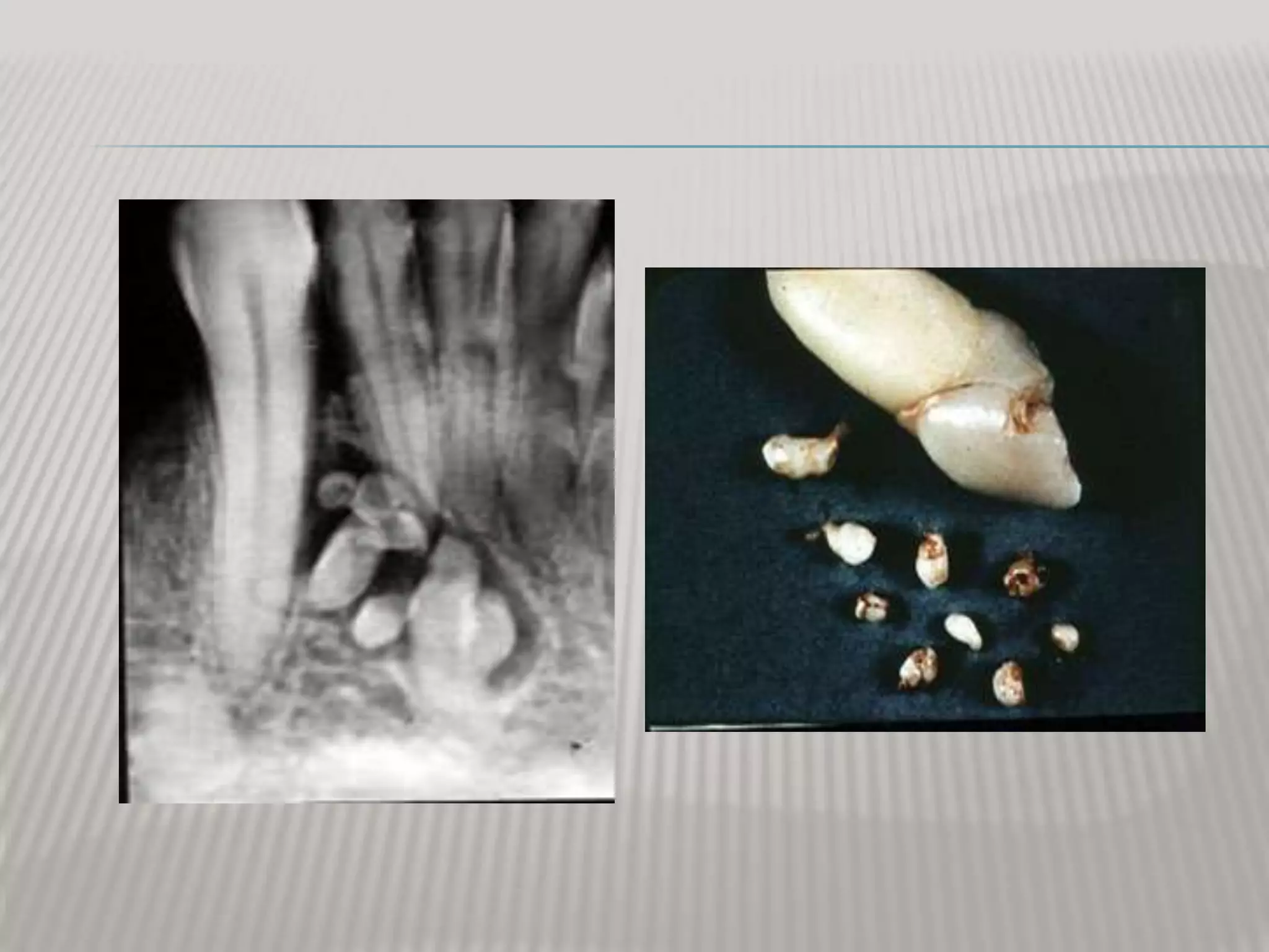


This document discusses odontomas, which are benign odontogenic tumors composed of dental tissue like enamel, dentin, and pulp. There are two main types: compound odontomas, which appear like small tooth structures, and complex odontomas, which have a disorganized appearance. Odontomas are usually asymptomatic and discovered incidentally on x-rays during dental exams. On x-rays, they appear as radiopaque masses surrounded by a radiolucent rim. Treatment involves simple surgical removal, with an excellent prognosis and no recurrence.