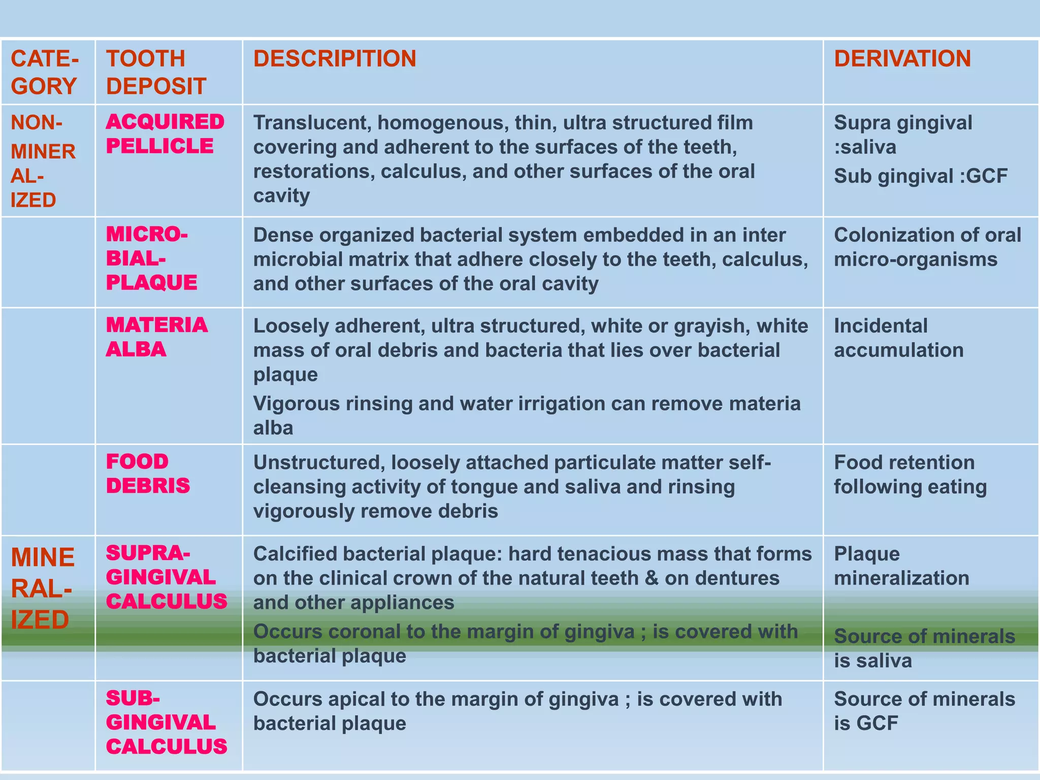
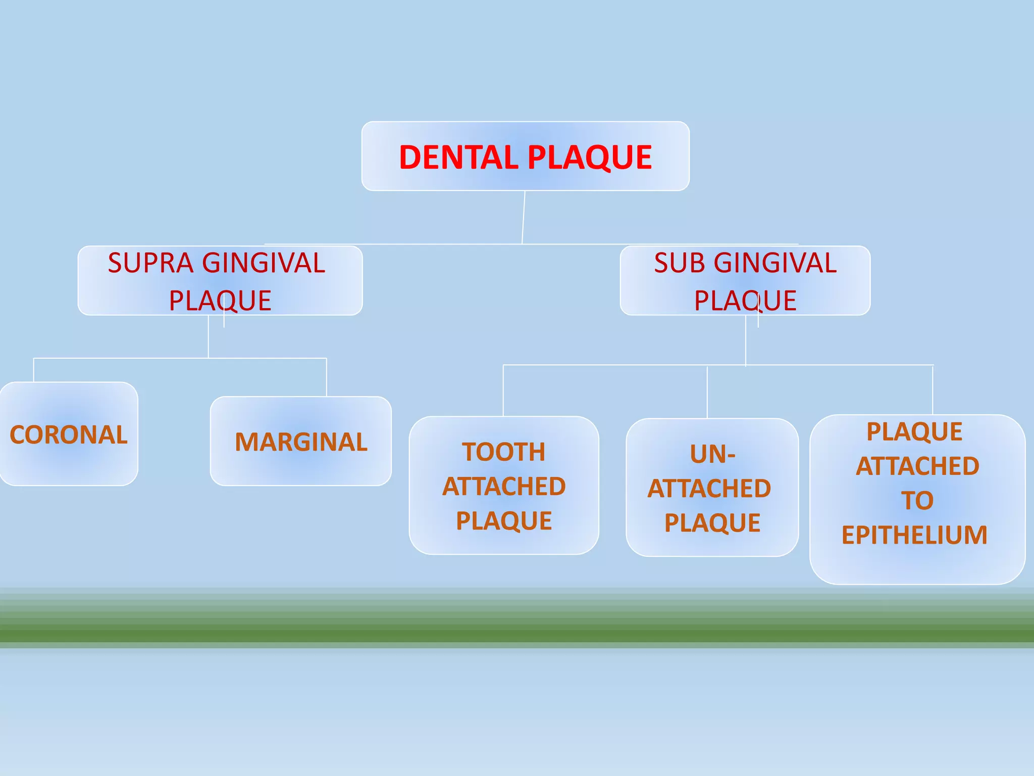
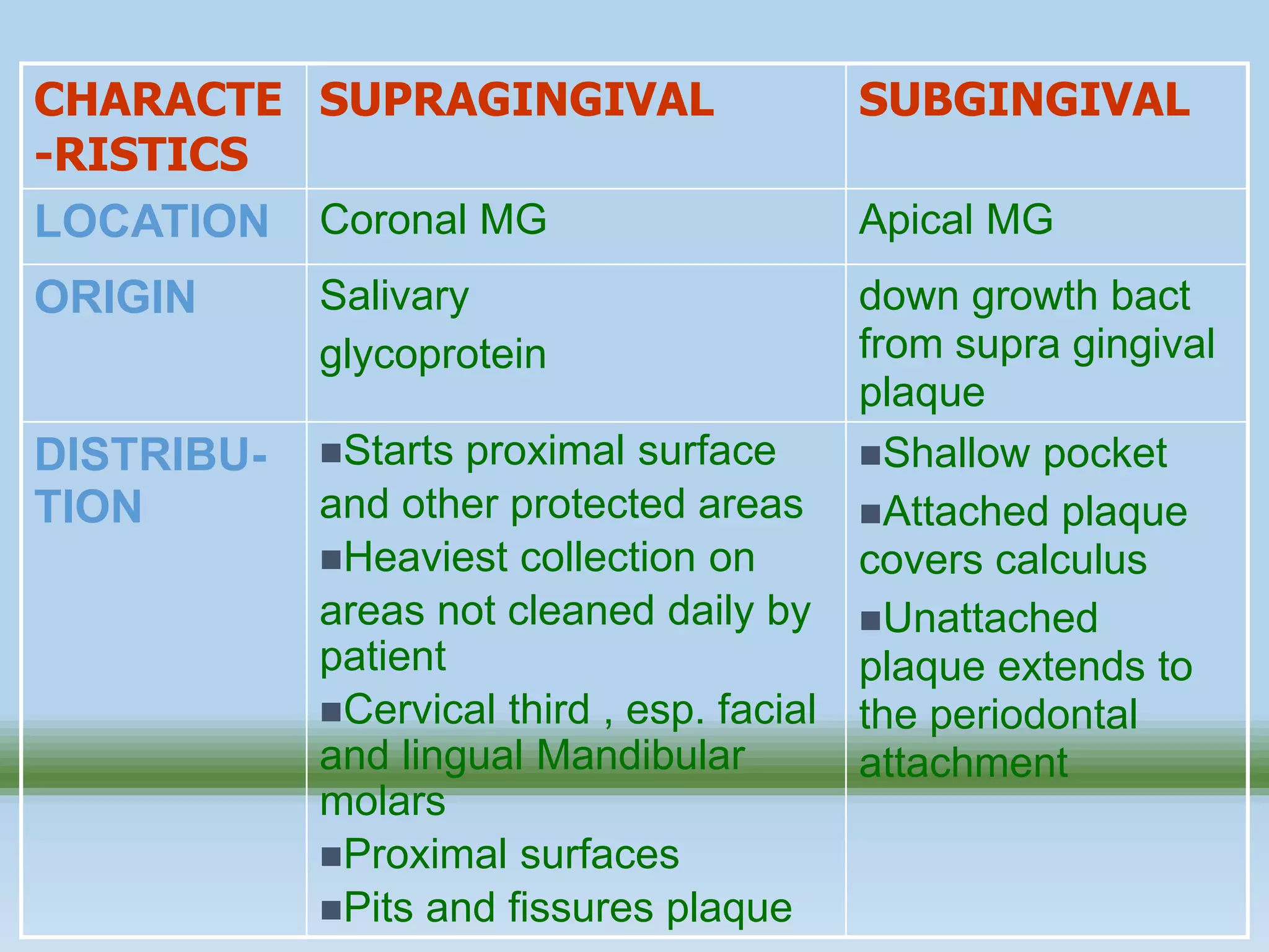
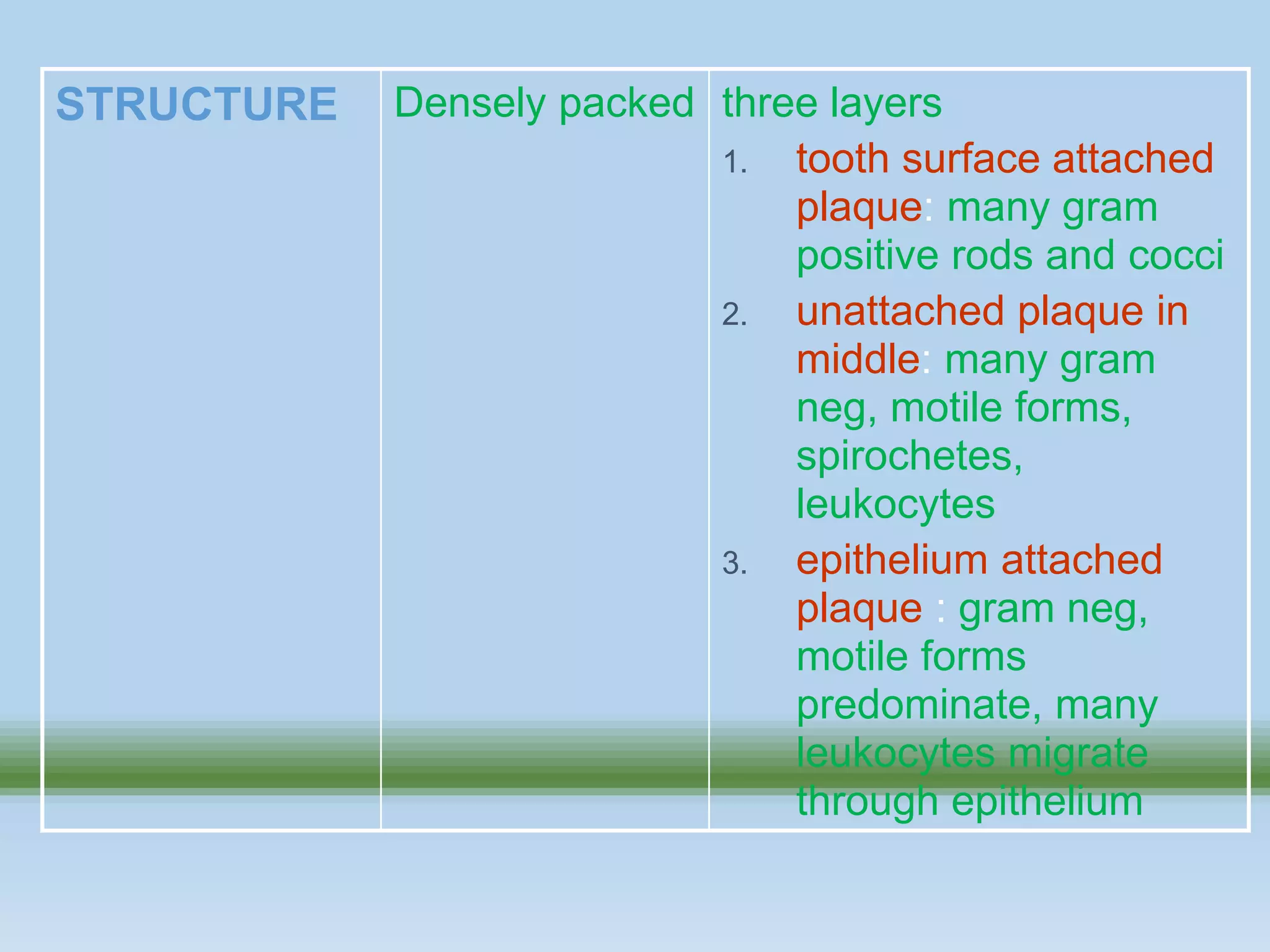
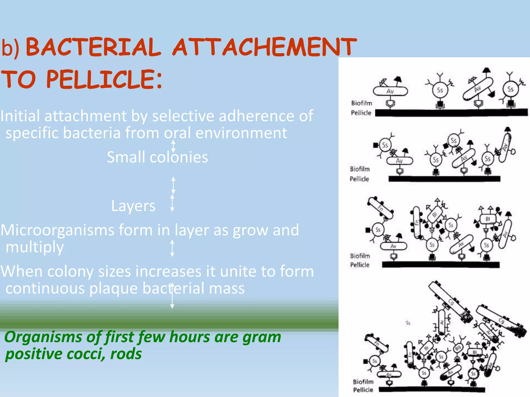
PRESENTED BY
ROSHNI MAURYA
2ND YR PG STUDENT
DENTAL PLAQUE
CONTENTS
• Introduction
• Definition
• History
• Composition
• Classification/types of dental plaque
• Formation/development
• Growth dynamics of the dental plaque
• Structural and physiological properties of dental
plaque
• Clinical significance of the plaque
( what makes plaque pathogenic?)
• Conclusion
INTRODUCTION :
• Dental plaque is the community of microorganisms
found on a tooth surface as a biofilm, embedded in
a matrix of polymers of host and bacterial origin
• Plaque is derived from French word “PLAQUER”
Which means “TO PLATE”
• WHO DEFINITION: plaque is a specific but highly variable
structural entity resulting from colonization of microorganisms
on tooth surfaces, restorations and other parts of oral cavity and
consists of salivary components like mucin, desquamated
epithelial cells, debris and microorgansims all embedded in a
gelatinous extracellular matrix.
• GLICKMAN: It is soft amorphous granular deposits which
accumulate on surface of teeth, dental restoration and dental
calculus. (Clinical periodontology 3rd ed.)
Definition
• A highly variable entity resulting from the colonization and growth of
m/o on the surfaces of the teeth and oral soft tissues and consisting
of a number of microbial species and strains embedded in an
extracellular matrix.
American Dental Association
• WH Boven, 1976. Dental plaque is defined clinically as a structured,
resilient, yellow-grayish substance that adheres tenaciously to the
intraoral hard surfaces, including removable and fixed restoration.
(Carrnaza 10th ed.)
• PD Marsh, 2004. Dental plaque can be defined as the diverse
community of micro-organisms found on the tooth surface as a
biofilm, embedded in an extracellular matrix of polymers of host and
microbial origin.
• Dental plaque must be differentiated from
other tooth deposits, like materia alba and
calculus.
• Materia Alba refers to soft accumulations of
bacteria and tissue cells that lack the organized
structure of dental plaque.
• Calculus is hard deposits that form by
mineralization of dental plaque and is
generally covered by a layer of unmineralised
plaque.
6
• Material alba • Calculus
7
Carranza 11th edition
8
CATE-
GORY
TOOTH
DEPOSIT
DESCRIPITION DERIVATION
NON-
MINER
AL-
IZED
ACQUIRED
PELLICLE
Translucent, homogenous, thin, ultra structured film
covering and adherent to the surfaces of the teeth,
restorations, calculus, and other surfaces of the oral
cavity
Supra gingival
:saliva
Sub gingival :GCF
MICRO-
BIAL-
PLAQUE
Dense organized bacterial system embedded in an inter
microbial matrix that adhere closely to the teeth, calculus,
and other surfaces of the oral cavity
Colonization of oral
micro-organisms
MATERIA
ALBA
Loosely adherent, ultra structured, white or grayish, white
mass of oral debris and bacteria that lies over bacterial
plaque
Vigorous rinsing and water irrigation can remove materia
alba
Incidental
accumulation
FOOD
DEBRIS
Unstructured, loosely attached particulate matter self-
cleansing activity of tongue and saliva and rinsing
vigorously remove debris
Food retention
following eating
MINE
RAL-
IZED
SUPRA-
GINGIVAL
CALCULUS
Calcified bacterial plaque: hard tenacious mass that forms
on the clinical crown of the natural teeth & on dentures
and other appliances
Occurs coronal to the margin of gingiva ; is covered with
bacterial plaque
Plaque
mineralization
Source of minerals
is saliva
SUB-
GINGIVAL
CALCULUS
Occurs apical to the margin of gingiva ; is covered with
bacterial plaque
Source of minerals
is GCF
History
• The descriptive term gelatinous microbial plaque was
first introduced by J.C William and G.V Black in 1898
to describe microbial colonies on tooth surfaces.
• More recently, Black’s terminology has been
shortened to the term ‘Plaque’ by Wild (1941) and its
usage has been restricted to denote only those
deposits that are predominantly microbial in nature.
Classification of Plaque
DENTAL PLAQUE
SUPRA GINGIVAL
PLAQUE
SUB GINGIVAL
PLAQUE
TOOTH
ATTACHED
PLAQUE
UN-
ATTACHED
PLAQUE
PLAQUE
ATTACHED
TO
EPITHELIUM
CORONAL MARGINAL
SUPRAGINGIVAL PLAQUE SUBGINGIVAL PLAQUE
CHARACTE
-RISTICS
SUPRAGINGIVAL SUBGINGIVAL
LOCATION Coronal MG Apical MG
ORIGIN Salivary
glycoprotein
down growth bact
from supra gingival
plaque
DISTRIBU-
TION
Starts proximal surface
and other protected areas
Heaviest collection on
areas not cleaned daily by
patient
Cervical third , esp. facial
and lingual Mandibular
molars
Proximal surfaces
Pits and fissures plaque
Shallow pocket
Attached plaque
covers calculus
Unattached
plaque extends to
the periodontal
attachment
ADHES-
ION
Firmly attached to acquired
pellicle
Surface bacteria
(unattached ) loose: washed
away by saliva
Adhere to
tooth surface
subgingival pellicle
 calculus
RETEN-
TION
Rough surface teeth
restoration , malpositioned
or carious teeth
Pocket hold plaque
against tooth and
overhanging margins
SHAPE
AND
SIZE
Friction of tongue lips
cheeks , cervical area
Thickness: thicker at
the cervical end and on
proximal surfaces
Healthy gingival: thin
plaque ,15 to 20 cells thick
Chronic gingivitis: thick
plaque,100 to 300 cells thick
Molded by pocket
wall, follows the form
created by
subgingival calculus
MICRO-
ORGANISM
Early plaque:formsGram+ve
cocci
Older plaque ( 3 to 4 days)
increased number of
filaments and fusiforms
4 to 9 days undistributed:
more complex flora with rods,
filamentous forms
7 to 12 days: vibrios,
spirochetes, more gram org
Anaerobic
population
Gram-neg ,
motile
spirochete and
rods
SOURCE
OF
NOURISH-
MENT
Saliva ingested food Tissue fluid
(sulcular fluid),
exudates,
leucocytes
STRUCTURE Densely packed three layers
1. tooth surface attached
plaque: many gram
positive rods and cocci
2. unattached plaque in
middle: many gram
neg, motile forms,
spirochetes,
leukocytes
3. epithelium attached
plaque : gram neg,
motile forms
predominate, many
leukocytes migrate
through epithelium
Structure:
• Early studies: plaque appears as a compacted
consortium of microorganisms
Confocal laser scanning microscopy has revealed
Supra gingival plaque can have a
• structured architecture polymer containing channel or pores
have been observed that link the plaque/oral environment
interface to the tooth surface ( Wood et al 2000,Auschillet al
2001,Zaura Arite et al 2001)
• the use of live /dead stains has indicated that
bacterial vitality varies throughout the biofilm, with
the most viable bacteria present in the central part of
plaque , and lining the voids and channels (Auschill et al
2001)
• this more open architecture should enable the
molecule to readily move in and out of the plaque ,
but the presence of a matrix comprised of a diverge
range of exo polymers creates a complex environment
for accurately predicting the penetration and
distribution of molecules with in plaque (Robinson et al
1997,Thurnheer et al 2003,Marcotte et al 2004).
Sub gingival plaque:
Histological sections of human sub gingival plaque viewed
by Conventional light microscopy suggested that:
• a complex organization of attached microorganisms in
which there can exist distinct tooth associated and
epithelial associated biofilms, with the possibility of a less
dense zone of organisms between the two (Socransky &
Haffajee 2002)
• Between sub gingival plaque and the tooth an
electron dense organic material is interposed ,
termed as cuticle.
• This cuticle probably contains the remains of the
epithelial attachment lamina originally connecting the
junctional epithelium to the tooth , with the
additional of material deposited from the gingival
exudates (Frank & Cimasoni 1970, Lie & Selvig 1975, Eide et
al 1983)
• It has been suggested that cuticle represents a secretory
product of the adjacent epithelial cells. (Schroeder & Lisgarten
1977)
Composition of plaque
COMPOSITION OF PLAQUE:
COMPOSED mainly of
• Microorganisms ,
• Intermicrobial matrix
(a) Bacterial portion:
• 70 to 80 % of total solid plaque volume.
• 1 g (WET WT)contains approx 1011 bacteria.
(B)Intermicrobial Matrix
• the material present between the bacteria in the dental
plaque is called the intermicrobial matrix.
• accounts for approx 25% of plaque volume.
Three sources may contribute to the intermicrobial
matrix:
•the plaque microorganisms
•the saliva
•gingival exudates
Degenerating or dead bacteria may also contribute to inter
microbial matrix.
Organic and inorganic SOLIDS approx 20 % and water
• WATER 70 to 80 % of solid matter, which higher in sub gingival
plaque than supra gingival.
INORGANIC:
• Main Calcium and Phosphorus(mostly) magnesium, sodium
fluoride(in small amount, higher than saliva)
• CALCIUM AND PHOSPHATE more on lingual surface of anterior
teeth.
ORGANIC:
• Surrounds microorganisms contains carbohydrates, proteins and
lipids
Inter bacterial matrix:
CARBOHYDRATES:
Fructans (Levans) :
• synthesized in plaque from dietary sucrose
• provide a storage of energy which may be utilized by the
microorganisms in time of low sugar supply
Glucans:
also synthesize from sucrose & of two types:
• Dextran: serve as energy storage
• Mutan: acts primarily as a skeleton in the matrix in much the
same way as collagen stabilizes the intercellular substances of
connective tissue.
• A fibrillar component is seen in the matrix between gram+ve cocci
• In parts of the plaque with the presence of gram –ve organisms, the
intermicrobial matrix is regularly characterized by the presence of
small vesicles surrounded by trilaminar membrane, which is similar in
structure to that of the outer envelope of the cell wall of gram -ve
microorganisms.
• Such vesicles may contain endotoxin and proteolytic enzymes, may
also be involved in adherence between bacteria ( hofstad et al 1972,
grenier and mayrand 1987)
• In other regions it appears granular or homogenous
Intermicrobial matrix in plaque varies
considerably from region to region
Non bacterial microorganisms that are found in plaque
include
• mycoplasma species
• yeast
• protozoa, and
• viruses
The micro organisms exist with in a intracellular matrix
that also contains
• a few host cells, such as epithelial cells ,
• macrophages and
• leukocytes
Selected Bacterial Species Found In Dental Plaque:
Bacteria Facultative Anerobic
Gram +ve Strep.mutans
Strep.sanguis
A.viscosus
Gram -ve A.actinomycetemcomitans
Capnocytophypa sp.
Ekinella corrodens
P.Gingivalis
F.nucleatum
P.intermedia
B.forsythus
C.rectus
Spirochetes  T.denticola
PROTINES:
• Supra gingival plaque contains proteins from saliva.
• Sub gingival contains Albumin – cervical fluid and
gingival exudates.
LIPIDS:
• lipopolysaccharides, endotoxins from gram negative
bacteria.
PLAQUE AS A BIOFILM
•As the bacteria attach to a surface and
to each other, they cluster together to
form sessile, mushroom-shaped
microcolonies that are attached to the
surface at a narrow base.
•Each microcolony is a tiny, independent
community containing thousands of
compatible bacteria.
•Different microcolonies may contain
different combinations of bacterial
species.
•Bacteria in the center of a microcolony
may live in a strict anaerobic
environment, while other bacteria at the
edges of the fluid channels may live in an
aerobic environment.
•Thus, the biofilm structure provides a
range of customized living environments
(with differing pHs, nutrient availability,
and oxygen concentrations) within which
bacteria with different physiological
needs can survive.
•The extracellular slime layer is a
protective barrier that surrounds the
mushroomshaped bacterial
microcolonies.
•The slime layer protects the
bacterial microcolonies from
antibiotics, antimicrobials, and host
defense mechanisms.
•A series of fluid channels penetrates
the extracellular slime layer.
• These fluid channels provide nutrients
and oxygen for the bacterial micro
colonies and facilitate movement of
bacterial metabolites, waste products,
and enzymes within the biofilm
structure.
• Each bacterial microcolony uses
chemical signals to create a primitive
communication system used to
communicate with other bacterial
microcolonies
Quorum sensing
Etiology and Pathogenesis of Periodontal Disease, 2010)
• The regulation of bacterial gene expression in response to
changes in cell density is known as quorum sensing. These
bacteria then proliferate and communicate by signals to each
other (Okuda et al. 2004).
• In order to maintain the ecosystem, various anaerobes, anchor
to each other by forming aggregated bacterial masses (Kigure
et al. 1995; Okuda et al. 2004).
• Quorum-sensing bacteria synthesize and secrete extracellular
signaling molecules called autoinducers, which accumulate in
the environment as the population increases (Okuda et al.
2004).
• Gram-positive bacteria generally communicate via small
diffusible Peptides, while many gram-negative bacteria secrete
Acyl Homoserine Lactones (AHLs) (Whitehead et al. 2001),
which vary in structure depending on the species of bacteria
that produce them.
• Another system involves the synthesis of autoinducer- 2 is LuxS
(Federle and Bassler 2003; Winzer et al. 2003). This system may
be involved in cross-communication among both gram-positive
and gram-negative bacteria. (Marsh 2004).
Plaque formation
stages of formation in
dental plaque
1. Formation of pellicle
2. Attachment of bacteria to
pellicle
3. Bacterial multiplication
and colonization
4. Plaque growth and
maturation
5. Matrix formation.
a) PELLICLE FORMATION:
• formed by adsorption of proteins on tooth surface
• composed of glycoprotein from saliva , adsorbed by
hydroxyapatite
• Have negative charged phosphate --interact +ve
charge salivary crevicular macromolecules
• This adsorbed material becomes highly insoluble
coating over teeth, calculus, and restoration, complete
and partial denture.
DERIVATION: Supra gingival from saliva
Sub gingival from gingival sulcus.
It is translucent, homogenous, thin ultra structured
film covering adherent tooth surface.
SIGNIFICANCE OF PELLICLE:
• PROTECTIVE: provide barrier against acids thus may
reduce dental caries attack.
• LUBRICATION: keep surface moist prevent drying.
• NIDUS FOR BACTERIA: Plaque formation by adherence
of microorganisms.
• ATTACHEMENT OF CALCULUS: A mode of calculus
attachment in acquired pellicle.
b) BACTERIAL ATTACHEMENT
TO PELLICLE:
Initial attachment by selective adherence of
specific bacteria from oral environment
Small colonies
Layers
Microorganisms form in layer as grow and
multiply
When colony sizes increases it unite to form
continuous plaque bacterial mass
Organisms of first few hours are gram
positive cocci, rods
c) MATRIX FORMATION:
• Intermicrobial substances derived mainly from
saliva for supra gingival plaque and
• gingival sulcus fluid and exudates for sub
gingival plaque
• other product produced by certain bacteria, dietary
source polysaccharide are sticky contribute
to adhesion of plaque to teeth.
CHANGE IN PLAQUE MICROORGANISMS
DAY 1 TO 2 gram +ve Cocci, Streptococci (dominate),
Strepto mutans, Strepto sanguis
DAY 2 TO 4 Cocci still dominate , increase gram+ve
filamentous form, slender rods , gradually
filamentous forms replaces cocci.
DAY 4 TO 7 Filaments increases in numbers, and a
more mixed flora begins to appear with
rods, filamentous forms and Fusobacteria .
 Plaque near margin thickens and
develops a more mature flora with gram
neg spirochetes and vibros .
 As plaque spreads coronally, the new
plaque has the characteristic coccal forms
DAY 7 TO 14 Vibros and spirochetes appear. No.
of WBC increases , as plaque
mature and thickens more,gram+ve
anaerbic org appear “during this
period sign of inflammation on
gingiva occurs”
DAY 14 TO 21 Vibros + spirochetes prevalent
along with cocci and filamentous
The densly packed filamentous
organisms arrange themselves
perpendicular to the tooth surface in
a palisade “gingivitis evident
clinically”.
Plaque formation at ultrastructual level
1. Formation of pellicle on the tooth surface
2. Initial adhesion and attachment of bacteria
Phase 1: Transport to the surface
Phase 2: Initial adhesion
Phase 3: Attachment
Phase 4: Colonization of surface and biofilm
formation
3. Colonization and plaque maturation
Formation of pellicle
• All surface of oral cavity (both hard and soft) are coated with a
pellicle.
• With in nanoseconds after a vigorously polishing the teeth, a
thin layer, called acquired pellicle, covers the tooth surface.
• Acquired pellicle term is misleading as it implies that bacteria
can colonize only when pellicle is in place for few hours.
• Bacteria can be part of early deposit, within seconds after
prophylaxis.
• Pellicle formation involves selective adsorption of
macromolecules (eg glycoprotein) to apatite surface.
Forces involved in enamel pellicle formation:
1.Electrostatic forces (between hydorxyapatite surfaces which has
negatively charged phosphate group, that interacts with opposite
charged groups in the salivary macromolecules).
2.van der Waals force
3.Hydrostatic force
(Detachment of adhering bacteria might occur through cohesive
failure in conditioning film between bacteria and surface ,i.e.
pellicle)
Initial Adhesion & Attachment of Bacteria
Phase 1: Transport to the surface
This include transport of bacteria to the tooth surface.
Random contact occurs through:
1. Brownian motion (avg displacement of 40 µm/hr).
2. Through sedimentation of microorganisms.
3. Through liquid flow.
4. Through active bacterial movement (chemotactic
movement).
PHASE 2: INITIAL ADHESION
• The second stage results in an initial, reversible adhesion
of bacterium, initiated by interaction between bacterium
and the surface, from a certain distance (50 nm),
through long range and short range forces, including van
der Waals attractive and electrostatic repulsive forces.
Derjaguin, Landau, Verwey and Overbeek (DLVO)
postulated that,
• above a separation of 1 nm, the summation of vander wal
(attaractive; GA) and electrostatic forces (repulsive; GE)
describes total long range interaction.
• If a particle reaches the primary minimum (<1nm from the
surface), a group of short range forces (eg hydrogen
bonding, ionic pair formation, steric interaction) dominates
the strength of adhesion.
PHASE 3: ATTACHMENT
• After initial adhesion, a firm anchorage will be established by
specific interactions (covalent, ionic, or hydrogen bonding).
• This follows direct contact or bridging true extra cellular
filamentous appendages (with length up to 10nm).
• On a rough surface, bacteria are better protected against shear
forces so that a change from reversible to irreversible bonding
occurs more easily and more frequently.
• Streptococci (mainly S. sanguis), bind to acidic proline-rich-
proteins and other receptors in pellicle as α-amylase and sialic
acid.
• Actinomyces can also function as primary colonizers: eg A.
viscosus possesses fimbrae that contain adhesins that
specifically bind to proline-rich proteins of dental pellicle
• A. viscosus reognises cryptic segments ( with lock &key mech.)
of proline rich proteins, which are only available in adsorbed
molecules.
( Mergenhagen et al 1987)
COLONIZATION & PLAQUE MATURATION
• When the firmly attached microorganisms start
growing and the newly formed bacterial clusters
remain attached, micro colonies or a biofilm can
develop.
• From this stage Intrabacterial connections occur
• at least 18 genera from the oral cavity have shown
some form of co-aggregation (cell-to-cell recognition
of genetically distinct partner cell types).
• All oral bacteria possess surface molecules that foster
some type of cell to cell interaction.
• This occurs primarily through the highly specific stereo
chemical interaction of protein and carbohydrate
molecules located on bacterial cell surfaces, in addition to
the less specific interactions resulting from hydrophobic,
electrostatic and van der waals forces.
• Coaggregation is mediated by lectin-like adhesion and can
be inhibited by lactose and other galactosides
Interactions of secondary colonizers with early colonizers include the coaggregation of
a. Fusobacterium nucleatum with Streptococcus sanguis
b. Prevotella loescheii with Actinomyces viscosus.
c. Capnocytophaga ochraceus with A. viscosus.
Coaggregation exist between
a. Different gram positive species.
b. Gram positive and gram negative species
c. Different gram negative species (predominate in later stage) eg F. nucleatum with P.
gingivalis or T. denticola.
Special examples of coaggregations are
a. “Corncob” formation
b. “Test-tube brush” formation.
c. Probiotics.
• “Corncob” formation have been observed
between rod-shaped bacterial cells eg.
Bacterionema matruchotii or Actinomyces sp. that
forms inner core of the structure and coccal cells eg.
Streptococci or P. gingivalis that attach along the
surface of the rod shaped cells.
• “Test-tube brush” composed of filamentous bacteria to
which gram negative rods adhere.
“Probiotics” : “literally meaning ‘for life”
WHO defined it as live microorganisms which when
administered in adequate amounts confer a health
benefit on the host.
BENEFITS OF COMMUNITY LIFE STYLE to micro
organisms:
• A broad habitat range for growth. e,.g. oxygen consuming
sp. create environmental conditions suitable for obligate
anaerobes
• A more efficient metabolism e.g. complex hast
macromolecule can only be degraded by consortia of oral
bacteria.
• Increased resistance to stress and antimicrobial agents
• Enhanced virulence
( pathogenic synergism) {Caldwell et al 1997, Shapiro ,1998, Marsh
and Bowen,2000}
Relationship of Specific Microorganisms to Periodontal
Diseases
• Our understanding of the relationship between the
microorganisms found in dental plaque and the common
dental disease of periodontitis has undergone numerous
phases historically
• Early in the 19th century, it was felt that, like the situation
with diseases such as tuberculosis, a specific bacterial
species was responsible for the disease processes.
• The criteria by which a given bacterial species was
associated with disease historically has been through the
application of Koch's Postulates.
These criteria were developed by Robert Koch in the late
1800's. The criteria are as follows:
• A specific organism can always be found in association
with a given disease.
• The organism can be isolated and grown in pure culture in
the laboratory.
• The pure culture will produce the disease when inoculated
into a susceptible animal.
• It is possible to recover the organism in pure culture from
the experimentally infected animal.
CURRENT HYPOTHESES ON THE ROLE OF PLAQUE
BACTERIA IN THE ETIOLOGY OF DENTAL DISEASES
• Non-specific plaque hypothesis
• Specific plaque hypothesis
• Ecological plaque hypothesis
• Both the Plaque and Non-plaque hypothesis were delineated in 1976 by
Walter Loesche, a researcher at the University of Michigan.
• According to non-specific plaque hypothesis periodontal disease results
from the “elaboration of noxious products by the entire plaque flora”.
• Specific plaque hypothesis states that “only certain plaque is
pathogenic, and its pathogenicity depends on the presence of or
increase in specific microrganisms”. For example role of Aactinobacillus
actinomycetemcomitans in aggressive periodontitis. Thus plaque
harboring specific pathogens results in a periodontal disease because
these organisms produce substances that mediate the destruction of
host tissues
Ecological plaque hypothesis:
(By Marsh and Bradshaw; 1997)
• Disease results from shifts in the balance of the resident plaque
microflora.
• A change in the nutrient status of a pocket or chemical and
physical changes to the habitat are thus considered the primary
cause for the overgrowth by pathogens
• Disease could be controlled not only by targeting putative
pathogens but also by interfering with the factors responsible
for driving the deleterious shifts in the microflora as by
reducing GCF rate , or the site could be made less anaerobic by
use of redox agents
Growth dynamics of dental
plaque
Ultra structural aspects:
• Important changes in the plaque growth rate can be
detected within first 24 hours.
• During the first 2 to 8 hour, the adhering pioneering
streptococci saturate the salivary pellicular binding sites
and thus cover 3% to 30% of enamel surface.
• Instead of steady growth during the next 20 hrs,a short
period of rapid growth for 4 to 6 hr (Loesche suggested it
as generation time)
• After 1 day, the term biofilm is fully deserved -because
organization takes place within it.
• As bacterial densities approach approximately 2- 6 million
bacteria/mm2 on enamel surface, a marked increase in
growth rate can be observed to 32 million bacteria/mm2
(doubling in 3 to 4 hrs.)
• This occurs due to multiplication of already adhering
microorganisms rather than by new colonizers.
• Thickness of plaque increases slowly with time,
increasing to 20 to 30 µm after 3 days
• During the first 24 hr, plaque growth is negligible from a clinical view point
(<3% coverage of vestibular tooth surface, an amount that is clinically
almost undetectable).
• During the following 3 days, plaque growth increases at a rapid rate, then
slows down from that point onward.
• After 4 days, on average, 30 % of total tooth crown area will be covered
with plaque.
• There is ecologic shift within biofilm, with transition from early aerobic
environment characterized by gram positive facultative species to a highly
oxygen deprived environment in which gram-negative anaerobic
microorganisms predominate.
• During night, plaque growth rate is reduced by about 50%. This may be
explained from the fact that supra gingival-plaque obtains its nutrients
mainly from the saliva
Clinical Aspects: Supra Gingival Plaque Formation:
Experimental gingivitis model
• From day 0 on, when mechanical plaque
control was stopped, plaque will slowly
form on the teeth.
• With time the plaque composition
changes, with a shift to more gram
negative species and more rods, filaments
and from day 7 on spirals and spirochetes.
• From day 3 on, the first symptoms of
gingival inflammation become visible.
• When proper plaque control is
reestablished, the plaque composition
returns to the initial situation and
symptoms of gingivitis disappear
Topography of supragingival plaque
Early plaque formation on teeth follows a
typical topographic pattern, with initial
growth along the gingival margin and
from the inter dental spaces (areas
protected against shear forces) to extend
further in coronal direction.
Early plaque formation on surface
irregularities of teeth
Surface micro roughness
Small plastic strip divided in half (a rough region, average
roughness Ra 2.0µm, located mesially, and a smooth region
Ra 0.1µm, located distally) was glued to the central upper
incisors of a patient who refrained from oral hygiene for 3 days
Disclosing agent used was : 0.5% neutral red
SMOOTH REGION
ROUGH PART
After removal, the strip
was cut in small slices
for microscopic
evaluation
Rough part contains a
thicker plaque layer than
smooth part .
Arrow shows border
between
rough and smooth
surface
Other factors determining supragingival plaque
formation are:
1. Individual variables influencing plaque formation: “heavy
or fast plaque” former and “light or slow plaque” former-
this depends on various factors as
• diet, chewing fibrous food,
• smoking,
• amalgam restorations,
• tongue and palate brushing,
• the colloidal stability in saliva,
• antimicrobial factors of GCF and saliva
• pellicle composition and
• retention depth of dento-gingival area
2. Variation within the dentition
In general early plaque formation occurs faster
1. In lower jaw compared to upper jaw
2. In molar areas
3. On buccal tooth surfaces compared to oral sites
(especially in upper jaw)
4. In the interdental regions compared to the strict
buccal or oral surfaces.
3. Impact of gingival inflammation
• Plaque formation is more rapid on tooth surfaces facing inflamed
gingival margins than on those adjacent to healthy gingiva.
Substances from GCF as mineral, protein, carbohydrate favors both
the initial adhesion and growth of early colonizing bacteria.
4. Impact of patient’s age
• Subjects age does not influence de novo plaque formation.
Developed plaque in older patient , resulted in more severe gingival
inflammation, this indicate an increased susceptibility gingivitis with
aging.
5. Spontaneous tooth cleaning
• Only negligible differences in plaque extension (plaque retaining
areas) areas is observed.
Changes in subgingival micro biota during 1st week after mechanical debridement
reported only partial reduction from 108 to 105 followed by a fast regrowth to almost
pretreatment level within 7 days. Smooth abutments (Ra<0.2 µm) were found to harbor
25 times less bacteria than rough ones, with a slight higher density of coccoid
(ie non-pathogenic) cells
Recent studies demonstrate
that a complex subgingival
microbiota, including
most periopathogens, is
established within 1 week after
abutment (transmucosal implant)
insertion
De novo subgingival plaque formation
Scanning electron microscope of
bacteria invading dentinal tubules
15000x
PHYSIOLOGIC PROPERTIES OF
DENTAL PLAQUE
Factor modifiers:
• Oxidation Reduction potential (Saocransky et. at.
1964)
• pH (Kleinbers and Hall 1969)
• Temperature (Haffagee et al 1992)
Factor antagonizes:
• Bacteriocins (Rogers et. al. 1979, Hammond et. al.
1987, Steven et. al. 1987)
• H2O2 (Holmberg and Hammond 1973, Hillman et. al.
1985 )
• Organic acids (Mashimo et. al. 1985)
•gingival sulcus and periodontal pocket provides the
area for growth
•mean temperature:
Averages about 35ºC
Ranges about 30ºC to 38ºC (Haffagee et al 1992)
•pH :
pH 7.0-8.5 ( Forscher et. at. 1954 , Kleinbers and
Hall 1969, Cimasoni 1983)
•Oxidation Reduction potential
-300 to + 310mv at pH 7.0 (Onisi et. at. 1960, Kenney
& Ash 1969)
• Early colonizers (e.g. Streptococci, Actinomyces) use
oxygen and lower the redox potential of the
environment, which then favours the growth of
anaerobic species.
Gram +ve sp. use
• sugars as an energy source and
• saliva as carbon source
• The bacteria that predominate in mature plaque are
ananerobic and use amino acid and small peptides as
energy source.
• Lactate and Formate byproducts of metabolism of
Streptococci and Actinomyces and may be used in
metabolism of other plaque microorganisms
The growth of P.gingivalis is enhanced by metabolic
byproducts produced by other microorganisms, such as
• succinate from Capnocytophaga ochraceus and
• protoheme from Campylobacter rectus
Factor antagonizes
For example:
• S. sanguis produces H2O2 which inhibits growth of
A.actinomycetemcomitans (Hillman et. al. 1985 )
• A.actinomycetemcomitans produces bacteriocins
that inhibits S. sanguis (Hammond et. al. 1987, Steven
et. al. 1987)
Sources of nutrients available:
Mainly 3 sources:
diet, host, and other sub gingival species
Certain nutrients are:
• Vit K analogues (Gibbons and Macdonald 1960)
• Certain growth factors like hemin (Evans 1951,
Gibbons and Macdonald 1960)
Host as source of nutrients:
• Bacterial enzymes that degrade host proteins result in the
release of ammonia, which may be used by bacteria as
nitrogen source
• Hemin iron from the breakdown of host Hb may be
important in the metabolism of P.gingivalis
• Increase in Steroid hormone are associated with significant
increase in the proportions of P.intrmedia found in sub
gingival plaque
Clinical significance of plaque
Suprgingival plaque
• Microbial aggregations on the tooth surface if prevented from
maturing may become compatible with gingival health.
• Supragingival plaque if allowed to grow and mature, may
induce gingivitis and can lead to the formation of a
microenvironment that permits the development of subgingival
plaque.
• Therefore, supragingival plaque strongly influence the growth,
accumulation and pathologic potential of sub gingival plaque,
especially in the early stages of gingivitis and periodontitis.
Subgingival plaque
• In association with the presence of supragingival plaque,
there are inflammatory changes that modify the anatomic
relationships of the gingival margin and tooth Surface.
• This results in enlarged gingiva, which increases the space
for bacterial colonization and also protects bacteria from
normal cleansing mechanisms. They derive from GCF.
• Many of these microorganisms lack the adherence ability
and utilizes plaque bacteria as a means of colonization
subgingival area.
conclusion
• Dental plaque biofilm cannot be eliminated.
However, the pathogenic nature of the dental
plaque biofilm can be reduced by reducing the
bioburden (total microbial load and different
pathogenic isolates within that dental plaque
biofilm) and maintaining a normal flora with
appropriate oral hygiene methods that include
daily brushing,flossing and rinsing with
antimicrobial mouthrinses.This can result in the
prevention or management of the associated
sequelae, including the development of periodontal
diseases and possibly the impact of periodontal
diseases on specific systemic disorders. JADA, Vol. 137
November 2006.

Dental plaque

  • 1.
    PRESENTED BY ROSHNI MAURYA 2NDYR PG STUDENT DENTAL PLAQUE
  • 2.
    CONTENTS • Introduction • Definition •History • Composition • Classification/types of dental plaque • Formation/development • Growth dynamics of the dental plaque • Structural and physiological properties of dental plaque • Clinical significance of the plaque ( what makes plaque pathogenic?) • Conclusion
  • 3.
    INTRODUCTION : • Dentalplaque is the community of microorganisms found on a tooth surface as a biofilm, embedded in a matrix of polymers of host and bacterial origin • Plaque is derived from French word “PLAQUER” Which means “TO PLATE”
  • 4.
    • WHO DEFINITION:plaque is a specific but highly variable structural entity resulting from colonization of microorganisms on tooth surfaces, restorations and other parts of oral cavity and consists of salivary components like mucin, desquamated epithelial cells, debris and microorgansims all embedded in a gelatinous extracellular matrix. • GLICKMAN: It is soft amorphous granular deposits which accumulate on surface of teeth, dental restoration and dental calculus. (Clinical periodontology 3rd ed.)
  • 5.
    Definition • A highlyvariable entity resulting from the colonization and growth of m/o on the surfaces of the teeth and oral soft tissues and consisting of a number of microbial species and strains embedded in an extracellular matrix. American Dental Association • WH Boven, 1976. Dental plaque is defined clinically as a structured, resilient, yellow-grayish substance that adheres tenaciously to the intraoral hard surfaces, including removable and fixed restoration. (Carrnaza 10th ed.) • PD Marsh, 2004. Dental plaque can be defined as the diverse community of micro-organisms found on the tooth surface as a biofilm, embedded in an extracellular matrix of polymers of host and microbial origin.
  • 6.
    • Dental plaquemust be differentiated from other tooth deposits, like materia alba and calculus. • Materia Alba refers to soft accumulations of bacteria and tissue cells that lack the organized structure of dental plaque. • Calculus is hard deposits that form by mineralization of dental plaque and is generally covered by a layer of unmineralised plaque. 6
  • 7.
    • Material alba• Calculus 7
  • 8.
  • 9.
    CATE- GORY TOOTH DEPOSIT DESCRIPITION DERIVATION NON- MINER AL- IZED ACQUIRED PELLICLE Translucent, homogenous,thin, ultra structured film covering and adherent to the surfaces of the teeth, restorations, calculus, and other surfaces of the oral cavity Supra gingival :saliva Sub gingival :GCF MICRO- BIAL- PLAQUE Dense organized bacterial system embedded in an inter microbial matrix that adhere closely to the teeth, calculus, and other surfaces of the oral cavity Colonization of oral micro-organisms MATERIA ALBA Loosely adherent, ultra structured, white or grayish, white mass of oral debris and bacteria that lies over bacterial plaque Vigorous rinsing and water irrigation can remove materia alba Incidental accumulation FOOD DEBRIS Unstructured, loosely attached particulate matter self- cleansing activity of tongue and saliva and rinsing vigorously remove debris Food retention following eating MINE RAL- IZED SUPRA- GINGIVAL CALCULUS Calcified bacterial plaque: hard tenacious mass that forms on the clinical crown of the natural teeth & on dentures and other appliances Occurs coronal to the margin of gingiva ; is covered with bacterial plaque Plaque mineralization Source of minerals is saliva SUB- GINGIVAL CALCULUS Occurs apical to the margin of gingiva ; is covered with bacterial plaque Source of minerals is GCF
  • 10.
  • 11.
    • The descriptiveterm gelatinous microbial plaque was first introduced by J.C William and G.V Black in 1898 to describe microbial colonies on tooth surfaces. • More recently, Black’s terminology has been shortened to the term ‘Plaque’ by Wild (1941) and its usage has been restricted to denote only those deposits that are predominantly microbial in nature.
  • 12.
  • 13.
    DENTAL PLAQUE SUPRA GINGIVAL PLAQUE SUBGINGIVAL PLAQUE TOOTH ATTACHED PLAQUE UN- ATTACHED PLAQUE PLAQUE ATTACHED TO EPITHELIUM CORONAL MARGINAL
  • 14.
  • 15.
    CHARACTE -RISTICS SUPRAGINGIVAL SUBGINGIVAL LOCATION CoronalMG Apical MG ORIGIN Salivary glycoprotein down growth bact from supra gingival plaque DISTRIBU- TION Starts proximal surface and other protected areas Heaviest collection on areas not cleaned daily by patient Cervical third , esp. facial and lingual Mandibular molars Proximal surfaces Pits and fissures plaque Shallow pocket Attached plaque covers calculus Unattached plaque extends to the periodontal attachment
  • 16.
    ADHES- ION Firmly attached toacquired pellicle Surface bacteria (unattached ) loose: washed away by saliva Adhere to tooth surface subgingival pellicle  calculus RETEN- TION Rough surface teeth restoration , malpositioned or carious teeth Pocket hold plaque against tooth and overhanging margins SHAPE AND SIZE Friction of tongue lips cheeks , cervical area Thickness: thicker at the cervical end and on proximal surfaces Healthy gingival: thin plaque ,15 to 20 cells thick Chronic gingivitis: thick plaque,100 to 300 cells thick Molded by pocket wall, follows the form created by subgingival calculus
  • 17.
    MICRO- ORGANISM Early plaque:formsGram+ve cocci Older plaque( 3 to 4 days) increased number of filaments and fusiforms 4 to 9 days undistributed: more complex flora with rods, filamentous forms 7 to 12 days: vibrios, spirochetes, more gram org Anaerobic population Gram-neg , motile spirochete and rods SOURCE OF NOURISH- MENT Saliva ingested food Tissue fluid (sulcular fluid), exudates, leucocytes
  • 18.
    STRUCTURE Densely packedthree layers 1. tooth surface attached plaque: many gram positive rods and cocci 2. unattached plaque in middle: many gram neg, motile forms, spirochetes, leukocytes 3. epithelium attached plaque : gram neg, motile forms predominate, many leukocytes migrate through epithelium
  • 19.
    Structure: • Early studies:plaque appears as a compacted consortium of microorganisms Confocal laser scanning microscopy has revealed Supra gingival plaque can have a • structured architecture polymer containing channel or pores have been observed that link the plaque/oral environment interface to the tooth surface ( Wood et al 2000,Auschillet al 2001,Zaura Arite et al 2001)
  • 20.
    • the useof live /dead stains has indicated that bacterial vitality varies throughout the biofilm, with the most viable bacteria present in the central part of plaque , and lining the voids and channels (Auschill et al 2001) • this more open architecture should enable the molecule to readily move in and out of the plaque , but the presence of a matrix comprised of a diverge range of exo polymers creates a complex environment for accurately predicting the penetration and distribution of molecules with in plaque (Robinson et al 1997,Thurnheer et al 2003,Marcotte et al 2004).
  • 21.
    Sub gingival plaque: Histologicalsections of human sub gingival plaque viewed by Conventional light microscopy suggested that: • a complex organization of attached microorganisms in which there can exist distinct tooth associated and epithelial associated biofilms, with the possibility of a less dense zone of organisms between the two (Socransky & Haffajee 2002)
  • 22.
    • Between subgingival plaque and the tooth an electron dense organic material is interposed , termed as cuticle. • This cuticle probably contains the remains of the epithelial attachment lamina originally connecting the junctional epithelium to the tooth , with the additional of material deposited from the gingival exudates (Frank & Cimasoni 1970, Lie & Selvig 1975, Eide et al 1983)
  • 23.
    • It hasbeen suggested that cuticle represents a secretory product of the adjacent epithelial cells. (Schroeder & Lisgarten 1977)
  • 24.
  • 25.
    COMPOSITION OF PLAQUE: COMPOSEDmainly of • Microorganisms , • Intermicrobial matrix (a) Bacterial portion: • 70 to 80 % of total solid plaque volume. • 1 g (WET WT)contains approx 1011 bacteria.
  • 26.
    (B)Intermicrobial Matrix • thematerial present between the bacteria in the dental plaque is called the intermicrobial matrix. • accounts for approx 25% of plaque volume.
  • 27.
    Three sources maycontribute to the intermicrobial matrix: •the plaque microorganisms •the saliva •gingival exudates Degenerating or dead bacteria may also contribute to inter microbial matrix.
  • 28.
    Organic and inorganicSOLIDS approx 20 % and water • WATER 70 to 80 % of solid matter, which higher in sub gingival plaque than supra gingival. INORGANIC: • Main Calcium and Phosphorus(mostly) magnesium, sodium fluoride(in small amount, higher than saliva) • CALCIUM AND PHOSPHATE more on lingual surface of anterior teeth. ORGANIC: • Surrounds microorganisms contains carbohydrates, proteins and lipids Inter bacterial matrix:
  • 29.
    CARBOHYDRATES: Fructans (Levans) : •synthesized in plaque from dietary sucrose • provide a storage of energy which may be utilized by the microorganisms in time of low sugar supply Glucans: also synthesize from sucrose & of two types: • Dextran: serve as energy storage • Mutan: acts primarily as a skeleton in the matrix in much the same way as collagen stabilizes the intercellular substances of connective tissue.
  • 30.
    • A fibrillarcomponent is seen in the matrix between gram+ve cocci • In parts of the plaque with the presence of gram –ve organisms, the intermicrobial matrix is regularly characterized by the presence of small vesicles surrounded by trilaminar membrane, which is similar in structure to that of the outer envelope of the cell wall of gram -ve microorganisms. • Such vesicles may contain endotoxin and proteolytic enzymes, may also be involved in adherence between bacteria ( hofstad et al 1972, grenier and mayrand 1987) • In other regions it appears granular or homogenous Intermicrobial matrix in plaque varies considerably from region to region
  • 31.
    Non bacterial microorganismsthat are found in plaque include • mycoplasma species • yeast • protozoa, and • viruses The micro organisms exist with in a intracellular matrix that also contains • a few host cells, such as epithelial cells , • macrophages and • leukocytes
  • 32.
    Selected Bacterial SpeciesFound In Dental Plaque: Bacteria Facultative Anerobic Gram +ve Strep.mutans Strep.sanguis A.viscosus Gram -ve A.actinomycetemcomitans Capnocytophypa sp. Ekinella corrodens P.Gingivalis F.nucleatum P.intermedia B.forsythus C.rectus Spirochetes  T.denticola
  • 33.
    PROTINES: • Supra gingivalplaque contains proteins from saliva. • Sub gingival contains Albumin – cervical fluid and gingival exudates. LIPIDS: • lipopolysaccharides, endotoxins from gram negative bacteria.
  • 34.
    PLAQUE AS ABIOFILM •As the bacteria attach to a surface and to each other, they cluster together to form sessile, mushroom-shaped microcolonies that are attached to the surface at a narrow base. •Each microcolony is a tiny, independent community containing thousands of compatible bacteria. •Different microcolonies may contain different combinations of bacterial species.
  • 35.
    •Bacteria in thecenter of a microcolony may live in a strict anaerobic environment, while other bacteria at the edges of the fluid channels may live in an aerobic environment. •Thus, the biofilm structure provides a range of customized living environments (with differing pHs, nutrient availability, and oxygen concentrations) within which bacteria with different physiological needs can survive.
  • 36.
    •The extracellular slimelayer is a protective barrier that surrounds the mushroomshaped bacterial microcolonies. •The slime layer protects the bacterial microcolonies from antibiotics, antimicrobials, and host defense mechanisms. •A series of fluid channels penetrates the extracellular slime layer.
  • 37.
    • These fluidchannels provide nutrients and oxygen for the bacterial micro colonies and facilitate movement of bacterial metabolites, waste products, and enzymes within the biofilm structure. • Each bacterial microcolony uses chemical signals to create a primitive communication system used to communicate with other bacterial microcolonies
  • 38.
    Quorum sensing Etiology andPathogenesis of Periodontal Disease, 2010) • The regulation of bacterial gene expression in response to changes in cell density is known as quorum sensing. These bacteria then proliferate and communicate by signals to each other (Okuda et al. 2004). • In order to maintain the ecosystem, various anaerobes, anchor to each other by forming aggregated bacterial masses (Kigure et al. 1995; Okuda et al. 2004). • Quorum-sensing bacteria synthesize and secrete extracellular signaling molecules called autoinducers, which accumulate in the environment as the population increases (Okuda et al. 2004).
  • 39.
    • Gram-positive bacteriagenerally communicate via small diffusible Peptides, while many gram-negative bacteria secrete Acyl Homoserine Lactones (AHLs) (Whitehead et al. 2001), which vary in structure depending on the species of bacteria that produce them. • Another system involves the synthesis of autoinducer- 2 is LuxS (Federle and Bassler 2003; Winzer et al. 2003). This system may be involved in cross-communication among both gram-positive and gram-negative bacteria. (Marsh 2004).
  • 40.
  • 43.
    stages of formationin dental plaque 1. Formation of pellicle 2. Attachment of bacteria to pellicle 3. Bacterial multiplication and colonization 4. Plaque growth and maturation 5. Matrix formation.
  • 44.
    a) PELLICLE FORMATION: •formed by adsorption of proteins on tooth surface • composed of glycoprotein from saliva , adsorbed by hydroxyapatite • Have negative charged phosphate --interact +ve charge salivary crevicular macromolecules • This adsorbed material becomes highly insoluble coating over teeth, calculus, and restoration, complete and partial denture. DERIVATION: Supra gingival from saliva Sub gingival from gingival sulcus. It is translucent, homogenous, thin ultra structured film covering adherent tooth surface.
  • 45.
    SIGNIFICANCE OF PELLICLE: •PROTECTIVE: provide barrier against acids thus may reduce dental caries attack. • LUBRICATION: keep surface moist prevent drying. • NIDUS FOR BACTERIA: Plaque formation by adherence of microorganisms. • ATTACHEMENT OF CALCULUS: A mode of calculus attachment in acquired pellicle.
  • 46.
    b) BACTERIAL ATTACHEMENT TOPELLICLE: Initial attachment by selective adherence of specific bacteria from oral environment Small colonies Layers Microorganisms form in layer as grow and multiply When colony sizes increases it unite to form continuous plaque bacterial mass Organisms of first few hours are gram positive cocci, rods
  • 47.
    c) MATRIX FORMATION: •Intermicrobial substances derived mainly from saliva for supra gingival plaque and • gingival sulcus fluid and exudates for sub gingival plaque • other product produced by certain bacteria, dietary source polysaccharide are sticky contribute to adhesion of plaque to teeth.
  • 48.
    CHANGE IN PLAQUEMICROORGANISMS DAY 1 TO 2 gram +ve Cocci, Streptococci (dominate), Strepto mutans, Strepto sanguis DAY 2 TO 4 Cocci still dominate , increase gram+ve filamentous form, slender rods , gradually filamentous forms replaces cocci. DAY 4 TO 7 Filaments increases in numbers, and a more mixed flora begins to appear with rods, filamentous forms and Fusobacteria .  Plaque near margin thickens and develops a more mature flora with gram neg spirochetes and vibros .  As plaque spreads coronally, the new plaque has the characteristic coccal forms
  • 49.
    DAY 7 TO14 Vibros and spirochetes appear. No. of WBC increases , as plaque mature and thickens more,gram+ve anaerbic org appear “during this period sign of inflammation on gingiva occurs” DAY 14 TO 21 Vibros + spirochetes prevalent along with cocci and filamentous The densly packed filamentous organisms arrange themselves perpendicular to the tooth surface in a palisade “gingivitis evident clinically”.
  • 50.
    Plaque formation atultrastructual level 1. Formation of pellicle on the tooth surface 2. Initial adhesion and attachment of bacteria Phase 1: Transport to the surface Phase 2: Initial adhesion Phase 3: Attachment Phase 4: Colonization of surface and biofilm formation 3. Colonization and plaque maturation
  • 51.
    Formation of pellicle •All surface of oral cavity (both hard and soft) are coated with a pellicle. • With in nanoseconds after a vigorously polishing the teeth, a thin layer, called acquired pellicle, covers the tooth surface. • Acquired pellicle term is misleading as it implies that bacteria can colonize only when pellicle is in place for few hours. • Bacteria can be part of early deposit, within seconds after prophylaxis. • Pellicle formation involves selective adsorption of macromolecules (eg glycoprotein) to apatite surface.
  • 52.
    Forces involved inenamel pellicle formation: 1.Electrostatic forces (between hydorxyapatite surfaces which has negatively charged phosphate group, that interacts with opposite charged groups in the salivary macromolecules). 2.van der Waals force 3.Hydrostatic force (Detachment of adhering bacteria might occur through cohesive failure in conditioning film between bacteria and surface ,i.e. pellicle)
  • 53.
    Initial Adhesion &Attachment of Bacteria Phase 1: Transport to the surface This include transport of bacteria to the tooth surface. Random contact occurs through: 1. Brownian motion (avg displacement of 40 µm/hr). 2. Through sedimentation of microorganisms. 3. Through liquid flow. 4. Through active bacterial movement (chemotactic movement).
  • 54.
    PHASE 2: INITIALADHESION • The second stage results in an initial, reversible adhesion of bacterium, initiated by interaction between bacterium and the surface, from a certain distance (50 nm), through long range and short range forces, including van der Waals attractive and electrostatic repulsive forces.
  • 55.
    Derjaguin, Landau, Verweyand Overbeek (DLVO) postulated that, • above a separation of 1 nm, the summation of vander wal (attaractive; GA) and electrostatic forces (repulsive; GE) describes total long range interaction. • If a particle reaches the primary minimum (<1nm from the surface), a group of short range forces (eg hydrogen bonding, ionic pair formation, steric interaction) dominates the strength of adhesion.
  • 56.
    PHASE 3: ATTACHMENT •After initial adhesion, a firm anchorage will be established by specific interactions (covalent, ionic, or hydrogen bonding). • This follows direct contact or bridging true extra cellular filamentous appendages (with length up to 10nm). • On a rough surface, bacteria are better protected against shear forces so that a change from reversible to irreversible bonding occurs more easily and more frequently.
  • 57.
    • Streptococci (mainlyS. sanguis), bind to acidic proline-rich- proteins and other receptors in pellicle as α-amylase and sialic acid. • Actinomyces can also function as primary colonizers: eg A. viscosus possesses fimbrae that contain adhesins that specifically bind to proline-rich proteins of dental pellicle • A. viscosus reognises cryptic segments ( with lock &key mech.) of proline rich proteins, which are only available in adsorbed molecules. ( Mergenhagen et al 1987)
  • 58.
    COLONIZATION & PLAQUEMATURATION • When the firmly attached microorganisms start growing and the newly formed bacterial clusters remain attached, micro colonies or a biofilm can develop. • From this stage Intrabacterial connections occur • at least 18 genera from the oral cavity have shown some form of co-aggregation (cell-to-cell recognition of genetically distinct partner cell types).
  • 59.
    • All oralbacteria possess surface molecules that foster some type of cell to cell interaction. • This occurs primarily through the highly specific stereo chemical interaction of protein and carbohydrate molecules located on bacterial cell surfaces, in addition to the less specific interactions resulting from hydrophobic, electrostatic and van der waals forces. • Coaggregation is mediated by lectin-like adhesion and can be inhibited by lactose and other galactosides
  • 60.
    Interactions of secondarycolonizers with early colonizers include the coaggregation of a. Fusobacterium nucleatum with Streptococcus sanguis b. Prevotella loescheii with Actinomyces viscosus. c. Capnocytophaga ochraceus with A. viscosus. Coaggregation exist between a. Different gram positive species. b. Gram positive and gram negative species c. Different gram negative species (predominate in later stage) eg F. nucleatum with P. gingivalis or T. denticola. Special examples of coaggregations are a. “Corncob” formation b. “Test-tube brush” formation. c. Probiotics.
  • 61.
    • “Corncob” formationhave been observed between rod-shaped bacterial cells eg. Bacterionema matruchotii or Actinomyces sp. that forms inner core of the structure and coccal cells eg. Streptococci or P. gingivalis that attach along the surface of the rod shaped cells. • “Test-tube brush” composed of filamentous bacteria to which gram negative rods adhere. “Probiotics” : “literally meaning ‘for life” WHO defined it as live microorganisms which when administered in adequate amounts confer a health benefit on the host.
  • 62.
    BENEFITS OF COMMUNITYLIFE STYLE to micro organisms: • A broad habitat range for growth. e,.g. oxygen consuming sp. create environmental conditions suitable for obligate anaerobes • A more efficient metabolism e.g. complex hast macromolecule can only be degraded by consortia of oral bacteria. • Increased resistance to stress and antimicrobial agents • Enhanced virulence ( pathogenic synergism) {Caldwell et al 1997, Shapiro ,1998, Marsh and Bowen,2000}
  • 63.
    Relationship of SpecificMicroorganisms to Periodontal Diseases • Our understanding of the relationship between the microorganisms found in dental plaque and the common dental disease of periodontitis has undergone numerous phases historically • Early in the 19th century, it was felt that, like the situation with diseases such as tuberculosis, a specific bacterial species was responsible for the disease processes. • The criteria by which a given bacterial species was associated with disease historically has been through the application of Koch's Postulates.
  • 64.
    These criteria weredeveloped by Robert Koch in the late 1800's. The criteria are as follows: • A specific organism can always be found in association with a given disease. • The organism can be isolated and grown in pure culture in the laboratory. • The pure culture will produce the disease when inoculated into a susceptible animal. • It is possible to recover the organism in pure culture from the experimentally infected animal.
  • 65.
    CURRENT HYPOTHESES ONTHE ROLE OF PLAQUE BACTERIA IN THE ETIOLOGY OF DENTAL DISEASES • Non-specific plaque hypothesis • Specific plaque hypothesis • Ecological plaque hypothesis
  • 66.
    • Both thePlaque and Non-plaque hypothesis were delineated in 1976 by Walter Loesche, a researcher at the University of Michigan. • According to non-specific plaque hypothesis periodontal disease results from the “elaboration of noxious products by the entire plaque flora”. • Specific plaque hypothesis states that “only certain plaque is pathogenic, and its pathogenicity depends on the presence of or increase in specific microrganisms”. For example role of Aactinobacillus actinomycetemcomitans in aggressive periodontitis. Thus plaque harboring specific pathogens results in a periodontal disease because these organisms produce substances that mediate the destruction of host tissues
  • 67.
    Ecological plaque hypothesis: (ByMarsh and Bradshaw; 1997) • Disease results from shifts in the balance of the resident plaque microflora. • A change in the nutrient status of a pocket or chemical and physical changes to the habitat are thus considered the primary cause for the overgrowth by pathogens • Disease could be controlled not only by targeting putative pathogens but also by interfering with the factors responsible for driving the deleterious shifts in the microflora as by reducing GCF rate , or the site could be made less anaerobic by use of redox agents
  • 69.
    Growth dynamics ofdental plaque
  • 70.
    Ultra structural aspects: •Important changes in the plaque growth rate can be detected within first 24 hours. • During the first 2 to 8 hour, the adhering pioneering streptococci saturate the salivary pellicular binding sites and thus cover 3% to 30% of enamel surface. • Instead of steady growth during the next 20 hrs,a short period of rapid growth for 4 to 6 hr (Loesche suggested it as generation time) • After 1 day, the term biofilm is fully deserved -because organization takes place within it.
  • 71.
    • As bacterialdensities approach approximately 2- 6 million bacteria/mm2 on enamel surface, a marked increase in growth rate can be observed to 32 million bacteria/mm2 (doubling in 3 to 4 hrs.) • This occurs due to multiplication of already adhering microorganisms rather than by new colonizers. • Thickness of plaque increases slowly with time, increasing to 20 to 30 µm after 3 days
  • 72.
    • During thefirst 24 hr, plaque growth is negligible from a clinical view point (<3% coverage of vestibular tooth surface, an amount that is clinically almost undetectable). • During the following 3 days, plaque growth increases at a rapid rate, then slows down from that point onward. • After 4 days, on average, 30 % of total tooth crown area will be covered with plaque. • There is ecologic shift within biofilm, with transition from early aerobic environment characterized by gram positive facultative species to a highly oxygen deprived environment in which gram-negative anaerobic microorganisms predominate. • During night, plaque growth rate is reduced by about 50%. This may be explained from the fact that supra gingival-plaque obtains its nutrients mainly from the saliva Clinical Aspects: Supra Gingival Plaque Formation:
  • 73.
    Experimental gingivitis model •From day 0 on, when mechanical plaque control was stopped, plaque will slowly form on the teeth. • With time the plaque composition changes, with a shift to more gram negative species and more rods, filaments and from day 7 on spirals and spirochetes. • From day 3 on, the first symptoms of gingival inflammation become visible. • When proper plaque control is reestablished, the plaque composition returns to the initial situation and symptoms of gingivitis disappear
  • 74.
    Topography of supragingivalplaque Early plaque formation on teeth follows a typical topographic pattern, with initial growth along the gingival margin and from the inter dental spaces (areas protected against shear forces) to extend further in coronal direction. Early plaque formation on surface irregularities of teeth
  • 75.
    Surface micro roughness Smallplastic strip divided in half (a rough region, average roughness Ra 2.0µm, located mesially, and a smooth region Ra 0.1µm, located distally) was glued to the central upper incisors of a patient who refrained from oral hygiene for 3 days Disclosing agent used was : 0.5% neutral red
  • 76.
    SMOOTH REGION ROUGH PART Afterremoval, the strip was cut in small slices for microscopic evaluation Rough part contains a thicker plaque layer than smooth part . Arrow shows border between rough and smooth surface
  • 77.
    Other factors determiningsupragingival plaque formation are: 1. Individual variables influencing plaque formation: “heavy or fast plaque” former and “light or slow plaque” former- this depends on various factors as • diet, chewing fibrous food, • smoking, • amalgam restorations, • tongue and palate brushing, • the colloidal stability in saliva, • antimicrobial factors of GCF and saliva • pellicle composition and • retention depth of dento-gingival area
  • 78.
    2. Variation withinthe dentition In general early plaque formation occurs faster 1. In lower jaw compared to upper jaw 2. In molar areas 3. On buccal tooth surfaces compared to oral sites (especially in upper jaw) 4. In the interdental regions compared to the strict buccal or oral surfaces.
  • 79.
    3. Impact ofgingival inflammation • Plaque formation is more rapid on tooth surfaces facing inflamed gingival margins than on those adjacent to healthy gingiva. Substances from GCF as mineral, protein, carbohydrate favors both the initial adhesion and growth of early colonizing bacteria. 4. Impact of patient’s age • Subjects age does not influence de novo plaque formation. Developed plaque in older patient , resulted in more severe gingival inflammation, this indicate an increased susceptibility gingivitis with aging. 5. Spontaneous tooth cleaning • Only negligible differences in plaque extension (plaque retaining areas) areas is observed.
  • 80.
    Changes in subgingivalmicro biota during 1st week after mechanical debridement reported only partial reduction from 108 to 105 followed by a fast regrowth to almost pretreatment level within 7 days. Smooth abutments (Ra<0.2 µm) were found to harbor 25 times less bacteria than rough ones, with a slight higher density of coccoid (ie non-pathogenic) cells Recent studies demonstrate that a complex subgingival microbiota, including most periopathogens, is established within 1 week after abutment (transmucosal implant) insertion De novo subgingival plaque formation Scanning electron microscope of bacteria invading dentinal tubules 15000x
  • 81.
  • 82.
    Factor modifiers: • OxidationReduction potential (Saocransky et. at. 1964) • pH (Kleinbers and Hall 1969) • Temperature (Haffagee et al 1992) Factor antagonizes: • Bacteriocins (Rogers et. al. 1979, Hammond et. al. 1987, Steven et. al. 1987) • H2O2 (Holmberg and Hammond 1973, Hillman et. al. 1985 ) • Organic acids (Mashimo et. al. 1985)
  • 83.
    •gingival sulcus andperiodontal pocket provides the area for growth •mean temperature: Averages about 35ºC Ranges about 30ºC to 38ºC (Haffagee et al 1992) •pH : pH 7.0-8.5 ( Forscher et. at. 1954 , Kleinbers and Hall 1969, Cimasoni 1983) •Oxidation Reduction potential -300 to + 310mv at pH 7.0 (Onisi et. at. 1960, Kenney & Ash 1969)
  • 84.
    • Early colonizers(e.g. Streptococci, Actinomyces) use oxygen and lower the redox potential of the environment, which then favours the growth of anaerobic species. Gram +ve sp. use • sugars as an energy source and • saliva as carbon source • The bacteria that predominate in mature plaque are ananerobic and use amino acid and small peptides as energy source.
  • 86.
    • Lactate andFormate byproducts of metabolism of Streptococci and Actinomyces and may be used in metabolism of other plaque microorganisms The growth of P.gingivalis is enhanced by metabolic byproducts produced by other microorganisms, such as • succinate from Capnocytophaga ochraceus and • protoheme from Campylobacter rectus
  • 87.
    Factor antagonizes For example: •S. sanguis produces H2O2 which inhibits growth of A.actinomycetemcomitans (Hillman et. al. 1985 ) • A.actinomycetemcomitans produces bacteriocins that inhibits S. sanguis (Hammond et. al. 1987, Steven et. al. 1987)
  • 88.
    Sources of nutrientsavailable: Mainly 3 sources: diet, host, and other sub gingival species Certain nutrients are: • Vit K analogues (Gibbons and Macdonald 1960) • Certain growth factors like hemin (Evans 1951, Gibbons and Macdonald 1960)
  • 89.
    Host as sourceof nutrients: • Bacterial enzymes that degrade host proteins result in the release of ammonia, which may be used by bacteria as nitrogen source • Hemin iron from the breakdown of host Hb may be important in the metabolism of P.gingivalis • Increase in Steroid hormone are associated with significant increase in the proportions of P.intrmedia found in sub gingival plaque
  • 91.
  • 92.
    Suprgingival plaque • Microbialaggregations on the tooth surface if prevented from maturing may become compatible with gingival health. • Supragingival plaque if allowed to grow and mature, may induce gingivitis and can lead to the formation of a microenvironment that permits the development of subgingival plaque. • Therefore, supragingival plaque strongly influence the growth, accumulation and pathologic potential of sub gingival plaque, especially in the early stages of gingivitis and periodontitis.
  • 93.
    Subgingival plaque • Inassociation with the presence of supragingival plaque, there are inflammatory changes that modify the anatomic relationships of the gingival margin and tooth Surface. • This results in enlarged gingiva, which increases the space for bacterial colonization and also protects bacteria from normal cleansing mechanisms. They derive from GCF. • Many of these microorganisms lack the adherence ability and utilizes plaque bacteria as a means of colonization subgingival area.
  • 94.
    conclusion • Dental plaquebiofilm cannot be eliminated. However, the pathogenic nature of the dental plaque biofilm can be reduced by reducing the bioburden (total microbial load and different pathogenic isolates within that dental plaque biofilm) and maintaining a normal flora with appropriate oral hygiene methods that include daily brushing,flossing and rinsing with antimicrobial mouthrinses.This can result in the prevention or management of the associated sequelae, including the development of periodontal diseases and possibly the impact of periodontal diseases on specific systemic disorders. JADA, Vol. 137 November 2006.