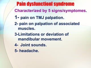
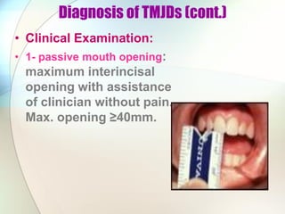
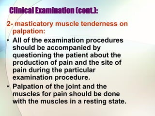
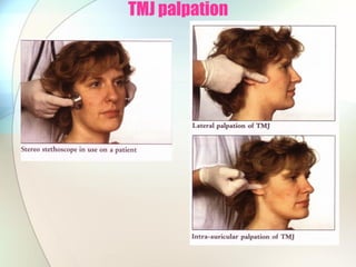
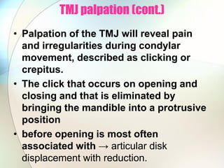
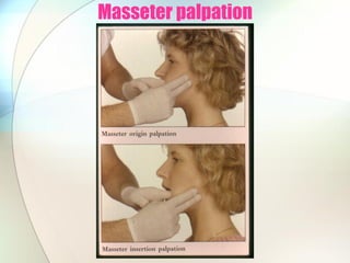
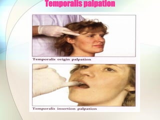
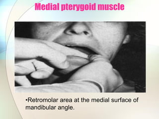
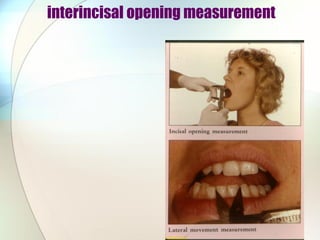
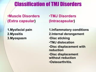
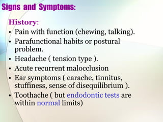
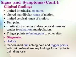
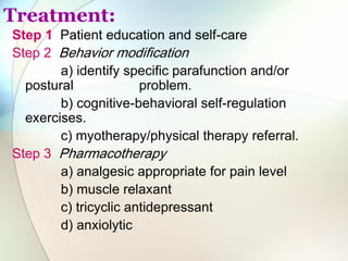
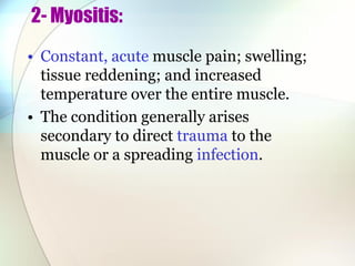
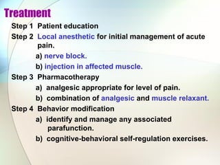
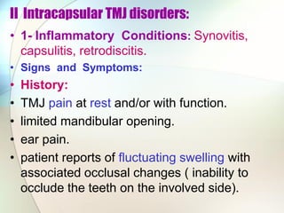
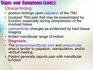
Tempromandibular Joint Diagnosis
By
Dr. Hassan M. Abouelkheir BDS, MSC, Phd.
Diagnosis of TMJDs
• History:
What are the Common Symptoms of TMJ
Disorders?
1- Headache: 80% of patients with a TMJ
disorder complain of headache, and 40%
report facial pain. Pain is often made worse
while opening and closing the jaw.
• 2- Ear pain: 50% of patients with a TMJ
disorder notice ear pain but do not have signs
of infection.
• 3- Sounds: Grinding, crunching, or popping
sounds, medically termed crepitus, are
common for patients with a TMJ disorder.
These sounds may or may not be accompanied
by increased pain.
Diagnosis of TMJDs (cont)
• 4- Dizziness: 40% of patients with a
TMJ disorder report a vague dizziness
or imbalance.
• 5- Fullness of the Ear: 33% of patients
with a TMJ disorder describe muffled,
clogged, or full ears. They may notice
ear fullness and pain during airplane
takeoffs and landings.
• 6- Ringing in the Ear - Tinnitus: For
unknown reasons, 33% of patients with
a TMJ disorder experience noise or
ringing (tinnitus).
Pain dysfunctionl syndrome
Characterized by 5 signs/symptomes.
1- pain on TMJ palpation.
2- pain on palpation of associated
muscles.
3-Limitations or deviation of
mandibular movement.
4- Joint sounds.
5- headache.
Diagnosis of TMJDs (cont.)
• Clinical Examination:
• 1- passive mouth opening:
maximum interincisal
opening with assistance
of clinician without pain.
Max. opening ≥40mm.
Clinical Examination (cont.):
2- masticatory muscle tenderness on
palpation:
• All of the examination procedures
should be accompanied by
questioning the patient about the
production of pain and the site of
pain during the particular
examination procedure.
• Palpation of the joint and the
muscles for pain should be done
with the muscles in a resting state.
TMJ palpation
TMJ palpation (cont.)
• Palpation of the TMJ will reveal pain
and irregularities during condylar
movement, described as clicking or
crepitus.
• The click that occurs on opening and
closing and that is eliminated by
bringing the mandible into a protrusive
position
• before opening is most often
associated with → articular disk
displacement with reduction.
Masseter palpation
Temporalis palpation
Lateral pterygoid muscle
•Behind the maxillary
tuberosity.
Medial pterygoid muscle
•Retromolar area at the medial surface of
mandibular angle.
interincisal opening measurement
Reduction in the vertical range of movements:
Due to conditions:
1- pain → muscular problem.
2- physical obstruction → Disc
displacement.
Deviation in movements:
Multifactorial;
A- Diagonal straight line from the beginning to
end point → Joint adhesion.
B- vertical until before the end of maximum
opening where there is deviation.→ anterior
disc displacement without reduction .
C- vertical with lateral movement at the meddle
of opening which then returns to the same
vertical plane→ Disc displacement with
reduction.
3- Computerized mandibular scan:
• Misalignment of the jaws
with upper & lower teeth
meeting in the wrong place
can be at the root of TMDs.
To trace this malocclusion
or unhealthy bite .
• The computerized
Mandibular Scan (CMS) is a
tracking device that records
in 3D the delicate
functioning movements of
the jaw with accuracy in the
tenths of a millimeter.
Radiology
• MRI is best technique for joint space
pathology
• CT is best technique for bony
pathology
• Plain films with arthrography
sometimes useful, although largely
replaced by MRI and CT
• Arthroscopy is also diagnostic
Assessment of Parafunctional Habits
• 1. Teeth grinding and teeth clenching
(bruxism) increase the wear on the
cartilage lining of the TMJ. Many patients
awaken in the morning with jaw or ear
pain.
• 2. Habitual gum chewing or fingernail
biting.
• 3- Dental problems and misalignment of
the teeth (malocclusion). Patients may
complain that it is difficult to find a
comfortable bite. Chewing on only one
side of the jaw can lead to or be a result of
TMJ problems.
Parafunctional Habits (cont.)
• 4. Trauma to the jaws. Previous history of
broken jaw or fractured facial bones.
• 5. Stress frequently leads to unreleased
nervous energy. They either consciously or
unconsciously grinding and clenching their
teeth
• 6. Occupational tasks such as holding the
telephone between the head and shoulder.
Classification of TMJ Disorders
•Muscle Disorders
(Extra capsular)
1.Myofacial pain
2.Myositis
3.Myospasm
•TMJ Disorders
(Intracapsular)
1.Inflammatory conditions
2.Internal derangement
•Disc sticking
•TMJ dislocation
•Disc displacement with
reduction
•Disc displacement
without reduction
•Osteoarthritis.
Muscle Disorders (Extracapsular):
1.Myofacial pain:
Myofacial pain as “ a regional, dull, aching
muscle pain with the presence of localized
tender sites (trigger point) in muscle, tendon,
or fascia”.
• TMD patients may have masticatory and/or
cervical myofacial pain.
Signs and Symptoms:
History:
• Pain with function (chewing, talking).
• Parafunctional habits or postural
problem.
• Headache ( tension type ).
• Acute recurrent malocclusion
• Ear symptoms ( earache, tinnitus,
stuffiness, sense of disequilibrium ).
• Toothache ( but endodontic tests are
within normal limits)
Signs and Symptoms (Cont.):
Clinical finding:
• limited interincisal opening.
• altered mandibular range of motion.
• limited cervical range of motion.
• Dull pain.
• masticatory muscles and/or cervical muscles
tender to palpation, manipulation.
• Trigger points referring pain to other sites.
• Diagnosis:
• History
• Generalized dull aching pain and trigger points
with pain referral are key findings for a myofacial
pain diagnosis.
Treatment:
Step 1 Patient education and self-care
Step 2 Behavior modification
a) identify specific parafunction and/or
postural problem.
b) cognitive-behavioral self-regulation
exercises.
c) myotherapy/physical therapy referral.
Step 3 Pharmacotherapy
a) analgesic appropriate for pain level
b) muscle relaxant
c) tricyclic antidepressant
d) anxiolytic
Treatment (cont.)
Step 4 Trigger-point management.
a) injection with local anesthetic.
b) physical therapy: vapocoolant
spray or ice and stretching.
Step 5 Orthopedic appliance therapy:
muscle relaxation splint.
2- Myositis:
• Constant, acute muscle pain; swelling;
tissue reddening; and increased
temperature over the entire muscle.
• The condition generally arises
secondary to direct trauma to the
muscle or a spreading infection.
Sign and Symptoms
History
• constant muscle pain that increases with function.
• limited mandibular opening.
• swelling and/or tissue reddening.
• history of trauma or infection.
• parafunction habit.
Clinical finding
• limited range of motion.
• tendonitis (inflammation of the tendinous
attachment of the muscle ).
• swelling and increased temperature over the
muscle.
Diagnosis:
• A localized, constant muscle pain secondary to
trauma, infection, or overuse of a muscle.
Treatment:
Step 1 If infection is diagnosed, it must be treated
with appropriate antibiotics and procedures to
eliminate the source of the infection.
Step 2 Patient education.
Step 3 Behavior modification: identify and manage
any parafunction that may be aggravating the
condition.
a) cognitive-behavioral self-regulation
exercises.
b) orthopedic appliance therapy.
Treatment (cont.):
Step 4 Pharmacotherapy: analgesics
appropriate for level pain.
a) sever pain: short-duration
narcotic analgesic.
b) moderate pain: NSAID for
analgesic and anti-inflammatory effect.
Step 5 Local anesthetic block to relieve
pain
Step 6 Local anesthetic with corticosteroid
for tendonitis
3- Myospasm:
• Acute muscle disorder characterized by a
sudden involuntary tonic muscle contraction.
• This condition is commonly referred to as
trismus.
• Myospasm is currently believed to be rare
and not a common cause of masticatory
muscle pain in patient with orofacial pain.
Signs and Symptoms
Clinical finding
• limited range of motion.
• significantly reduced interincisal opening.
• acute malocclusion.
• increased surface electromyography (EMG)
activity.
• trismus found secondary to odontogenic
infection.
• Diagnosis
• Acute pain and a significant reduction in
mandibular range of motion are key findings.
Treatment
Step 1 Patient education
Step 2 Local anesthetic for initial management of acute
pain.
a) nerve block.
b) injection in affected muscle.
Step 3 Pharmacotherapy
a) analgesic appropriate for level of pain.
b) combination of analgesic and muscle relaxant.
Step 4 Behavior modification
a) identify and manage any associated
parafunction.
b) cognitive-behavioral self-regulation exercises.
II Intracapsular TMJ disorders:
• 1- Inflammatory Conditions: Synovitis,
capsulitis, retrodiscitis.
• Signs and Symptoms:
• History:
• TMJ pain at rest and/or with function.
• limited mandibular opening.
• ear pain.
• patient reports of fluctuating swelling with
associated occlusal changes ( inability to
occlude the teeth on the involved side).
Signs and Symptoms (cont.):
Clinical finding:
• positive findings upon palpation of the TMJ
• localized TMJ pain that may be exacerbated by
function, especially during compression of the
involved tissue
• osteoarthritis changes as evidenced by hard tissue
imaging
• limited mandibular range of motion
• Diagnosis:
• The temporomandibular joint and preauricular
area is tender to palpation, manipulation, and/or
vertical loading.
• Patient generally reports pain with mandibular
function.
Treatment
Step 1 Patient education and self-care
Step 2 Pharmacotherapy
a) analgesic/NSAID for pain and inflammation
b) muscle relaxant, if muscle splinting is
determined
Step 3 Control of parafunctional behavioral activities
a) Cognitive-behavioral self-regulation exercises
Step 4 Physical therapy directed at enhancing reduction
of inflammation and ridding the area of
inflammatory mediators or by-
products
Step 5 Orthopedic appliance therapy:Stabilization
appliance.
2- Internal derangement:
• a -Disk Sticking:
• Disc sticking is an alteration in normal, smooth,
harmonious movement of the TMJ articular disc
without frank displacement.
• This condition may be secondary to an alteration in
quality or availability of the synovial fluid, resulting in
impaired lubrication of the joint.
• Altered synovial lubrication via bruxing/clenching may
cause repeated microtrauma.
• If overt trauma or macrotrauma occurs, concerns
about the potential for bleeding within the TMJ and the
development of adhesions must be addressed.
Signs and Symptoms:
History:
• occasional popping or clicking.
• jaw feels stiff in the morning, until I pop it.
• inability to open as wide as before.
Clinical finding
• intermittent painless or painful clicking ( usually on
opening only ).
• Usually experienced after a period of stasis, such as
upon awakening
• If painful, patient may exhibit limited mandibular
range of motion
Diagnosis:
• Based on intermittent, asymptomatic popping,
clicking, or stiffness in the temporomandibular joint.
• Treatment
Step 1 Patient education and self-care
Step 2 Reduction of parafunctional behavior:
cognitive- behavioral self-regulation
exercises
Step 3 If painful, analgesic/NSAID
Step 4 Physical therapy referral
a) gentle ROM exercise
b) gentle distraction and mobilization
step 5 Orthopedic appliance therapy: stabilization
appliance
b-TMJ Dislocation:
• An anatomical relationship in
which the lateral pterygoid
muscle advances the condyle
anterior and superior to the
crest of the articular eminence,
and due to muscle spasm in
the elevator muscles and/or
specific anatomical
relationships, the patient is
unable to return to a closed
position.
Signs and Symptoms:
History
• inability to close mouth without manipulation
• possible pain during the dislocation
• complaints of residual pain following return to closed
position.
Clinical finding
• radiographic evidence that the condyle is anterior and
superior to the crest of the articular eminence
• joint sounds near maximum opening, prior to
subluxation
• decreased mandibular range of motion due to
residual pain
Diagnosis
• Diagnosis of a temporomandibular dislocation is
made when the patient has opened wide and is
unable to return to a closed position.
Treatment:
Step 1 Patient education
Step 2 Manual reduction
Step 3 Pharmacotherapy
a) if pain is intolerable, use an auriculotemporal
nerve block to alleviate TMJ pain and reduce
muscle splinting.
b) intravenous sedation, if manual reduction
with nerve block is unsuccessful.
c) muscle relaxant.
d) analgesic/NSAID for residual pain.
Step 4 Avoidance training.
C- Disc Displacement with Reduction
Signs and Symptoms
History:
• reproducible joint noise (clicking) that occurs at variable
positions during mandibular opening and closing
• pain, if present, which may be constant or
precipitated/aggravated by mandibular movement
• reduced mandibular opening
Clinical finding:
• deviation to the affected side prior to click, with a return
toward midline following the click
• episodic and momentary “catching” of smooth
mandibular movement during opening that self-reduces
• limited mandibular range of motion secondary to
joint/muscle pain.
Diagnosis:
• The patient displays joint noise (clicking or popping)
on opening and closing, with or without pain, or
change in range of motion.
• Treatment:
Step 1 Patient education and self-care.
Step 2 Control of parafunctional behavior: cognitive-
behavioral self-regulation exercises.
Step 3 Restriction of mandibular function during
painful episodes.
step 4 Pharmacotherapy
a) analgesic/NSAID.
b) muscle relaxant if significant muscle
involvement is identified.
Step 5 Orthopedic appliance therapy: stabilization.
Step 6 Monitoring of patient response; assess
progression.
D-Disc Displacement Without Reduction:
Signs and Symptoms
History
• sudden onset of pain in the temporomandibular joint,
but no joint sounds
• limited mandibular opening
• prior history of TMJ clicking if not associated with
overt trauma.
Clinical finding:
• persistent, marked limited mouth opening (less than
35 mm) with a history of sudden onset
• mandibular deviation to the affected side on opening
• pain precipitated or exacerbated by forced mandibular
movement, and associated with palpation of the
affected joint
• hyperocclusion on the affected side if condition is
acute.
Diagnosis:
• Sudden, painful onset of persistent, marked limited
interincisal opening with loss of previous TMJ
sounds.
Treatment:
Step 1 Patient education and self-care
Step 2 Control of parafunctional behavior: cognitive-
behavioral self-regulation exercises
Step 3 Manual reduction/mobilization
Step 4 Pharmacotherapy
Step 5 Restriction of mandibular movement
Step 6 Orthopedic appliance therapy
a) stabilization appliance is treatment of first
choice.
b) anterior repositioning appliance if
stabilization orthodontic is not effect in
reducing pain.
e- Osteoarthritis:
DJD is non inflammatory disorder of
joints characterized by joint
deterioration and proliferation.
Signs and Symptoms:
History:
• pain with function.
• crepitus or multiple joint noises.
• trauma to the temporomandibular
joint.
• TMJ infection.
• active systemic arthritis.
Clinical finding:
• pain tenderness with palpation.
• limited range of motion with
mandibular deviation to the affected
side on opening.
• radiographic evidence of hard tissue
osteoarthritic change.
Diagnosis:
• Pain in the temporomandibular joint with palpation
and function, crepitus and radiographic evidence of
hard tissue osteoarthritic change.
• Treatment:
Step 1 Patient education and self-care
Step 2 Restriction of mandibular function
Step 3 Pharmacotherapy
a) analgesic/NSAID
b) muscle relaxant
Step 4 Control of parafunctional behavior
a) cognitive-behavioral self-reduction exercises
Step 5 Orthopedic appliance therapy
Step 6 Physical therapy
a) gentle ROM exercise
b) iontophoresis.
TMJ diagnosis

TMJ diagnosis

  • 1.
    Tempromandibular Joint Diagnosis By Dr.Hassan M. Abouelkheir BDS, MSC, Phd.
  • 2.
    Diagnosis of TMJDs •History: What are the Common Symptoms of TMJ Disorders? 1- Headache: 80% of patients with a TMJ disorder complain of headache, and 40% report facial pain. Pain is often made worse while opening and closing the jaw. • 2- Ear pain: 50% of patients with a TMJ disorder notice ear pain but do not have signs of infection. • 3- Sounds: Grinding, crunching, or popping sounds, medically termed crepitus, are common for patients with a TMJ disorder. These sounds may or may not be accompanied by increased pain.
  • 3.
    Diagnosis of TMJDs(cont) • 4- Dizziness: 40% of patients with a TMJ disorder report a vague dizziness or imbalance. • 5- Fullness of the Ear: 33% of patients with a TMJ disorder describe muffled, clogged, or full ears. They may notice ear fullness and pain during airplane takeoffs and landings. • 6- Ringing in the Ear - Tinnitus: For unknown reasons, 33% of patients with a TMJ disorder experience noise or ringing (tinnitus).
  • 4.
    Pain dysfunctionl syndrome Characterizedby 5 signs/symptomes. 1- pain on TMJ palpation. 2- pain on palpation of associated muscles. 3-Limitations or deviation of mandibular movement. 4- Joint sounds. 5- headache.
  • 5.
    Diagnosis of TMJDs(cont.) • Clinical Examination: • 1- passive mouth opening: maximum interincisal opening with assistance of clinician without pain. Max. opening ≥40mm.
  • 6.
    Clinical Examination (cont.): 2-masticatory muscle tenderness on palpation: • All of the examination procedures should be accompanied by questioning the patient about the production of pain and the site of pain during the particular examination procedure. • Palpation of the joint and the muscles for pain should be done with the muscles in a resting state.
  • 7.
  • 8.
    TMJ palpation (cont.) •Palpation of the TMJ will reveal pain and irregularities during condylar movement, described as clicking or crepitus. • The click that occurs on opening and closing and that is eliminated by bringing the mandible into a protrusive position • before opening is most often associated with → articular disk displacement with reduction.
  • 9.
  • 10.
  • 11.
    Lateral pterygoid muscle •Behindthe maxillary tuberosity.
  • 12.
    Medial pterygoid muscle •Retromolararea at the medial surface of mandibular angle.
  • 13.
  • 14.
    Reduction in thevertical range of movements: Due to conditions: 1- pain → muscular problem. 2- physical obstruction → Disc displacement. Deviation in movements: Multifactorial; A- Diagonal straight line from the beginning to end point → Joint adhesion. B- vertical until before the end of maximum opening where there is deviation.→ anterior disc displacement without reduction . C- vertical with lateral movement at the meddle of opening which then returns to the same vertical plane→ Disc displacement with reduction.
  • 15.
    3- Computerized mandibularscan: • Misalignment of the jaws with upper & lower teeth meeting in the wrong place can be at the root of TMDs. To trace this malocclusion or unhealthy bite . • The computerized Mandibular Scan (CMS) is a tracking device that records in 3D the delicate functioning movements of the jaw with accuracy in the tenths of a millimeter.
  • 16.
    Radiology • MRI isbest technique for joint space pathology • CT is best technique for bony pathology • Plain films with arthrography sometimes useful, although largely replaced by MRI and CT • Arthroscopy is also diagnostic
  • 17.
    Assessment of ParafunctionalHabits • 1. Teeth grinding and teeth clenching (bruxism) increase the wear on the cartilage lining of the TMJ. Many patients awaken in the morning with jaw or ear pain. • 2. Habitual gum chewing or fingernail biting. • 3- Dental problems and misalignment of the teeth (malocclusion). Patients may complain that it is difficult to find a comfortable bite. Chewing on only one side of the jaw can lead to or be a result of TMJ problems.
  • 18.
    Parafunctional Habits (cont.) •4. Trauma to the jaws. Previous history of broken jaw or fractured facial bones. • 5. Stress frequently leads to unreleased nervous energy. They either consciously or unconsciously grinding and clenching their teeth • 6. Occupational tasks such as holding the telephone between the head and shoulder.
  • 19.
    Classification of TMJDisorders •Muscle Disorders (Extra capsular) 1.Myofacial pain 2.Myositis 3.Myospasm •TMJ Disorders (Intracapsular) 1.Inflammatory conditions 2.Internal derangement •Disc sticking •TMJ dislocation •Disc displacement with reduction •Disc displacement without reduction •Osteoarthritis.
  • 20.
    Muscle Disorders (Extracapsular): 1.Myofacialpain: Myofacial pain as “ a regional, dull, aching muscle pain with the presence of localized tender sites (trigger point) in muscle, tendon, or fascia”. • TMD patients may have masticatory and/or cervical myofacial pain.
  • 21.
    Signs and Symptoms: History: •Pain with function (chewing, talking). • Parafunctional habits or postural problem. • Headache ( tension type ). • Acute recurrent malocclusion • Ear symptoms ( earache, tinnitus, stuffiness, sense of disequilibrium ). • Toothache ( but endodontic tests are within normal limits)
  • 22.
    Signs and Symptoms(Cont.): Clinical finding: • limited interincisal opening. • altered mandibular range of motion. • limited cervical range of motion. • Dull pain. • masticatory muscles and/or cervical muscles tender to palpation, manipulation. • Trigger points referring pain to other sites. • Diagnosis: • History • Generalized dull aching pain and trigger points with pain referral are key findings for a myofacial pain diagnosis.
  • 23.
    Treatment: Step 1 Patienteducation and self-care Step 2 Behavior modification a) identify specific parafunction and/or postural problem. b) cognitive-behavioral self-regulation exercises. c) myotherapy/physical therapy referral. Step 3 Pharmacotherapy a) analgesic appropriate for pain level b) muscle relaxant c) tricyclic antidepressant d) anxiolytic
  • 24.
    Treatment (cont.) Step 4Trigger-point management. a) injection with local anesthetic. b) physical therapy: vapocoolant spray or ice and stretching. Step 5 Orthopedic appliance therapy: muscle relaxation splint.
  • 25.
    2- Myositis: • Constant,acute muscle pain; swelling; tissue reddening; and increased temperature over the entire muscle. • The condition generally arises secondary to direct trauma to the muscle or a spreading infection.
  • 26.
    Sign and Symptoms History •constant muscle pain that increases with function. • limited mandibular opening. • swelling and/or tissue reddening. • history of trauma or infection. • parafunction habit. Clinical finding • limited range of motion. • tendonitis (inflammation of the tendinous attachment of the muscle ). • swelling and increased temperature over the muscle.
  • 27.
    Diagnosis: • A localized,constant muscle pain secondary to trauma, infection, or overuse of a muscle. Treatment: Step 1 If infection is diagnosed, it must be treated with appropriate antibiotics and procedures to eliminate the source of the infection. Step 2 Patient education. Step 3 Behavior modification: identify and manage any parafunction that may be aggravating the condition. a) cognitive-behavioral self-regulation exercises. b) orthopedic appliance therapy.
  • 28.
    Treatment (cont.): Step 4Pharmacotherapy: analgesics appropriate for level pain. a) sever pain: short-duration narcotic analgesic. b) moderate pain: NSAID for analgesic and anti-inflammatory effect. Step 5 Local anesthetic block to relieve pain Step 6 Local anesthetic with corticosteroid for tendonitis
  • 29.
    3- Myospasm: • Acutemuscle disorder characterized by a sudden involuntary tonic muscle contraction. • This condition is commonly referred to as trismus. • Myospasm is currently believed to be rare and not a common cause of masticatory muscle pain in patient with orofacial pain.
  • 30.
    Signs and Symptoms Clinicalfinding • limited range of motion. • significantly reduced interincisal opening. • acute malocclusion. • increased surface electromyography (EMG) activity. • trismus found secondary to odontogenic infection. • Diagnosis • Acute pain and a significant reduction in mandibular range of motion are key findings.
  • 31.
    Treatment Step 1 Patienteducation Step 2 Local anesthetic for initial management of acute pain. a) nerve block. b) injection in affected muscle. Step 3 Pharmacotherapy a) analgesic appropriate for level of pain. b) combination of analgesic and muscle relaxant. Step 4 Behavior modification a) identify and manage any associated parafunction. b) cognitive-behavioral self-regulation exercises.
  • 32.
    II Intracapsular TMJdisorders: • 1- Inflammatory Conditions: Synovitis, capsulitis, retrodiscitis. • Signs and Symptoms: • History: • TMJ pain at rest and/or with function. • limited mandibular opening. • ear pain. • patient reports of fluctuating swelling with associated occlusal changes ( inability to occlude the teeth on the involved side).
  • 33.
    Signs and Symptoms(cont.): Clinical finding: • positive findings upon palpation of the TMJ • localized TMJ pain that may be exacerbated by function, especially during compression of the involved tissue • osteoarthritis changes as evidenced by hard tissue imaging • limited mandibular range of motion • Diagnosis: • The temporomandibular joint and preauricular area is tender to palpation, manipulation, and/or vertical loading. • Patient generally reports pain with mandibular function.
  • 34.
    Treatment Step 1 Patienteducation and self-care Step 2 Pharmacotherapy a) analgesic/NSAID for pain and inflammation b) muscle relaxant, if muscle splinting is determined Step 3 Control of parafunctional behavioral activities a) Cognitive-behavioral self-regulation exercises Step 4 Physical therapy directed at enhancing reduction of inflammation and ridding the area of inflammatory mediators or by- products Step 5 Orthopedic appliance therapy:Stabilization appliance.
  • 36.
    2- Internal derangement: •a -Disk Sticking: • Disc sticking is an alteration in normal, smooth, harmonious movement of the TMJ articular disc without frank displacement. • This condition may be secondary to an alteration in quality or availability of the synovial fluid, resulting in impaired lubrication of the joint. • Altered synovial lubrication via bruxing/clenching may cause repeated microtrauma. • If overt trauma or macrotrauma occurs, concerns about the potential for bleeding within the TMJ and the development of adhesions must be addressed.
  • 37.
    Signs and Symptoms: History: •occasional popping or clicking. • jaw feels stiff in the morning, until I pop it. • inability to open as wide as before. Clinical finding • intermittent painless or painful clicking ( usually on opening only ). • Usually experienced after a period of stasis, such as upon awakening • If painful, patient may exhibit limited mandibular range of motion
  • 38.
    Diagnosis: • Based onintermittent, asymptomatic popping, clicking, or stiffness in the temporomandibular joint. • Treatment Step 1 Patient education and self-care Step 2 Reduction of parafunctional behavior: cognitive- behavioral self-regulation exercises Step 3 If painful, analgesic/NSAID Step 4 Physical therapy referral a) gentle ROM exercise b) gentle distraction and mobilization step 5 Orthopedic appliance therapy: stabilization appliance
  • 39.
    b-TMJ Dislocation: • Ananatomical relationship in which the lateral pterygoid muscle advances the condyle anterior and superior to the crest of the articular eminence, and due to muscle spasm in the elevator muscles and/or specific anatomical relationships, the patient is unable to return to a closed position.
  • 40.
    Signs and Symptoms: History •inability to close mouth without manipulation • possible pain during the dislocation • complaints of residual pain following return to closed position. Clinical finding • radiographic evidence that the condyle is anterior and superior to the crest of the articular eminence • joint sounds near maximum opening, prior to subluxation • decreased mandibular range of motion due to residual pain
  • 41.
    Diagnosis • Diagnosis ofa temporomandibular dislocation is made when the patient has opened wide and is unable to return to a closed position. Treatment: Step 1 Patient education Step 2 Manual reduction Step 3 Pharmacotherapy a) if pain is intolerable, use an auriculotemporal nerve block to alleviate TMJ pain and reduce muscle splinting. b) intravenous sedation, if manual reduction with nerve block is unsuccessful. c) muscle relaxant. d) analgesic/NSAID for residual pain. Step 4 Avoidance training.
  • 42.
    C- Disc Displacementwith Reduction
  • 43.
    Signs and Symptoms History: •reproducible joint noise (clicking) that occurs at variable positions during mandibular opening and closing • pain, if present, which may be constant or precipitated/aggravated by mandibular movement • reduced mandibular opening Clinical finding: • deviation to the affected side prior to click, with a return toward midline following the click • episodic and momentary “catching” of smooth mandibular movement during opening that self-reduces • limited mandibular range of motion secondary to joint/muscle pain.
  • 44.
    Diagnosis: • The patientdisplays joint noise (clicking or popping) on opening and closing, with or without pain, or change in range of motion. • Treatment: Step 1 Patient education and self-care. Step 2 Control of parafunctional behavior: cognitive- behavioral self-regulation exercises. Step 3 Restriction of mandibular function during painful episodes. step 4 Pharmacotherapy a) analgesic/NSAID. b) muscle relaxant if significant muscle involvement is identified. Step 5 Orthopedic appliance therapy: stabilization. Step 6 Monitoring of patient response; assess progression.
  • 45.
  • 46.
    Signs and Symptoms History •sudden onset of pain in the temporomandibular joint, but no joint sounds • limited mandibular opening • prior history of TMJ clicking if not associated with overt trauma. Clinical finding: • persistent, marked limited mouth opening (less than 35 mm) with a history of sudden onset • mandibular deviation to the affected side on opening • pain precipitated or exacerbated by forced mandibular movement, and associated with palpation of the affected joint • hyperocclusion on the affected side if condition is acute.
  • 47.
    Diagnosis: • Sudden, painfulonset of persistent, marked limited interincisal opening with loss of previous TMJ sounds. Treatment: Step 1 Patient education and self-care Step 2 Control of parafunctional behavior: cognitive- behavioral self-regulation exercises Step 3 Manual reduction/mobilization Step 4 Pharmacotherapy Step 5 Restriction of mandibular movement Step 6 Orthopedic appliance therapy a) stabilization appliance is treatment of first choice. b) anterior repositioning appliance if stabilization orthodontic is not effect in reducing pain.
  • 48.
    e- Osteoarthritis: DJD isnon inflammatory disorder of joints characterized by joint deterioration and proliferation. Signs and Symptoms: History: • pain with function. • crepitus or multiple joint noises. • trauma to the temporomandibular joint. • TMJ infection. • active systemic arthritis. Clinical finding: • pain tenderness with palpation. • limited range of motion with mandibular deviation to the affected side on opening. • radiographic evidence of hard tissue osteoarthritic change.
  • 49.
    Diagnosis: • Pain inthe temporomandibular joint with palpation and function, crepitus and radiographic evidence of hard tissue osteoarthritic change. • Treatment: Step 1 Patient education and self-care Step 2 Restriction of mandibular function Step 3 Pharmacotherapy a) analgesic/NSAID b) muscle relaxant Step 4 Control of parafunctional behavior a) cognitive-behavioral self-reduction exercises Step 5 Orthopedic appliance therapy Step 6 Physical therapy a) gentle ROM exercise b) iontophoresis.