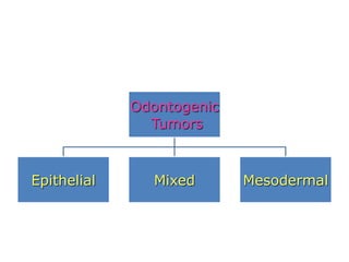
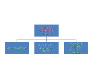
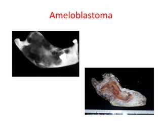
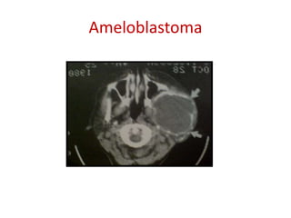
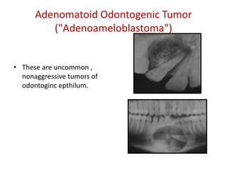
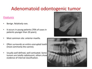
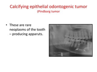
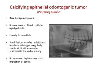
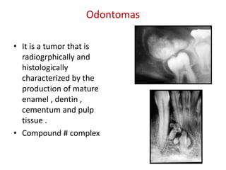
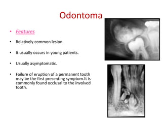
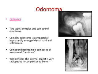
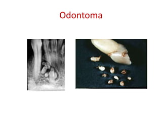
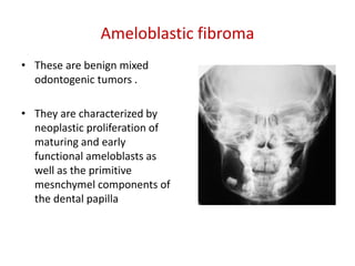
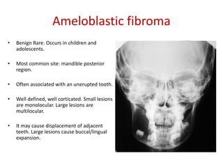
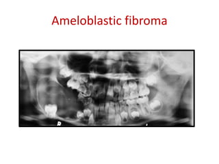
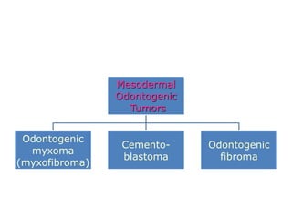
This document summarizes several odontogenic tumors including: ameloblastoma (benign, locally aggressive tumor of odontogenic epithelium that commonly occurs in the mandible), adenomatoid odontogenic tumor (uncommon, nonaggressive tumor that often surrounds an unerupted tooth), calcifying epithelial odontogenic tumor (rare benign neoplasm that usually occurs in the mandible), odontoma (tumor characterized by production of dental tissues that commonly occurs in young patients), ameloblastic fibroma (benign mixed odontogenic tumor that occurs in children/adolescents), odontogenic myxoma (benign tumor arising from dental papilla mesenchyme), cementoblast
Central Giant Cell Granuloma :
WHO has defined it as an intraosseous lesion consisting of cellular and fibrous tissue that contains multiple foci of hemorrhage, aggregation of multinucleated giant cells and occasionally trabaculae of woven bone
Etiology JAFFE (1953): considered this lesion to be a local reparative reaction of bone, possibly to intramedullary hemorrhage or trauma, hence the term reparative giant cell granuloma was accepted.
Charles A Waldron & W G Shafer (1966) suggested trauma be an important etiological factor in the initiation of the CGCG of the jaws
Thoma K H (1986) suggested that the lesion may be due to capillary injury caused by defective wall due to some type of trauma
J V Soames and J C Southam (1997) suggested that it could be a reaction to some form of hemodynamic disturbance in bone marrow, perhaps associated with trauma and hemorrhage REGEZI AND SCIUBBA(1999) :
Suggested that
Response to previous traumatic or inflammatory episodes.
This lesion is charecterised by proliferation of fibroblasts and multinucleated giant cells, in a densely packed stromaThe CGCG is a benign process that occurs almost exclusively within the jaw bones
CLINICAL PRESENTATION
Found predominantly in children and young adult
It has a female predilection (2:1)
Most commonly affected site is the anterior portion of the jaws, with an increased frequency of occurrence in the mandible
Majority of the CGCG of jaws are painless, expansion of bone is detected on routine examination
Few cases may be associated with pain, paresthesia or perforation of a cortical bone plate, occasionally resulting in the ulceration of the mucosal surface by the underlying lesion
Radiographic featuresCentral giant cell lesions present as radiolucent defects. Which may be unilocular or multilocular.
The defect is usually well delineated
The lesion may vary from a 5×5mm incidental radiographic findings to a destructive lesion greater than 10cm in size.
radiographic findings
A small unilocular lesion may be confused with periapical granuloma or cysts.
multilocular giant cell lesions cannot be radiographically distinguished from ameloblastomas or other multilocular lesions. Based on clinical and radiological features CGCG may be divided into two categories
- Non-aggressive lesion
- Aggressive lesion
The non aggressive lesion makes up most cases and exhibit few or no symptoms, they demonstrate slow growth and do not show cortical perforation or root resorption of teeth involved in the lesion. The aggressive lesions are characterized by pain, rapid growth, cortical perforation and root resorption and show marked tendency to recur when compared with non aggressive typeSoft spongy, brownish to reddish friable tissue of various size.
A specimen is usually coated with fresh or coagulated blood. Giant cell lesions of the jaws show a variety of features. Common to all is the presence of few to many multinucleated
A cyst is an epithelium-lined sac containing fluid or semisolid material. In the formation of a cyst, the epithelial cells first proliferate and later undergo degeneration and liquefaction. The liquefied material exerts equal pressure on the walls of the cyst from within. Cysts grow by expansion and thus displace the adjacent teeth by pressure. May can produce expansion of the cortical bone. On a radiograph, the radiolucency of a cyst is usually bordered by a radiopaque periphery of dense sclerotic bone. The radiolucency may be unilocular or multilocular. Odontogenic cysts are those which arise from the epithelium associated with the development of teeth. The source of epithelium is from the enamel organ, the reduced enamel epithelium, the cell rests of Malassez or the remnants of the dental lamina.
Fibro-osseous lesions of the jaws
Fibrous dysplasia
Cemento-osseous dysplasia
Focal cemento-osseous dysplasia
Periapical cemento-osseous dysplasia
Florid cemento-osseous dysplasia
Ossifying fibroma
Juvenile aggressive ossifying fibroma
Cherubism
Fibro-osseous lesions (FOL) are characterized by replacement of normal bone architecture by collagen fibers and fibroblasts containing calcified tissue.
They include a wide variety of lesions of developmental, dysplastic and neoplastic origins with clinical and radiographic presentation and behavior.
Because of the histological similarities between diverse diseases, proper diagnosis requires correlation of history, clinical and radiographic findings.Fibrous Dysplasia
2. Reactive (dysplastic lesions arising in the tooth-bearing area (presumably of periodontal origin).
a. Periapical cemento-osseous dysplasia
b. Focal cemento-osseous dysplasia
c. Florid cemento-osseous dysplasia
3. Fibro-osseous neoplasms (widely designated as cementifying fibroma, ossifying fibroma or cemento-ossifying fibroma.Bone dysplasias
a. Fibrous dyspla i. Monostoticii. Polyostotic
iii. Polyostotic with endocrinopathy (McCune-Albright)
iv Osteofibrous dysplasia
b. Osteitis deformansc. Pagetoid heritable bone dysplasias of childhood
d. Segmental odontomaxillary dysplasia
2. Cemento-osseous dysplasias
a. Focal cemento-osseous dysplasia b. Florid cemento-osseous dysplasia
3.Inflammatory/reactive processes
a. Focal sclerosing osteomyelitisb. Diffuse sclerosing osteomyelitis
c. Proliferative periostitis
4. Metabolic Disease: hyperparathyroidism
5. Neoplastic lesions (Ossifying fibromas)
a. Ossifying fibromab. Hyperparathyroidism jaw lesion syndrome
c. Juvenile ossifying fibroma i. Trabecular typeii. Psammomatoid type
d. Gigantiform cementomas
Odontogenic keratocyst (OKC) is the cyst arising from the cell rests of dental lamina. It can occur anywhere in the jaw, but commonly seen in the posterior part of the mandible. Radiographically, most OKCs are unilocular when presented at the periapex and can be mistaken for radicular or lateral periodontal cyst.
Odontogenic tumors v / dental implant courses by Indian dental academy Indian dental academy
The Indian Dental Academy is the Leader in continuing dental education , training dentists in all aspects of dentistry and
offering a wide range of dental certified courses in different formats.for more details please visit
www.indiandentalacademy.com
Odontogenic tumors are growths that develop in the jawbones or soft tissues of the mouth, arising from the tissues that form teeth. These tumors can be benign or malignant and vary widely in their presentation and behavior. Benign tumors include ameloblastoma, odontoma, and cementoblastoma, while malignant tumors include ameloblastic carcinoma and odontogenic sarcoma. Treatment typically involves surgical removal, and prognosis depends on the type and stage of the tumor.
Cementoblastoma is defined as a neoplasm characterized by formation of sheets of cementum like tissue containing a large number of reversal lines and lack of mineralization at the periphery of the mass or in the more active growth area. Locally aggressive resulting in bony expansion, root resorption, displacement of adjacent teeth, and jaw deformity.
Jaw lesion radiology ppt ppt . This powerpoint presentation includes important anatomy, radiographs and important pathology of jaw lesion with its imaging feature as well as its Xray ct mri image. This will help alot. this will help for radiology resident as well as ent resident and event dentist.
Odontogenic tumors vi/certified fixed orthodontic courses by Indian dental ac...Indian dental academy
The Indian Dental Academy is the Leader in continuing dental education , training dentists in all aspects of dentistry and
offering a wide range of dental certified courses in different formats.for more details please visit
www.indiandentalacademy.com
Benig tumors of jaw/certified fixed orthodontic courses by Indian dental acad...Indian dental academy
The Indian Dental Academy is the Leader in continuing dental education , training dentists in all aspects of dentistry and
offering a wide range of dental certified courses in different formats.
4. Ameloblastoma
• This a true neoplasm of
odontogenic epithelium
• It is an aggressive neoplasm
the arises from the
remnants of the dental
lamina and dental organ(
odontogenic epithelium)
5. Ameloblastoma
• Benign, locally aggressive odontogenic
tumor. Usually it slowly grows as
painless swelling of the affected site.
• It can occur at any age.
• Localized invasion into the
surrounding bone.
• 80-95% in the mandible (posterior
body, ramus region). In the maxilla
mostly in the premolar-molar region.
6. Ameloblastoma
• Unilocular (small lesions). Multilocular
(large discrete areas or honeycomb
appearance)
• Smooth, well-defined, well-corticated
margins
• Adjacent teeth are often displaced
and resorbed.
• It causes extensive bone expansion.
• Incomplete removal can result in
recurrence.
9. Adenomatoid Odontogenic Tumor
("Adenoameloblastoma")
• These are uncommon ,
nonaggressive tumors of
odontoginc epthilum.
10. Adenomatoid odontogenic tumor
Features
• Benign. Relatively rare.
• It occurs in young patients (70% of cases in
patients younger than 20 years).
• Most common site: anterior maxilla.
• Often surrounds an entire unerupted tooth
(most commonly the canine).
• Usually well defined, well corticated. Some
tumors are totally radiolucent; others show
evidence of internal classification.
12. Calcifying epithelial odontogenic tumor
(Pindborg tumor
• Rare benign neoplasm.
• It occurs more often in middle-
aged patients.
• Usually in mandible.
• Small lesions may be radiolucent.
In advanced stages irregularly
sized calcifications may be
scattered in the radiolucency.
• It can cause displacement and
impaction of teeth.
13. Odontomas
• It is a tumor that is
radiogrphically and
histologically
characterized by the
production of mature
enamel , dentin ,
cementum and pulp
tissue .
• Compound # complex
14. Odontoma
• Features
• Relatively common lesion.
• It usually occurs in young patients.
• Usually asymptomatic.
• Failure of eruption of a permanent tooth
may be the first presenting symptom.It is
commonly found occlusal to the involved
tooth.
15. Odontoma
• Features
• Two types: complex and compound
odontoma.
• Complex odontoma is composed of
haphazardly arranged dental hard and
soft tissues.
• Compound odontoma is composed of
many small "denticles" .
• Well defined. The internal aspect is very
radiopaque in comparison to bone.
17. Ameloblastic fibroma
• These are benign mixed
odontogenic tumors .
• They are characterized by
neoplastic proliferation of
maturing and early
functional ameloblasts as
well as the primitive
mesnchymel components of
the dental papilla
18. Ameloblastic fibroma
• Benign Rare. Occurs in children and
adolescents.
• Most common site: mandible posterior
region.
• Often associated with an unerupted tooth.
• Well defined, well corticated. Small lesions
are monolocular. Large lesions are
multilocular.
• It may cause displacement of adjacent
teeth. Large lesions cause buccal/lingual
expansion.
20. Ameloblastic
fibro-odontoma
• This is an extremely rare lesion. It consists of
elements of ameloblastic fibroma with small
segments of enamel and dentin.
22. Odontogenic myxoma (myxofibroma)
• They are benign,
intraosseous neoplasms
that arise from the
mesenchymal portion
of the dental papilla.
23. Odontogenic myxoma (myxofibroma)
• Features
• It represents approximately 3 - 6% of
all odontogenic tumors. It is painless
and grows slowly.
• It can occur at any age but most
commonly in the second and third
decades of life.
• More often affect the mandible
(molar/premolar region).
24. Odontogenic myxoma (myxofibroma)
• Features
• Typically multilocular (internal septa-
strings of a tennis racket or
honeycomb appearance). Large
lesions can have the sun ray
appearance of an osteosarcoma.
• Often well-defined.
• Adjacent teeth can be displaced but
rarely resorbed. It causes less bone
expansion than in other benign
tumors.
27. Cementoblastoma
• Features
• Benign neoplasm. Most commonly in
the second and third decade.
• Site: usually mandibular premolar
and molar regions.
• Attached to the root of the affected
tooth. Tooth displacement,
resorption are common.
• Pain in 50% of the cases, swelling.
• When radiopaque is usually
surrounded by a thin radiolucent
halo.
28. Radiographic Features
• Location:
• Periphery: well defined RO with
RL hallo surrounding the calcified
mass.
• Internal structure: mixed RL-RO
leseions may be amorphous
• Effect on surrounding tissues:
expansion, external root
resorption
30. Odontogenic
fibroma
• Features
• Rare neoplasm. More often between
the ages 10 and 40 years.
• Asymptomatic or swelling and tooth
mobility
• More common sites: mandible
(premolar-molar region), maxilla
(anterior region)
• Small lesions are usually unilocular, and
larger lesions multilocular.
• Well-defined margins.
• Adjacent teeth: often displaced,
impaction, root resorption.