Cemento osseus dysplasia (Doctor Faris Alabeedi MSc, MMedSc, PgDip, BDS.)
•Download as PPTX, PDF•
19 likes•7,065 views

.

Recommended
More Related Content
What's hot
What's hot (20)
Similar to Cemento osseus dysplasia (Doctor Faris Alabeedi MSc, MMedSc, PgDip, BDS.)
Similar to Cemento osseus dysplasia (Doctor Faris Alabeedi MSc, MMedSc, PgDip, BDS.) (20)
More from Doctor Faris Alabeedi
More from Doctor Faris Alabeedi (15)
Recently uploaded
Recently uploaded (20)
Cemento osseus dysplasia (Doctor Faris Alabeedi MSc, MMedSc, PgDip, BDS.)
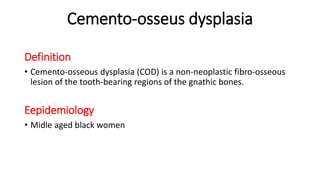
- 1. Cemento-osseus dysplasia Definition • Cemento-osseous dysplasia (COD) is a non-neoplastic fibro-osseous lesion of the tooth-bearing regions of the gnathic bones. Eepidemiology • Midle aged black women
- 2. 1-Periapical cemento-osseous dysplasia (PCOD) 2-Focal cemento-osseous dysplasia (FCOD) two different terms for the same reactive lesion, that represent the most common fibro-osseous lesions of the jaws All of the cemento-osseous dysplasias have similar/same microscopic appearance
- 3. 3-Florid cemento-osseous dysplasia (Fl.COD) denotes an extensive process with multifocal involvement of the jaws by lesional tissue • Bbasically When lesions with radiologic and microscopic features similar to FCOD/PCOD extend to two or more quadrants of the jaw, the disease is termed florid cemento-osseous dysplasia
- 4. divided into three variants (largely on the basis on anatomical location): periapical COD is associated with the apical areas of mandibular anterior teeth. Anterior mandibule focal COD is associated with a single tooth. Posterior mandibule florid COD has multifocal (multiquadrant) involvement. Posterior mandibule
- 5. Clinically • Asymptomatic. Except, Florid if inflamed lead to pain and discharge • Associated with vital teeth ‘might be found in edentulous areas’ • All are non-expansive. Except, Florid variant • Tooth bearing region of jaw • 1.5 cm • Mandible 86% • Bilateral in FlCOD
- 6. Radiograph ‘essential’ A focus of COD is generally well defined and demonstrates a thin radiolucent rim. The periodontal ligament should appear intact, and the lesion should not be fused to the roots • If identified clinically and radiographically, biopsy might not be needed
- 7. Three stages of Radiographic appearance • Early stage: well-defined radiolucency at the apices of mandibular teeth • intermediate stage: mixed radiolucent-opaque pattern with a well defined radiolucent rim around the radiopacity • late stage: diffuse radiopacity often with ill-defined borders.
- 8. (a) Periapical Cemental Dysplasia (PACD), a subtype of focal cemento- osseous dysplasia of the anterior mandible begins as a periapical radiolucency. (b) PACD opacifies forming aradiographic ‘‘target lesion’’ in later stages of the disease.
- 9. (a) Multiple confluent opacities in all four jaw quadrants. (b). Bilateral radiolucent and mixed lucent/opaque
- 10. Histopathology • characterized by a cellular fibrous stroma that have swirling and/or loose collagen and areas of vascularity • the stroma are mineralizing tissues: 1-osteoid trabeculae that is occasionally surrounded by osteoblasts , 2-bone 3-cementum like material “cementicles’’ or ‘‘bonicles” • As the lesions mature, they become increasingly calcified displaying very little fibrotic stroma with thick curvilinear trabeculae (‘‘ginger root’’ pattern) or irregularly shaped cementum-like masses
- 11. fibrous tissue to dense mineralized bone trabeculae with peripheral osteoblasts and cementum-like deposits
- 12. (a) Early stage lesion with hemorrhagic foci (b) Early region with fibroosseous pattern (c) Mid stage lesion with progressively more trabeculae
- 13. mature lesion with a trabecular bone pattern predominantly fibroblastic proliferative stroma associated with sparse bone deposition Florid osseous dysplasia: area of dense bone, inflammation and hemorrhage
- 14. Changes might occur in florid • hypocellular sclerotic masses may form • Inflammation • Cystic changes resembling simple bone cyst may occur
- 15. Refrances: • (Eversole, Su and ElMofty, 2008; Zegalie, Speight and Martin, 2015)Eversole, R., Su, L. and ElMofty, S. (2008) ‘Benign fibro-osseous lesions of the craniofacial complex a review’, Head and Neck Pathology, 2(3), pp. 177–202. doi: 10.1007/s12105-008-0057-2. • De Noronha Santos Netto, J. et al. (2013) ‘Benign fibro-osseous lesions: Clinicopathologic features from 143 cases diagnosed in an oral diagnosis setting’, Oral Surgery, Oral Medicine, Oral Pathology and Oral Radiology. Elsevier, 115(5), pp. e56–e65. doi: 10.1016/j.oooo.2012.05.022. • Zegalie, N., Speight, P. M. and Martin, L. (2015) ‘Ossifying fibromas of the jaws and craniofacial bones’, Diagnostic Histopathology. Elsevier Ltd, 21(9), pp. 351–358. doi: 10.1016/j.mpdhp.2015.07.004. • WHO Classification of Head and Neck Tumours, 4ed, (2017)