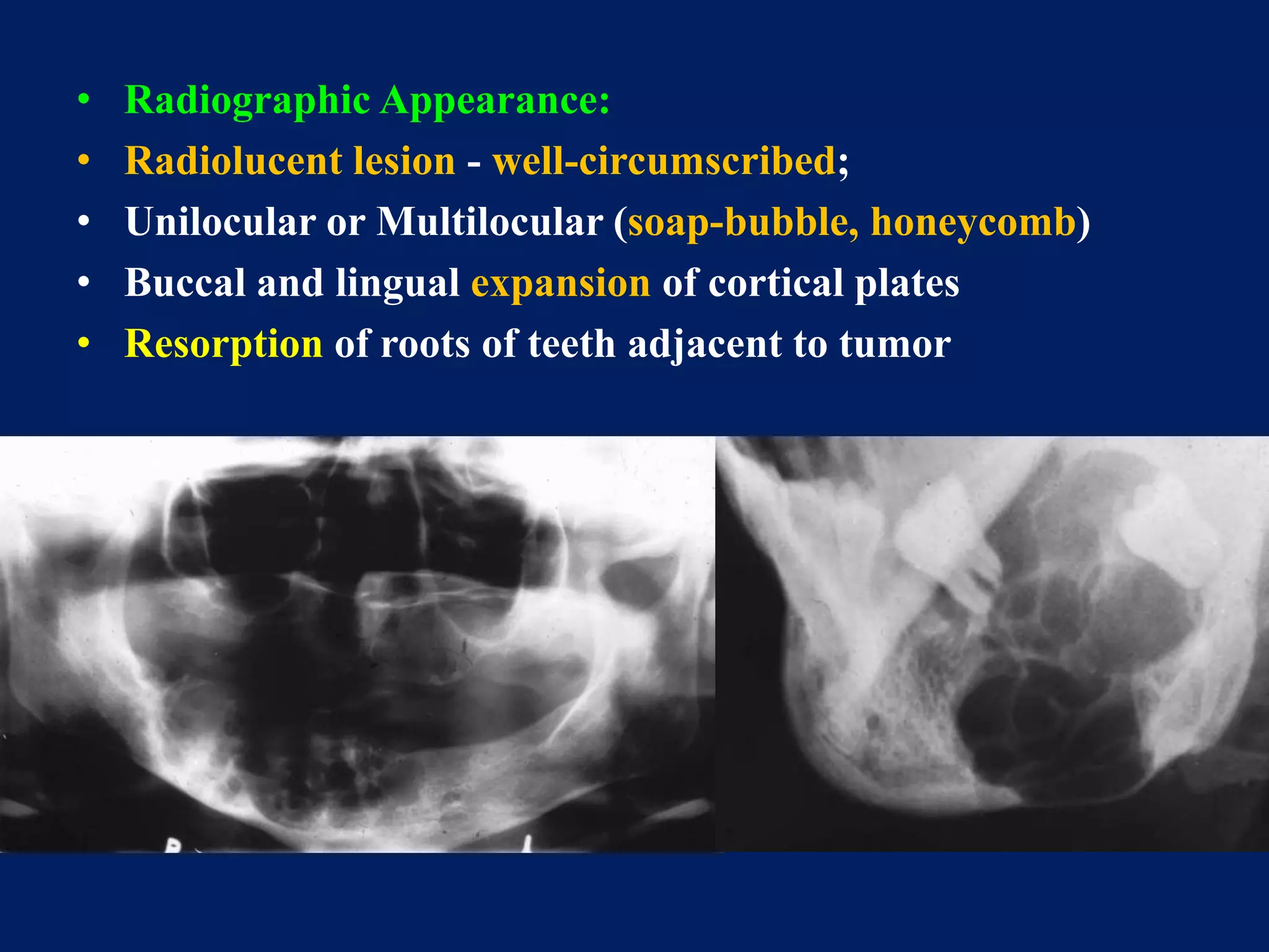
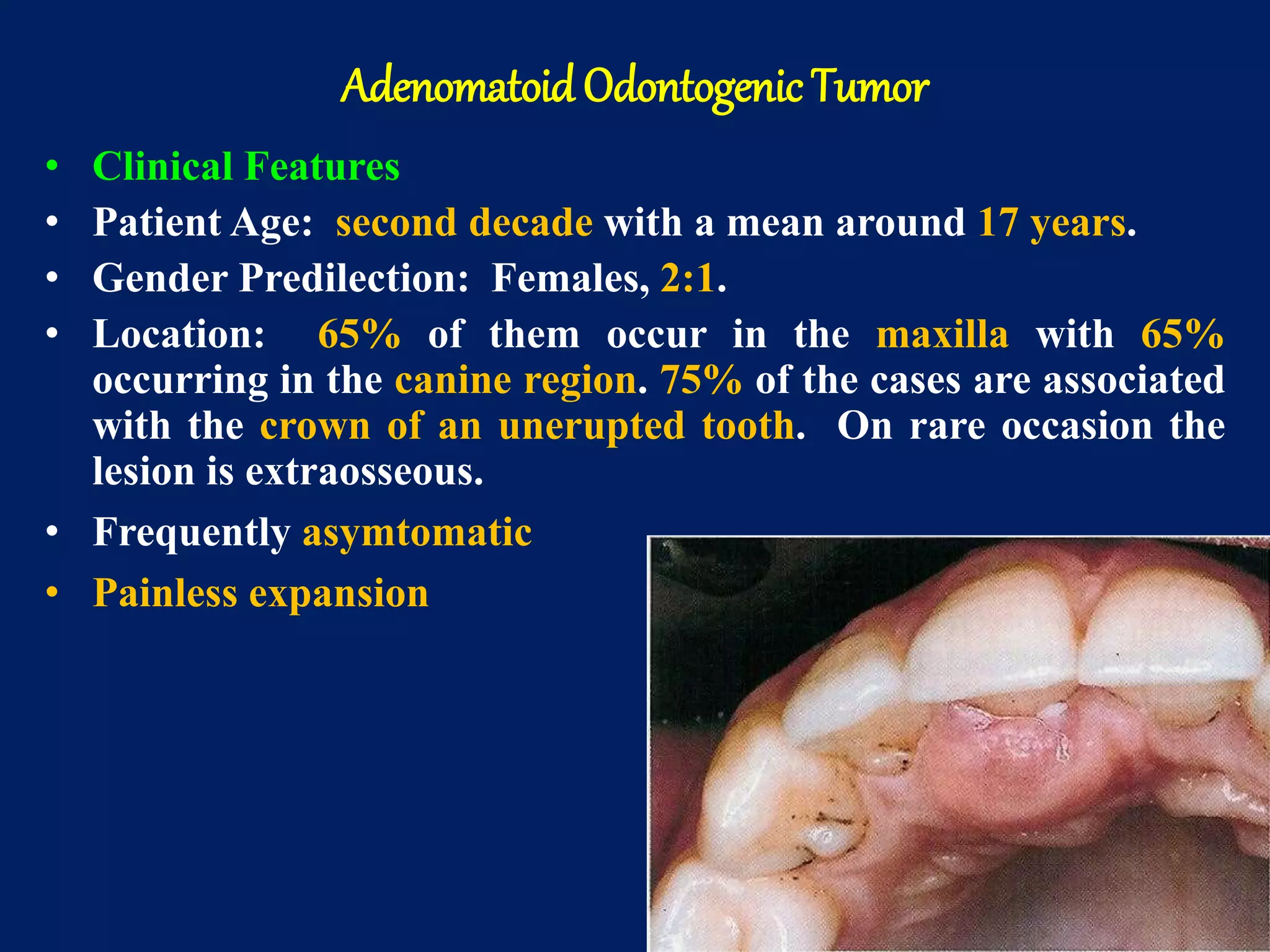
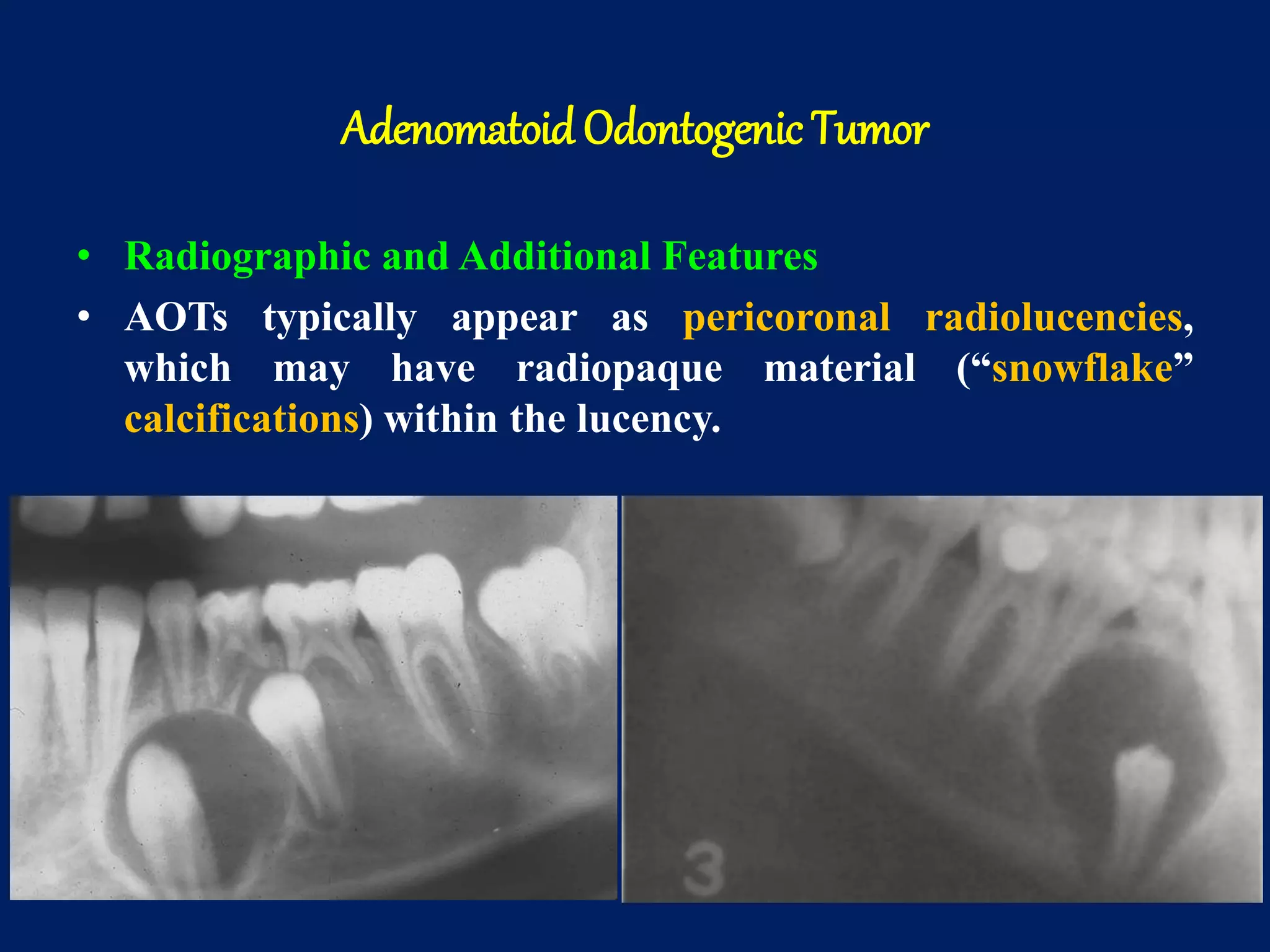
Odontogenic Tumors
DR.MADHUSUDHANREDDY
Armycollege of dentalsciences
secundrabad
Classification of Odontogenic Tumors
– I. Tumors of odontogenic epithelium
» A. Ameloblastoma: 1. Malignant ameloblastoma
2. Ameloblastic carcinoma
» B. Clear cell odontogenic carcinoma
» C. Adenomatoid odontogenic tumor
» D. Calcifying epithelial odontogenic tumor
» E. Squamous odontogenic tumor
– II. Mixed odontogenic tumors
» A. Ameloblastic fibroma
» B. Ameloblastic fibro-odontoma
» C. Ameloblastic fibrosarcoma
» D. Odontoameloblastoma
» E. Compound odontoma
» F. Complex odontoma
– III. Tumors of odontogenic ectomesenchyme
» A. Odontogenic fibroma
» B. Granular cell odontogenic tumor
» C. Odontogenic myxoma
» D. Cementoblastoma
• * From Neville, et al.
Tumors of Odontogenic Epithelium
• Tumors composed of
– Odontogenic epithelium
– Mature connective tissue
– Absence of odontogenic ectomesenchyme
• This group contains
– Ameloblastoma
– Adenomatoid odontogenic tumor (AOT)
– Calcifying epithelial odontogenic tumor (CEOT)
– Squamous odontogenic tumor (SOT)
Ameloblastoma
• Other names
– Adamantinoma
– Adamantoblastoma
• Second most common odontogenic tumor
• Ameloblastoma is the tumor of odontogenic epithelial
origin. It may arise from
– Rests of dental lamina
– Developing enamel organ
– Epithelial lining of an odontogenic cyst
– Basal cells of oral mucosa
Ameloblastoma
• It is typically slow-growing, locally invasive and runs a
benign course.
• H.G.B. Robinson described it as being tumor that is
“usually unicentric, non-functional, intermittent in growth,
anatomically benign and clinically persistent.”
Ameloblastoma
• Ameloblastomas occur in 3 different clinico-radiographic
situations requiring different therapeutic considerations
and having different prognoses.
– Conventional Solid/Multicystic (86 % of all cases)
– Unicystic (13 % of all cases)
– Peripheral or Extraosseous (1 % of all cases)
Solid or Multicystic Ameloblastoma
• Clinical Features
• Patient Age: 3rd to 7th decades.
• Sex Predilection: Approximately equal.
• Location: 80 % in mandible; 70 % in posterior regions.
• Greater frequency in blacks
• Pain less swelling or expansion of jaw
• Radiographic Appearance:
• Radiolucent lesion - well-circumscribed;
• Unilocular or Multilocular (soap-bubble, honeycomb)
• Buccal and lingual expansion of cortical plates
• Resorption of roots of teeth adjacent to tumor
Solid or Multicystic Ameloblastoma
• Histologic Features
• There are several microscopic subtypes
• Plexiform ameloblastoma
• Follicular ameloblastoma
• Acanthomatous variant
• Granular cell variant
• Desmoplastic
• Basal cell variant
Solid or Multicystic Ameloblastoma
 Histologic Features
 The follicular type is composed of islands of epithelium
which resemble the enamel organ in a mature fibrous
connective tissue stoma.
Solid or Multicystic Ameloblastoma
• Histologic Features
• The Acanthomatous type shows evidence of extensive
squamous metaplasia with keratin formation in the island
of odontogenic epithelium.
Solid or Multicystic Ameloblastoma
• Histologic Features
• In the granular cell type there is transformation of groups
of epithelial cells to granular cells;
• Common in young patients
• Clinically aggressive.
Solid or Multicystic Ameloblastoma
• Histologic Features
• The desmoplastic - islands/cords of odontogenic epithelium
in a very dense collagenous stroma.
• Predilection for the anterior maxilla
• May appear as a radiolucent-radiopaque lesion.
Solid or Multicystic Ameloblastoma
 Histologic Features
 The basal cell type is the least common and is composed of
uniform basaloid cells with no stellate reticulum.
Solid or Multicystic Ameloblastoma
• Histologic Features
• The plexiform type is composed of long, anastomosing
cords or larger sheets of odontogenic epithelium.
• Its stroma tends to be loose and more vascular.
Solid or Multicystic Ameloblastoma
• Treatment
• Simple enucleation
• Curettage
• En bloc resection.
• Marginal resection - most widely used method of treatment
with the least recurrences reported (up to 15 %).
• Most surgeons advocate a margin of at least 1.0 cm beyond
the radiographic limits of the tumor as the tumor often
extends beyond the apparent radiologic/clinical margins.
Solid or Multicystic Ameloblastoma
• Prognosis
• Curettage has resulted in recurrence rates ranging from
55-90 %.
• Marginal resection has resulted in approximately a 15 %
recurrence rate.
• Ameloblastomas arising in the maxilla are particularly
dangerous as it is often difficult in getting adequate
margins.
Unicystic Ameloblastoma
• Clinical Features
• Patient Age: younger than those with the solid/multicystic
form.
• 50% are diagnosed during the second decade of life.
• Sex Predilection: Same as for the SMA
• Location: 90 % occur in the mandible usually in the
posterior region.
Unicystic Ameloblastoma
 Radiographic Appearance
 Typically appears as a radiolucency around the crown of
an unerupted tooth (most commonly a mandibular third
molar).
Unicystic Ameloblastoma
• Histologic Features
• Fibrous cystic wall
• Lining epithelium
– Basal columnar or cuboidal cells with resembling
ameloblast like cells
• Hyperchromatic nucleus with reversal of polarity
• Cytoplasmic vacuolization
• Nuclear palisading
– Overlying epithelial cells are loosely cohesive
resembling stellate reticulum
Unicystic Ameloblastoma
• Histologic Features
• Three histopathologic variants are recognized:
– Luminal: the tumor is confined to the luminal surface of
the cyst.
– Intraluminal/plexiform: the tumor projects from the cystic
lining; sometimes resembles the plexiform type of
solid/multicystic ameloblastoma.
– Mural: the tumor infiltrates the fibrous cystic wall.
Unicystic Ameloblastoma
• Treatment and Prognosis
• Enucleation of the cyst is probably adequate for the
luminal and intraluminal/plexiform types.
• Treatment of the mural type is controversial with some
surgeons believing that local resection is best.
• 10-20 % recurrence after enucleation and curettage with
all unicystic ameloblastomas.
Peripheral Ameloblastoma
• These tumors are extraosseous and therefore occupy the
lamina propria underneath the surface epithelium but
outside of the bone.
• Histologically, these lesions have the same features as the
intraosseous forms of the tumor.
Peripheral Ameloblastoma
• Clinical Features
• Patient Age: Wide age range but most occur during
middle-age.
• Gender Predilection: This is not known.
• Location: Posterior gingival/alveolar mucosa is involved
most frequently.
• There is a slight predilection for the mandible.
Peripheral Ameloblastoma
• Radiographic Appearance: Although not in bone, a few
cases have shown superficial erosion of the alvelolar bone.
• Histologic Appearance: Islands of ameloblastic epithelium
are observed in the lamina propria; plexiform and
follicular patterns are the most common; in 50 % of the
cases the tumor connects with the basal cell layer of the
surface epithelium.
Peripheral Ameloblastoma
• Treatment and Prognosis
• Unlike its intraosseous counterpart, this tumor has an
innocuous clinical behavior.
• Patients respond well to local surgical excision.
• Some reports indicate a 25 % recurrence rate but in these
cases as second surgical procedure results in cure.
• There has been a rare malignant change reported.
Adenomatoid Odontogenic Tumor (AOT)
• Formerly called an adenoameloblastoma
• AOT represents about 3-7% of all odontogenic tumors.
• This epithelial tumor has an inductive effect on the
odontogenic ectomesenchyme with dentinoid frequently
being produced.
Adenomatoid Odontogenic Tumor
• Tumor classified as hemartoma
• Tumor cells are derieved from
– Enamel organ epithelium
– Remnants of dental lamina
• AOT is called as 2/3rd tumor
– 2/3rd cases occurs in age group pf 10-19 yrs
– 2/3rd cases seen in females
– 2/3rd in the anterior jaws
– 2/3rd in maxilla
Adenomatoid Odontogenic Tumor
• Clinical Features
• Patient Age: second decade with a mean around 17 years.
• Gender Predilection: Females, 2:1.
• Location: 65% of them occur in the maxilla with 65%
occurring in the canine region. 75% of the cases are associated
with the crown of an unerupted tooth. On rare occasion the
lesion is extraosseous.
• Frequently asymtomatic
• Painless expansion
• These lesions are frequently asymptomatic and therefore
are discovered upon routine radiographic examination.
AOTs may also block the eruption of a permanent tooth
and be discovered when radiographs are taken to “search
for” the unerupted tooth.
Adenomatoid Odontogenic Tumor
• Radiographic and Additional Features
• AOTs typically appear as pericoronal radiolucencies,
which may have radiopaque material (“snowflake”
calcifications) within the lucency.
Adenomatoid Odontogenic Tumor
• Histologic Features
• The lesion is usually surrounded by a thick, fibrous
capsule.
• The tumor is composed of spindle-shaped epithelial cells
that form sheets, strands or whorled masses with little
connective tissue.
• The epithelial cells may form rosette-like structures,
tubular or duct-like structures may be prominent or
absent.
• Calcifications may be observed in the tumor mass.
Adenomatoid Odontogenic Tumor
• Treatment and Prognosis
• Enucleation is the treatment of choice as the tumor is easily
removed from the bone.
• AOTs seldom recur.
Calcifying Epithelial Odontogenic Tumor (CEOT)
• Pindborg Tumor
• Pindborg tumor accounts for < 1 % of all odontogenic
tumors.
• It is clearly of odontogenic origin but its histogenesis is
uncertain.
• The tumor cells are said to resemble
– Stratum intermedium.
– Enamel organ
– Dental lamina
Calcifying Epithelial Odontogenic Tumor
• Clinical Features
• Patient Age: 2nd to 10th decades (mean age 40 years).
• Gender Predilection: no reported sex predilection.
• Location: 75 % of the CEOTs occur in the mandible with
most occurring in the posterior region. A rare peripheral
CEOT does occur.
• Painless swelling slow growing
• Peripheral – sessile gingival mass
• Anterior gingiva
Calcifying Epithelial Odontogenic Tumor
• Bony lesions most commonly present as painless, slow-
growing swellings.
• Peripheral lesions typically appear as non-specific sessile
gingival masses.
• Radiographic Features
• CEOTs occur as radiolucent lesions with/without opaque
foci.
• They are usually well-circumscribed and may be
unilocular or multilocular.
• Slightly over 50% of the CEOTs are associated with an
unerupted tooth.
Calcifying Epithelial Odontogenic Tumor
• Histologic Features
• This lesion is typically composed of islands, sheets or strands of
polyhedral epithelial cells in a fibrous stroma.
• Areas of amorphous, eosinophilic, hyalinized extracellular
material may be scattered throughout.
• Cells outlines are distinct and intercellular bridges may be
seen.
• Nuclei show considerable variation with giant nuclei and
pleomorphism observed.
• Calcifications may be noted as well as amyloid-like
material. Liesegang rings also may be present.
Calcifying Epithelial Odontogenic Tumor
• Treatment and Prognosis
• Conservative local resection is the treatment of choice as
these lesions are typically less aggressive than the
ameloblastoma.
• With this treatment the recurrence rate is approximately
15 % and the overall prognosis is good.
Squamous Odontogenic Tumor (SOT)
• Clinical Features
• Patient Age: Second through the seventh decades (mean
40 years).
• Gender Predilection: None
• Location: SOTs occur with about equal frequency in
maxilla and mandible. They are more common in the
anterior regions of the jaws than in the posterior. The
lesions occur in the alveolar process.
Squamous Odontogenic Tumor
• Radiographic Features
• SOTs appear as non-specific radiolucent lesions. They may
be well-circumscribed or ill-defined. They often appear
triangular in shape and lateral to the tooth root.
Squamous Odontogenic Tumor
• Histologic Features
• Islands of bland-appearing squamous epithelium
• Mature fibrous connective tissue stroma.
• The peripheral cells do not show the characteristic
polarization seen in the ameloblastoma.
Squamous Odontogenic Tumor
• Treatment and Prognosis
• SOTs often present as painless gingival swellings associated
with tooth mobility. Approximately 25 % are
asymptomatic.
• Conservative local excision or curettage appears to be
effective treatment and there have only be a few
recurrences reported.
Mixed Odontogenic Tumors
This group of tumors is composed of
proliferating odontogenic epithelium in a
cellular ectomesenchyme resembling the dental
papilla.
Ameloblastic Fibroma
• Clinical Features
• This true mixed odontogenic tumor is more common in
patients in the first and second decades of life with a mean
of 14 years.
• It is slightly more common in males than females.
• Approximately 70 % of the ameloblastic fibromas occur in
the posterior mandible.
Ameloblastic Fibroma
• Radiographic Features
• Generally, these lesions appear as either a unilocular or
multilocular radiolucency.
• They tend to be well-defined and may have a sclerotic
border.
• Approximately, 50 % are associated with an unerupted
tooth.
Ameloblastic Fibroma
• Histologic Features
• The tumor is composed of a cell-rich mesenchymal tissue
resembling the primitive dental papilla admixed with
proliferating odontogenic epithelium.
Ameloblastic Fibroma
• Additional Features, Treatment and Prognosis
• The tumor is often encapsulated with small tumors usually
being asymptomatic. Larger tumors produce swelling,
which can expand the cortex and be quite pronounced.
• Most ameloblastic fibromas are treated by conservative
surgical excision; however, a 20 % recurrence rate has led
some surgeons to recommend a more aggressive approach.
Odontoma
• The odontoma is the most common odontogenic tumor.
• It is not a true neoplasm but rather is considered to be a
developmental anomaly (hamartoma).
• Two types of odontomas are recognized:
– Compound: this type of odontoma is composed of
multiple small tooth-like structures.
– Complex: this lesion is composed of a conglomerate
mass of enamel and dentin, which bears no anatomic
resemblance to a tooth.
Odontoma
• Clinical Features
• Patient Age: Most cases are recognized during the second
decade of life with a mean of 14 years.
• Gender Predilection: Approximately equal.
• Location: Some what more common in the maxilla. The
compound type is more often in the anterior maxilla while
the complex type occurs more often in the posterior regions
of either jaw.
Odontoma
• Radiographic Features
• Early lesions are radiolucent with smooth, well-defined
contours.
• Later a well-defined radiopaque appearance develops.
• The compound type shows apparent tooth shapes while the
complex type appears as a uniform opaque mass with no
apparent tooth shapes present.
Odontoma
• Additional Features
• Most odontomas are small and do not exceed the size of a
normal tooth in the region.
• However, large ones do occur and these may cause
expansion of the jaw.
• Most odontomas are asymptomatic and as a result are
discovered upon routine radiographic examination.
• Odontomas may block the eruption of a permanent tooth
and in these cases are often discovered when “searching
for” the “missing” tooth radiographically.
Odontoma
• Histologic Features
• The compound odontoma is composed of enamel, dentin
and cementum arrange in recognizable tooth forms; some
enamel matrix may be retained in immature and
hypomineralized specimens.
• The complex odontoma is composed of enamel, dentin and
cementum but these tissues are arranged in a random
manner that bears no morphological resemblance to a
tooth.
Complexodontome
Compoundodontome
Odontoma
• Treatment and Prognosis
• Odontomas are treated by simple local excision and the
prognosis is excellent.
Tumors of Odontogenic Ectomesenchyme
Odontogenic Fibroma
• Clinical Features
• Fewer than 70 cases have been reported in the English
literature.
• Patient Age: Age from 9-80 years old with a mean of 40
years.
• Gender Predilection: Females, 7.4:1 in one study.
• Location: 60% occur in the maxilla - located anterior to
the 1st molar.
• Mandible - approximately 50 % occur in the posterior jaw.
Odontogenic Fibroma
• Radiographic Appearance
• The odontogenic fibroma
– Well-defined
– Unilocular radiolucency
– It is often associated with the apical area of an erupted
tooth.
• Larger lesions - multilocular
• Many odontogenic fibromas have sclerotic borders.
• Root resorption is common.
Odontogenic Fibroma
• Additional Features
• Small odontogenic fibromas are usually asymptomatic.
• The larger lesions may be associated with localized bony
expansion of the jaw or with the loosening of adjacent
teeth.
Odontogenic Fibroma
• Histologic Features
• Some authors have described two separate types of
odontogenic fibromas.
• The simple odontogenic fibroma is composed of stellate
fibroblasts arranged in a whorled pattern with fine
collagen fibrils and a lot of ground substance.
• Foci of odontogenic epithelium may or may not be present.
• Occasionally, foci of dystrophic calcification may be
present.
Odontogenic Fibroma
• Histologic Features
• The WHO type odontogenic fibroma appears as a fairly
cellular fibrous connective tissue with collagen fibers
arranged in interlacing bundles.
• Odontogenic epithelium in the form of long strands or
isolated nests is present throughout the lesion.
• Calcifications composed of cementoid and/or dentinoid
may be present.
Odontogenic Fibroma
• Treatment and Prognosis
• The odontogenic fibroma is usually treated by enucleation
and curettage.
• There have been few recurrences, this the prognosis is
good.
Odontogenic Myxoma
• Clinical and Radiographic Features
• Patient Age: 10-50 years with a mean around 30 years.
• Gender Predilection: Reported to be about equal.
• Location: May occur in any area of the jaws but more
common in the mandible.
• Radiographic Appearance: Radiolucent lesion often with a
multilocular appearance. The borders may be indistinct.
Odontogenic Myxoma
• Histologic Features
• The tumor is composed of loosely arranged
– Stellate
– Spindle-shaped
– Round cells in - loose myxoid stroma with few collagen
bundles.
• Epithelial cells are not required for diagnosis.
• The odontogenic myxoma may be confused with a
chrondromyxoid fibroma or with myxoid change in an
enlarged dental follicle or papilla.
Odontogenic Myxoma
• Treatment and Prognosis
• Small odontogenic myxomas are treated by curettage,
while larger lesions may require surgical resection.
• Odontogenic myxomas are not encapsulated and tend to
infiltrate adjacent tissues.
• Recurrence rates of up to 25 % are reported.
• Overall, the prognosis is good for most odontogenic
myxomas.
Cementoblastoma (True Cementoma)
• Clinical Features
• Patient Age: This lesion is most commonly occurs in the
2nd and 3rd decades.
• Gender Predilection: Approximately equal.
• Location: The cementoblastoma is associated with the
roots of posterior teeth and is more common in the
mandible than the maxilla.
Cementoblastoma
• Radiographic
• Radiographically, the lesion appears as an opaque lesion
attached to and replacing the root of the involved tooth.
• Opaque spicules radiate from the central mass.
• Histologic Features
• The lesion is composed of sheets or thick trabeculae of
mineralized material with irregularly placed lacunae and
prominent basophilic reversal lines. Multinucleated giant
cells are often present.
• This lesion closely resembles the osteoblastoma.
Cementoblastoma
• Additional Features, Treatment and Prognosis
• The cementoblastoma is a slow-growing lesion that may
cause local expansion of the jaw.
• Pain less and asymptomatic.
• Treatment - Surgical extraction of the tooth together with
the attached mass.
• Prognosis is excellent as the lesion does not recur.

Odontogenic tumors ppt

  • 1.
  • 8.
    Classification of OdontogenicTumors – I. Tumors of odontogenic epithelium » A. Ameloblastoma: 1. Malignant ameloblastoma 2. Ameloblastic carcinoma » B. Clear cell odontogenic carcinoma » C. Adenomatoid odontogenic tumor » D. Calcifying epithelial odontogenic tumor » E. Squamous odontogenic tumor – II. Mixed odontogenic tumors » A. Ameloblastic fibroma » B. Ameloblastic fibro-odontoma » C. Ameloblastic fibrosarcoma » D. Odontoameloblastoma » E. Compound odontoma » F. Complex odontoma – III. Tumors of odontogenic ectomesenchyme » A. Odontogenic fibroma » B. Granular cell odontogenic tumor » C. Odontogenic myxoma » D. Cementoblastoma • * From Neville, et al.
  • 9.
    Tumors of OdontogenicEpithelium • Tumors composed of – Odontogenic epithelium – Mature connective tissue – Absence of odontogenic ectomesenchyme • This group contains – Ameloblastoma – Adenomatoid odontogenic tumor (AOT) – Calcifying epithelial odontogenic tumor (CEOT) – Squamous odontogenic tumor (SOT)
  • 10.
    Ameloblastoma • Other names –Adamantinoma – Adamantoblastoma • Second most common odontogenic tumor • Ameloblastoma is the tumor of odontogenic epithelial origin. It may arise from – Rests of dental lamina – Developing enamel organ – Epithelial lining of an odontogenic cyst – Basal cells of oral mucosa
  • 11.
    Ameloblastoma • It istypically slow-growing, locally invasive and runs a benign course. • H.G.B. Robinson described it as being tumor that is “usually unicentric, non-functional, intermittent in growth, anatomically benign and clinically persistent.”
  • 12.
    Ameloblastoma • Ameloblastomas occurin 3 different clinico-radiographic situations requiring different therapeutic considerations and having different prognoses. – Conventional Solid/Multicystic (86 % of all cases) – Unicystic (13 % of all cases) – Peripheral or Extraosseous (1 % of all cases)
  • 13.
    Solid or MulticysticAmeloblastoma • Clinical Features • Patient Age: 3rd to 7th decades. • Sex Predilection: Approximately equal. • Location: 80 % in mandible; 70 % in posterior regions.
  • 14.
    • Greater frequencyin blacks • Pain less swelling or expansion of jaw
  • 15.
    • Radiographic Appearance: •Radiolucent lesion - well-circumscribed; • Unilocular or Multilocular (soap-bubble, honeycomb) • Buccal and lingual expansion of cortical plates • Resorption of roots of teeth adjacent to tumor
  • 16.
    Solid or MulticysticAmeloblastoma • Histologic Features • There are several microscopic subtypes • Plexiform ameloblastoma • Follicular ameloblastoma • Acanthomatous variant • Granular cell variant • Desmoplastic • Basal cell variant
  • 17.
    Solid or MulticysticAmeloblastoma  Histologic Features  The follicular type is composed of islands of epithelium which resemble the enamel organ in a mature fibrous connective tissue stoma.
  • 19.
    Solid or MulticysticAmeloblastoma • Histologic Features • The Acanthomatous type shows evidence of extensive squamous metaplasia with keratin formation in the island of odontogenic epithelium.
  • 21.
    Solid or MulticysticAmeloblastoma • Histologic Features • In the granular cell type there is transformation of groups of epithelial cells to granular cells; • Common in young patients • Clinically aggressive.
  • 22.
    Solid or MulticysticAmeloblastoma • Histologic Features • The desmoplastic - islands/cords of odontogenic epithelium in a very dense collagenous stroma. • Predilection for the anterior maxilla • May appear as a radiolucent-radiopaque lesion.
  • 23.
    Solid or MulticysticAmeloblastoma  Histologic Features  The basal cell type is the least common and is composed of uniform basaloid cells with no stellate reticulum.
  • 24.
    Solid or MulticysticAmeloblastoma • Histologic Features • The plexiform type is composed of long, anastomosing cords or larger sheets of odontogenic epithelium. • Its stroma tends to be loose and more vascular.
  • 25.
    Solid or MulticysticAmeloblastoma • Treatment • Simple enucleation • Curettage • En bloc resection. • Marginal resection - most widely used method of treatment with the least recurrences reported (up to 15 %). • Most surgeons advocate a margin of at least 1.0 cm beyond the radiographic limits of the tumor as the tumor often extends beyond the apparent radiologic/clinical margins.
  • 26.
    Solid or MulticysticAmeloblastoma • Prognosis • Curettage has resulted in recurrence rates ranging from 55-90 %. • Marginal resection has resulted in approximately a 15 % recurrence rate. • Ameloblastomas arising in the maxilla are particularly dangerous as it is often difficult in getting adequate margins.
  • 27.
    Unicystic Ameloblastoma • ClinicalFeatures • Patient Age: younger than those with the solid/multicystic form. • 50% are diagnosed during the second decade of life. • Sex Predilection: Same as for the SMA • Location: 90 % occur in the mandible usually in the posterior region.
  • 28.
    Unicystic Ameloblastoma  RadiographicAppearance  Typically appears as a radiolucency around the crown of an unerupted tooth (most commonly a mandibular third molar).
  • 29.
    Unicystic Ameloblastoma • HistologicFeatures • Fibrous cystic wall • Lining epithelium – Basal columnar or cuboidal cells with resembling ameloblast like cells • Hyperchromatic nucleus with reversal of polarity • Cytoplasmic vacuolization • Nuclear palisading – Overlying epithelial cells are loosely cohesive resembling stellate reticulum
  • 30.
    Unicystic Ameloblastoma • HistologicFeatures • Three histopathologic variants are recognized: – Luminal: the tumor is confined to the luminal surface of the cyst. – Intraluminal/plexiform: the tumor projects from the cystic lining; sometimes resembles the plexiform type of solid/multicystic ameloblastoma. – Mural: the tumor infiltrates the fibrous cystic wall.
  • 33.
    Unicystic Ameloblastoma • Treatmentand Prognosis • Enucleation of the cyst is probably adequate for the luminal and intraluminal/plexiform types. • Treatment of the mural type is controversial with some surgeons believing that local resection is best. • 10-20 % recurrence after enucleation and curettage with all unicystic ameloblastomas.
  • 34.
    Peripheral Ameloblastoma • Thesetumors are extraosseous and therefore occupy the lamina propria underneath the surface epithelium but outside of the bone. • Histologically, these lesions have the same features as the intraosseous forms of the tumor.
  • 35.
    Peripheral Ameloblastoma • ClinicalFeatures • Patient Age: Wide age range but most occur during middle-age. • Gender Predilection: This is not known. • Location: Posterior gingival/alveolar mucosa is involved most frequently. • There is a slight predilection for the mandible.
  • 36.
    Peripheral Ameloblastoma • RadiographicAppearance: Although not in bone, a few cases have shown superficial erosion of the alvelolar bone. • Histologic Appearance: Islands of ameloblastic epithelium are observed in the lamina propria; plexiform and follicular patterns are the most common; in 50 % of the cases the tumor connects with the basal cell layer of the surface epithelium.
  • 41.
    Peripheral Ameloblastoma • Treatmentand Prognosis • Unlike its intraosseous counterpart, this tumor has an innocuous clinical behavior. • Patients respond well to local surgical excision. • Some reports indicate a 25 % recurrence rate but in these cases as second surgical procedure results in cure. • There has been a rare malignant change reported.
  • 42.
    Adenomatoid Odontogenic Tumor(AOT) • Formerly called an adenoameloblastoma • AOT represents about 3-7% of all odontogenic tumors. • This epithelial tumor has an inductive effect on the odontogenic ectomesenchyme with dentinoid frequently being produced.
  • 43.
    Adenomatoid Odontogenic Tumor •Tumor classified as hemartoma • Tumor cells are derieved from – Enamel organ epithelium – Remnants of dental lamina • AOT is called as 2/3rd tumor – 2/3rd cases occurs in age group pf 10-19 yrs – 2/3rd cases seen in females – 2/3rd in the anterior jaws – 2/3rd in maxilla
  • 44.
    Adenomatoid Odontogenic Tumor •Clinical Features • Patient Age: second decade with a mean around 17 years. • Gender Predilection: Females, 2:1. • Location: 65% of them occur in the maxilla with 65% occurring in the canine region. 75% of the cases are associated with the crown of an unerupted tooth. On rare occasion the lesion is extraosseous. • Frequently asymtomatic • Painless expansion
  • 45.
    • These lesionsare frequently asymptomatic and therefore are discovered upon routine radiographic examination. AOTs may also block the eruption of a permanent tooth and be discovered when radiographs are taken to “search for” the unerupted tooth.
  • 46.
    Adenomatoid Odontogenic Tumor •Radiographic and Additional Features • AOTs typically appear as pericoronal radiolucencies, which may have radiopaque material (“snowflake” calcifications) within the lucency.
  • 48.
    Adenomatoid Odontogenic Tumor •Histologic Features • The lesion is usually surrounded by a thick, fibrous capsule. • The tumor is composed of spindle-shaped epithelial cells that form sheets, strands or whorled masses with little connective tissue. • The epithelial cells may form rosette-like structures, tubular or duct-like structures may be prominent or absent. • Calcifications may be observed in the tumor mass.
  • 52.
    Adenomatoid Odontogenic Tumor •Treatment and Prognosis • Enucleation is the treatment of choice as the tumor is easily removed from the bone. • AOTs seldom recur.
  • 53.
    Calcifying Epithelial OdontogenicTumor (CEOT) • Pindborg Tumor • Pindborg tumor accounts for < 1 % of all odontogenic tumors. • It is clearly of odontogenic origin but its histogenesis is uncertain. • The tumor cells are said to resemble – Stratum intermedium. – Enamel organ – Dental lamina
  • 54.
    Calcifying Epithelial OdontogenicTumor • Clinical Features • Patient Age: 2nd to 10th decades (mean age 40 years). • Gender Predilection: no reported sex predilection. • Location: 75 % of the CEOTs occur in the mandible with most occurring in the posterior region. A rare peripheral CEOT does occur. • Painless swelling slow growing • Peripheral – sessile gingival mass • Anterior gingiva
  • 55.
    Calcifying Epithelial OdontogenicTumor • Bony lesions most commonly present as painless, slow- growing swellings. • Peripheral lesions typically appear as non-specific sessile gingival masses. • Radiographic Features • CEOTs occur as radiolucent lesions with/without opaque foci. • They are usually well-circumscribed and may be unilocular or multilocular. • Slightly over 50% of the CEOTs are associated with an unerupted tooth.
  • 57.
    Calcifying Epithelial OdontogenicTumor • Histologic Features • This lesion is typically composed of islands, sheets or strands of polyhedral epithelial cells in a fibrous stroma.
  • 58.
    • Areas ofamorphous, eosinophilic, hyalinized extracellular material may be scattered throughout. • Cells outlines are distinct and intercellular bridges may be seen.
  • 59.
    • Nuclei showconsiderable variation with giant nuclei and pleomorphism observed. • Calcifications may be noted as well as amyloid-like material. Liesegang rings also may be present.
  • 60.
    Calcifying Epithelial OdontogenicTumor • Treatment and Prognosis • Conservative local resection is the treatment of choice as these lesions are typically less aggressive than the ameloblastoma. • With this treatment the recurrence rate is approximately 15 % and the overall prognosis is good.
  • 61.
    Squamous Odontogenic Tumor(SOT) • Clinical Features • Patient Age: Second through the seventh decades (mean 40 years). • Gender Predilection: None • Location: SOTs occur with about equal frequency in maxilla and mandible. They are more common in the anterior regions of the jaws than in the posterior. The lesions occur in the alveolar process.
  • 62.
    Squamous Odontogenic Tumor •Radiographic Features • SOTs appear as non-specific radiolucent lesions. They may be well-circumscribed or ill-defined. They often appear triangular in shape and lateral to the tooth root.
  • 63.
    Squamous Odontogenic Tumor •Histologic Features • Islands of bland-appearing squamous epithelium • Mature fibrous connective tissue stroma. • The peripheral cells do not show the characteristic polarization seen in the ameloblastoma.
  • 66.
    Squamous Odontogenic Tumor •Treatment and Prognosis • SOTs often present as painless gingival swellings associated with tooth mobility. Approximately 25 % are asymptomatic. • Conservative local excision or curettage appears to be effective treatment and there have only be a few recurrences reported.
  • 67.
    Mixed Odontogenic Tumors Thisgroup of tumors is composed of proliferating odontogenic epithelium in a cellular ectomesenchyme resembling the dental papilla.
  • 68.
    Ameloblastic Fibroma • ClinicalFeatures • This true mixed odontogenic tumor is more common in patients in the first and second decades of life with a mean of 14 years. • It is slightly more common in males than females. • Approximately 70 % of the ameloblastic fibromas occur in the posterior mandible.
  • 69.
    Ameloblastic Fibroma • RadiographicFeatures • Generally, these lesions appear as either a unilocular or multilocular radiolucency. • They tend to be well-defined and may have a sclerotic border. • Approximately, 50 % are associated with an unerupted tooth.
  • 71.
    Ameloblastic Fibroma • HistologicFeatures • The tumor is composed of a cell-rich mesenchymal tissue resembling the primitive dental papilla admixed with proliferating odontogenic epithelium.
  • 74.
    Ameloblastic Fibroma • AdditionalFeatures, Treatment and Prognosis • The tumor is often encapsulated with small tumors usually being asymptomatic. Larger tumors produce swelling, which can expand the cortex and be quite pronounced. • Most ameloblastic fibromas are treated by conservative surgical excision; however, a 20 % recurrence rate has led some surgeons to recommend a more aggressive approach.
  • 75.
    Odontoma • The odontomais the most common odontogenic tumor. • It is not a true neoplasm but rather is considered to be a developmental anomaly (hamartoma). • Two types of odontomas are recognized: – Compound: this type of odontoma is composed of multiple small tooth-like structures. – Complex: this lesion is composed of a conglomerate mass of enamel and dentin, which bears no anatomic resemblance to a tooth.
  • 76.
    Odontoma • Clinical Features •Patient Age: Most cases are recognized during the second decade of life with a mean of 14 years. • Gender Predilection: Approximately equal. • Location: Some what more common in the maxilla. The compound type is more often in the anterior maxilla while the complex type occurs more often in the posterior regions of either jaw.
  • 77.
    Odontoma • Radiographic Features •Early lesions are radiolucent with smooth, well-defined contours. • Later a well-defined radiopaque appearance develops. • The compound type shows apparent tooth shapes while the complex type appears as a uniform opaque mass with no apparent tooth shapes present.
  • 79.
    Odontoma • Additional Features •Most odontomas are small and do not exceed the size of a normal tooth in the region. • However, large ones do occur and these may cause expansion of the jaw. • Most odontomas are asymptomatic and as a result are discovered upon routine radiographic examination. • Odontomas may block the eruption of a permanent tooth and in these cases are often discovered when “searching for” the “missing” tooth radiographically.
  • 80.
    Odontoma • Histologic Features •The compound odontoma is composed of enamel, dentin and cementum arrange in recognizable tooth forms; some enamel matrix may be retained in immature and hypomineralized specimens. • The complex odontoma is composed of enamel, dentin and cementum but these tissues are arranged in a random manner that bears no morphological resemblance to a tooth.
  • 81.
  • 82.
  • 83.
    Odontoma • Treatment andPrognosis • Odontomas are treated by simple local excision and the prognosis is excellent.
  • 84.
    Tumors of OdontogenicEctomesenchyme
  • 85.
    Odontogenic Fibroma • ClinicalFeatures • Fewer than 70 cases have been reported in the English literature. • Patient Age: Age from 9-80 years old with a mean of 40 years. • Gender Predilection: Females, 7.4:1 in one study. • Location: 60% occur in the maxilla - located anterior to the 1st molar. • Mandible - approximately 50 % occur in the posterior jaw.
  • 87.
    Odontogenic Fibroma • RadiographicAppearance • The odontogenic fibroma – Well-defined – Unilocular radiolucency – It is often associated with the apical area of an erupted tooth. • Larger lesions - multilocular • Many odontogenic fibromas have sclerotic borders. • Root resorption is common.
  • 89.
    Odontogenic Fibroma • AdditionalFeatures • Small odontogenic fibromas are usually asymptomatic. • The larger lesions may be associated with localized bony expansion of the jaw or with the loosening of adjacent teeth.
  • 90.
    Odontogenic Fibroma • HistologicFeatures • Some authors have described two separate types of odontogenic fibromas. • The simple odontogenic fibroma is composed of stellate fibroblasts arranged in a whorled pattern with fine collagen fibrils and a lot of ground substance. • Foci of odontogenic epithelium may or may not be present. • Occasionally, foci of dystrophic calcification may be present.
  • 91.
    Odontogenic Fibroma • HistologicFeatures • The WHO type odontogenic fibroma appears as a fairly cellular fibrous connective tissue with collagen fibers arranged in interlacing bundles. • Odontogenic epithelium in the form of long strands or isolated nests is present throughout the lesion. • Calcifications composed of cementoid and/or dentinoid may be present.
  • 94.
    Odontogenic Fibroma • Treatmentand Prognosis • The odontogenic fibroma is usually treated by enucleation and curettage. • There have been few recurrences, this the prognosis is good.
  • 95.
    Odontogenic Myxoma • Clinicaland Radiographic Features • Patient Age: 10-50 years with a mean around 30 years. • Gender Predilection: Reported to be about equal. • Location: May occur in any area of the jaws but more common in the mandible. • Radiographic Appearance: Radiolucent lesion often with a multilocular appearance. The borders may be indistinct.
  • 97.
    Odontogenic Myxoma • HistologicFeatures • The tumor is composed of loosely arranged – Stellate – Spindle-shaped – Round cells in - loose myxoid stroma with few collagen bundles. • Epithelial cells are not required for diagnosis. • The odontogenic myxoma may be confused with a chrondromyxoid fibroma or with myxoid change in an enlarged dental follicle or papilla.
  • 99.
    Odontogenic Myxoma • Treatmentand Prognosis • Small odontogenic myxomas are treated by curettage, while larger lesions may require surgical resection. • Odontogenic myxomas are not encapsulated and tend to infiltrate adjacent tissues. • Recurrence rates of up to 25 % are reported. • Overall, the prognosis is good for most odontogenic myxomas.
  • 100.
    Cementoblastoma (True Cementoma) •Clinical Features • Patient Age: This lesion is most commonly occurs in the 2nd and 3rd decades. • Gender Predilection: Approximately equal. • Location: The cementoblastoma is associated with the roots of posterior teeth and is more common in the mandible than the maxilla.
  • 101.
    Cementoblastoma • Radiographic • Radiographically,the lesion appears as an opaque lesion attached to and replacing the root of the involved tooth. • Opaque spicules radiate from the central mass.
  • 103.
    • Histologic Features •The lesion is composed of sheets or thick trabeculae of mineralized material with irregularly placed lacunae and prominent basophilic reversal lines. Multinucleated giant cells are often present. • This lesion closely resembles the osteoblastoma.
  • 104.
    Cementoblastoma • Additional Features,Treatment and Prognosis • The cementoblastoma is a slow-growing lesion that may cause local expansion of the jaw. • Pain less and asymptomatic. • Treatment - Surgical extraction of the tooth together with the attached mass. • Prognosis is excellent as the lesion does not recur.