Downloaded 265 times
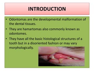

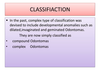
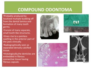
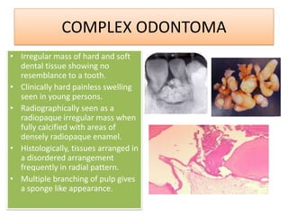
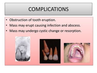
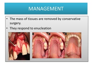


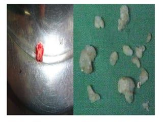


This document discusses odontomas, which are benign developmental malformations of dental tissues that have the basic histological structure of teeth but in a disorganized pattern. Odontomas are classified as compound or complex. Compound odontomas consist of separate small tooth-like structures, while complex odontomas are irregular hard and soft dental tissues showing no tooth resemblance. Odontomas typically present in people aged 10-20 as painless swellings and can cause complications like eruption issues or cyst formation if not removed by conservative surgery.


