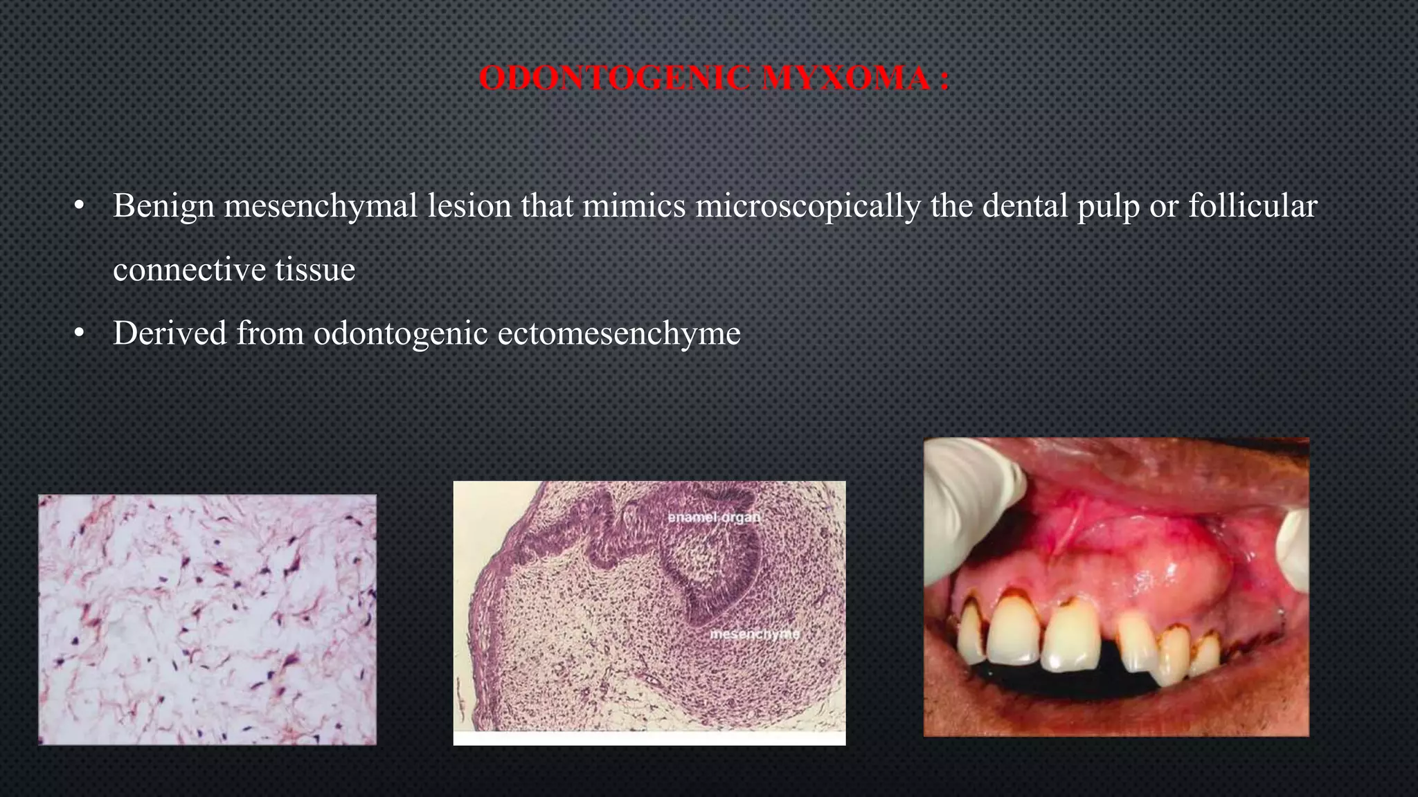
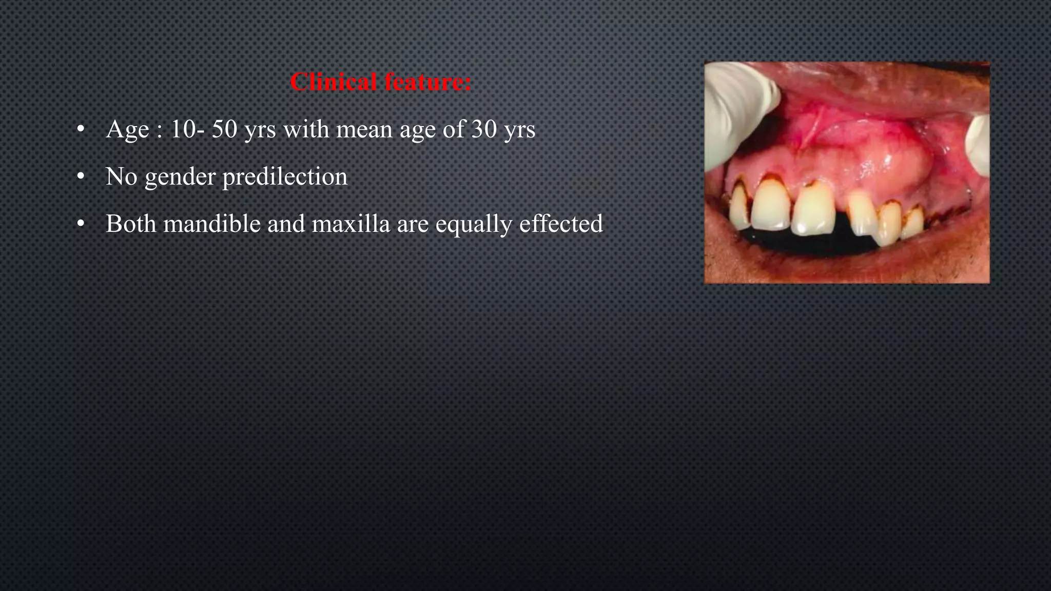
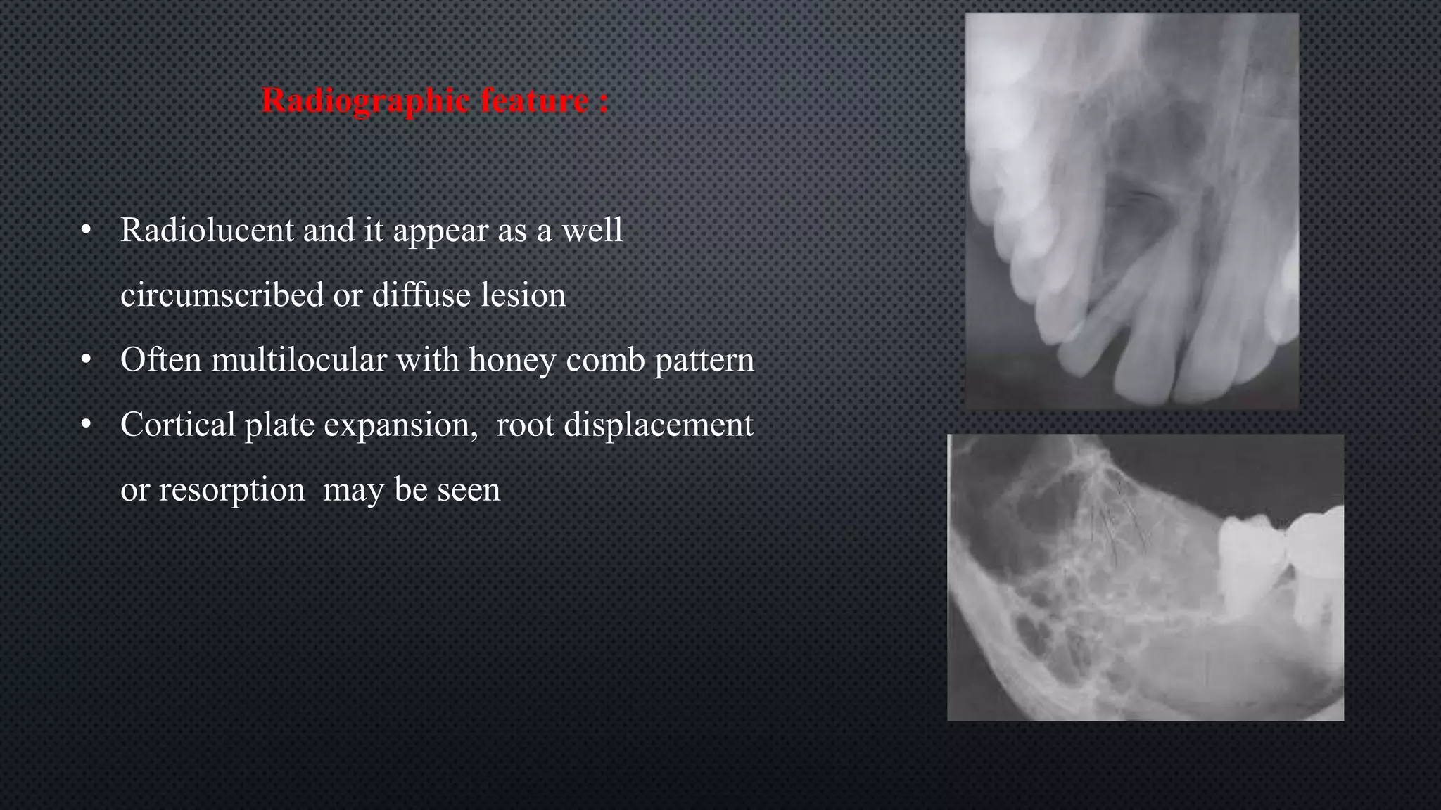
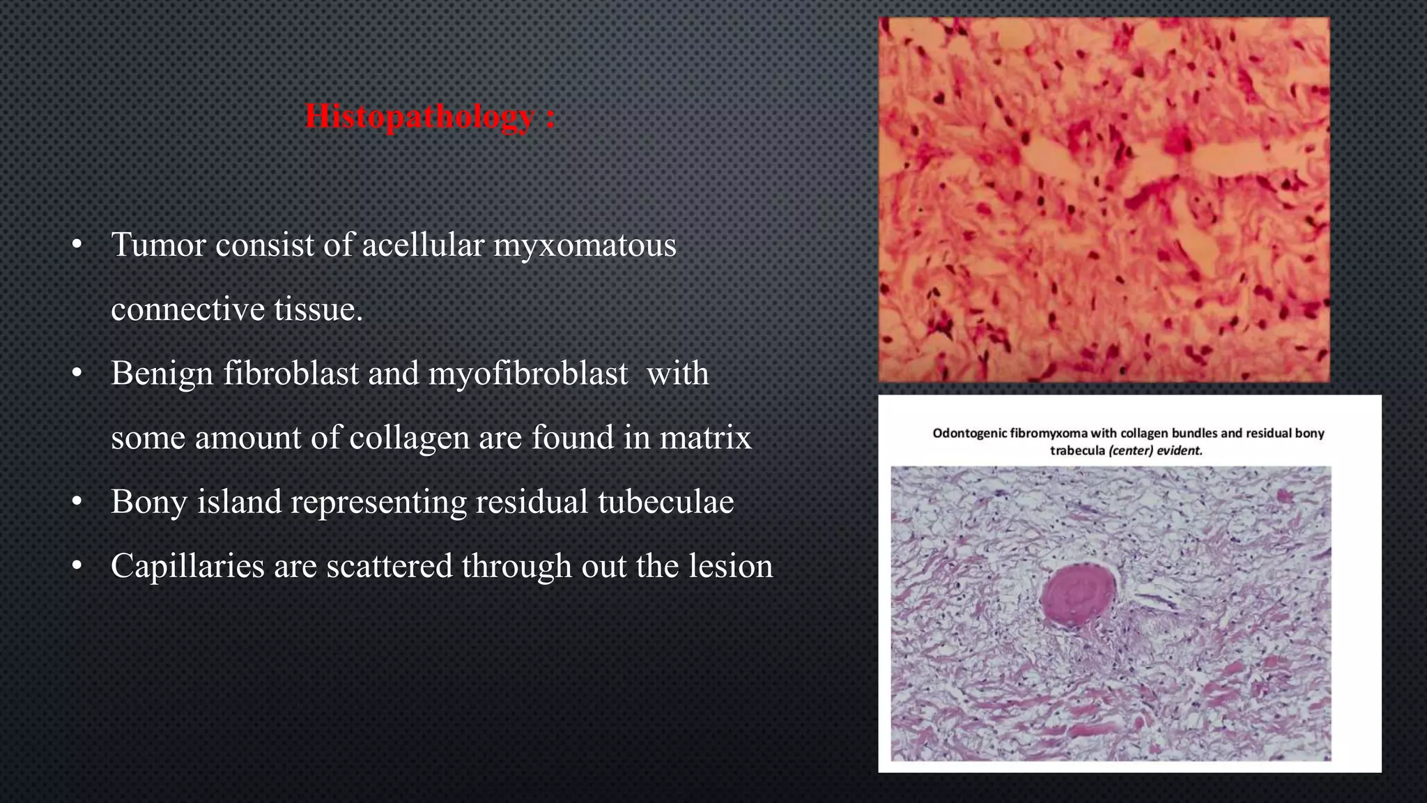
ODONTOGENIC
MYXOMA
DR AMITHA G
DEPT OF ORAL AND MAXILLO FACIAL PATHOLOGY
ODONTOGENIC MYXOMA :
• Benign mesenchymal lesion that mimics microscopically the dental pulp or follicular
connective tissue
• Derived from odontogenic ectomesenchyme
Clinical feature:
• Age : 10- 50 yrs with mean age of 30 yrs
• No gender predilection
• Both mandible and maxilla are equally effected
Radiographic feature :
• Radiolucent and it appear as a well
circumscribed or diffuse lesion
• Often multilocular with honey comb pattern
• Cortical plate expansion, root displacement
or resorption may be seen
Histopathology :
• Tumor consist of acellular myxomatous
connective tissue.
• Benign fibroblast and myofibroblast with
some amount of collagen are found in matrix
• Bony island representing residual tubeculae
• Capillaries are scattered through out the lesion

Odontogenic Myxoma

  • 1.
    ODONTOGENIC MYXOMA DR AMITHA G DEPTOF ORAL AND MAXILLO FACIAL PATHOLOGY
  • 2.
    ODONTOGENIC MYXOMA : •Benign mesenchymal lesion that mimics microscopically the dental pulp or follicular connective tissue • Derived from odontogenic ectomesenchyme
  • 3.
    Clinical feature: • Age: 10- 50 yrs with mean age of 30 yrs • No gender predilection • Both mandible and maxilla are equally effected
  • 4.
    Radiographic feature : •Radiolucent and it appear as a well circumscribed or diffuse lesion • Often multilocular with honey comb pattern • Cortical plate expansion, root displacement or resorption may be seen
  • 5.
    Histopathology : • Tumorconsist of acellular myxomatous connective tissue. • Benign fibroblast and myofibroblast with some amount of collagen are found in matrix • Bony island representing residual tubeculae • Capillaries are scattered through out the lesion

Editor's Notes

  • #6 Pic : abundance of fusiform, stellate and round cells in myxoid tissue Large amount of collagen are evedent- odontogenic fibromyxoma