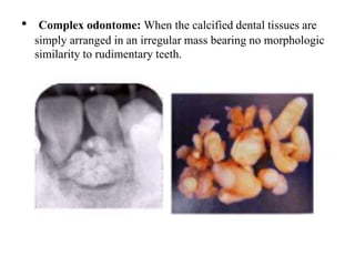
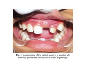
Complex Odontome associated
with Maxillary Impacted
Permanent Central Incisor: A
Case Report
Raghavendra M Shetty, Sangamesh Halawar, Hanumanth Reddy, Sujata Rath,
Sunaina Shetty, Anushka Deoghare
Presented by:-
Khushboo Sinhmar
M.D.S. 2ND YEAR
CONTENTS
• INTRODUCTION
• CLINICAL FEATURES
• ETIOLOGY
• CASE REPORT
• INTRA ORAL EXAMINATION
• RADIOGRAPHIC FINDINGS
• TREATMENT
• HISTOPATHOLOGICAL EXAMINATIONDISCUSSION
• CONCLUSION
• REFERENCE
INTRODUCTION
• ODONTOMA
• The term odontome was coined by Paul Broca in 1867
• Odontomas by definition alone refers to any tumor of
odontogenic origin. This is because odontomas result from the
growth of completely differentiated epithelial and mesenchymal
cells that give rise to ameloblasts and odontoblasts.
• TYPES:-
Acc to WHO classification Odontomes can be divided into three
types:
1)- Complex Odontome
2)- Compound Odontome
3)- Ameloblastic fibro Odontome
Compound odontome: Composed of all odontogenic tissues in
an orderly pattern that results in many teeth like structures, but
without morphologic resemblance to normal teeth.
• Compound odontome was classified as acc to Gravey et al
• Denticulo type : Composed of two or more of tissues separate
denticles, having crown and root, dental hard tissues
resembling to that of tooth
• Particulate type :Composed of two or more separate masses or
particles, bearing no resemblance to tooth, consists of hard
dental tissues
• Denticulo-particulate type: In this type, denticles and particles
are present together
• Complex odontome: When the calcified dental tissues are
simply arranged in an irregular mass bearing no morphologic
similarity to rudimentary teeth.
• Ameloblastic fibro-odontome:
Consists of varying amounts of calcified dental tissue and
dental papilla-like tissue, the later component resembling an
ameloblastic fibroma. The ameloblastic fibro-odontome is
considered as an immature precursor of complex odontome.
2)- On the basis of their developmental origin in 1914, Gabell,
James and Payne grouped odontome into three types:
• a. Epithelial
• b. Composite (epithelial and mesodermal) and
• c. Connective tissue.
3) According to their position within the jaws:
• 1) Intra osseous
• 2) Extra osseous
4) According to Thoma and Goldman (1946):
• Germinated composite odontomes
• Compound composite odontomes
• Complex composite odontomes
• Dilated odontomes
• Cystic odontomes
CLINICAL FEATURES
1) SHAPE-
Compound Odontome is regularly shaped solitary or multiple
denticles
Complesx odontome amorphous conglomeration of dental tissues
2) APPEARANCE-
Compound Odontome Bizzare peg shaped teeth show anatomic
resemblance
Complesx odontome- an irregular mass
3) COMPOSITION- Both compound and complex tumours are
formed of enamel dentin , also have variable amounts of
cementum and pulpal tissue
4) INCIDENCE:- Compound odontomes – 9-37%
Complex odontome- 5-30%
5) age- both complex and compound found in yoinger patients
with mean age 14 to 20 years
6) SIGNS AND SYMPTOMS
• Asymptomatic, although occasionally signs and symptoms
relating to their presence do occur
• These generally consist of
– Unerupted or impacted teeth,
– Impacted tooth,
– Retained deciduous teeth,
– Swelling and evidence of infection,
– Displacement of teeth and malocclusion.
• Compound odontomes seldom cause bony expansion but
complex odontome often cause slight or even marked bony
expansion.
ETIOLOGY
• Local trauma
In 1979, Shteyer, Taicher and Marmary reported a case of
odontome occurring at the subcondylar region associated
with a sinus tract linking to the third molar region. They
deduced that the missing third molar had migrated to the
subcondylar region followed by the occurrence of trauma
or infection which led to the development of the odontome.
• Inflammatory and/or infectious processes,
• cell rests of serres (dental lamina remnants)
• due to hereditary anomalies (Gardner’s syndrome, Hermanns
syndrome)
• Alterations in the genetic component responsible for
controlling dental development.
• Hitchin suggested that odontomes are inherited or are
due to a mutagene or interference, possibly postnatal, with
the genetic control of tooth development.
CASE REPORT
-12 years old female patient
-Chief complaints- Missing tooth in the upper front region of the
jaw
-Past, family and medical history- not relevant
-General physical examination- not significant
Intra oral examination
• Permanent dentition with unerupted left maxillary permanent
central incisor
• Inspection- swelling on the labial side of unerupted tooth
• Palpation- firm nodule of 2 cm in diameter
RADIOGRAPHIC FINDINGS
• Intraoral periapical and panoramic radiographs
revealed that radiopaque structures were present
obstructing the eruption of left maxillary permanent
central incisor.
• On basis of clinical and radiographic findings, case
was provisionally diagnosed as odontome
• Surgical removal of the odontome under local anesthesia.
• Full thickness mucoperiosteal flap was reflected from the
labial surface of right maxillary permanent lateral incisor to
left maxillary permanent canine.
• Layer of bone overlying the mass was removed and the
calcified masses were exposed
TREATMENT
• Four to five calcified irregular masses were removed without
disturbing the underlying tooth
• Curettage was done and the area was irrigated with povidineiodine
solution and normal saline (0.9%).
• Unerupted left maxillary permanent central incisor was located
• After hemostasis, begg’s bracket with a twisted ligature wire in a hook
form tied to it was bonded on the labial surface of the impacted incisor.
• The flap was repositioned and sutured, keeping the ligature wire hook
suspended in the oral cavity making sure the occlusion was not disturbed
HISTOPATHOLOGICAL
EXAMINATION
• Microscopically, hematoxylin and eosin-stained
(H&E) section showed structures exhibiting an
irregular arrangement of dentin, mesenchymal tissue
resembling pulp and a small area of cementum-like
material
POSTOP PANORMIC RADIOGRAPHY
DISCUSSION
• In clinical setting, dentists often encounter the problem of
tooth impaction, which has been defined as a situation where a
tooth fails to erupt into a normal functional position by the
expected times
• The treatment for odontomes in both primary and permanent
dentition is their surgical removal.
• If odontomes are extirpated early without disturbing the
underlying tooth germ, the eruption of the impacted teeth can
then be expected spontaneously or after orthodontic traction
• However, underlying impacted teeth are sometimes extracted
in association with the removal of odontomas
• In this case, the overlying odontomes were surgically removed
and the impacted central incisor has been kept under
observation to monitor its eruption.
• If the root of the impacted tooth is still developing, the tooth
may erupt normally; but, once the root apex has closed, the
tooth has lost its potential to erupt.
Conclusion
• Radiographic examination of all pediatric patients that present
clinical evidence of delayed permanent tooth eruption or
temporary tooth displacement, with or without history of
previous dental trauma should be performed.
• Early diagnosis of odontomes allows adoption of a less
complex and less expensive treatment and ensures normal
eruption pattern of permanent teeth which may otherwise get
impacted.
REFERENCES
1) Shetty RM, Halawar S, Reddy H, Rath S, Shetty S, Deoghare
A. Complex Odontome associated with Maxillary Impacted
Permanent Central Incisor: A Case Report. Int J Clin Pediatr
Dent 2013;6(1):58-61
2) V Satish, Maganur C Prabhadevi, Rajesh Sharma Odontome:
A Brief Overview International Journal of Clinical Pediatric
Dentistry, September-December 2011;4(3):177-185
3) Shteryer A, Taicher S, Marmary T. Odontoma in the
subcondylar region. Br J Oral Surg 1979 Nov;17(2):161-165.

Complex odontome associated with maxillary impacted

  • 1.
    Complex Odontome associated withMaxillary Impacted Permanent Central Incisor: A Case Report Raghavendra M Shetty, Sangamesh Halawar, Hanumanth Reddy, Sujata Rath, Sunaina Shetty, Anushka Deoghare Presented by:- Khushboo Sinhmar M.D.S. 2ND YEAR
  • 2.
    CONTENTS • INTRODUCTION • CLINICALFEATURES • ETIOLOGY • CASE REPORT • INTRA ORAL EXAMINATION • RADIOGRAPHIC FINDINGS • TREATMENT • HISTOPATHOLOGICAL EXAMINATIONDISCUSSION • CONCLUSION • REFERENCE
  • 3.
    INTRODUCTION • ODONTOMA • Theterm odontome was coined by Paul Broca in 1867 • Odontomas by definition alone refers to any tumor of odontogenic origin. This is because odontomas result from the growth of completely differentiated epithelial and mesenchymal cells that give rise to ameloblasts and odontoblasts.
  • 4.
    • TYPES:- Acc toWHO classification Odontomes can be divided into three types: 1)- Complex Odontome 2)- Compound Odontome 3)- Ameloblastic fibro Odontome
  • 5.
    Compound odontome: Composedof all odontogenic tissues in an orderly pattern that results in many teeth like structures, but without morphologic resemblance to normal teeth.
  • 6.
    • Compound odontomewas classified as acc to Gravey et al • Denticulo type : Composed of two or more of tissues separate denticles, having crown and root, dental hard tissues resembling to that of tooth • Particulate type :Composed of two or more separate masses or particles, bearing no resemblance to tooth, consists of hard dental tissues • Denticulo-particulate type: In this type, denticles and particles are present together
  • 8.
    • Complex odontome:When the calcified dental tissues are simply arranged in an irregular mass bearing no morphologic similarity to rudimentary teeth.
  • 9.
    • Ameloblastic fibro-odontome: Consistsof varying amounts of calcified dental tissue and dental papilla-like tissue, the later component resembling an ameloblastic fibroma. The ameloblastic fibro-odontome is considered as an immature precursor of complex odontome.
  • 10.
    2)- On thebasis of their developmental origin in 1914, Gabell, James and Payne grouped odontome into three types: • a. Epithelial • b. Composite (epithelial and mesodermal) and • c. Connective tissue. 3) According to their position within the jaws: • 1) Intra osseous • 2) Extra osseous
  • 11.
    4) According toThoma and Goldman (1946): • Germinated composite odontomes • Compound composite odontomes • Complex composite odontomes • Dilated odontomes • Cystic odontomes
  • 12.
    CLINICAL FEATURES 1) SHAPE- CompoundOdontome is regularly shaped solitary or multiple denticles Complesx odontome amorphous conglomeration of dental tissues 2) APPEARANCE- Compound Odontome Bizzare peg shaped teeth show anatomic resemblance Complesx odontome- an irregular mass
  • 13.
    3) COMPOSITION- Bothcompound and complex tumours are formed of enamel dentin , also have variable amounts of cementum and pulpal tissue 4) INCIDENCE:- Compound odontomes – 9-37% Complex odontome- 5-30% 5) age- both complex and compound found in yoinger patients with mean age 14 to 20 years
  • 14.
    6) SIGNS ANDSYMPTOMS • Asymptomatic, although occasionally signs and symptoms relating to their presence do occur • These generally consist of – Unerupted or impacted teeth, – Impacted tooth, – Retained deciduous teeth, – Swelling and evidence of infection, – Displacement of teeth and malocclusion. • Compound odontomes seldom cause bony expansion but complex odontome often cause slight or even marked bony expansion.
  • 15.
    ETIOLOGY • Local trauma In1979, Shteyer, Taicher and Marmary reported a case of odontome occurring at the subcondylar region associated with a sinus tract linking to the third molar region. They deduced that the missing third molar had migrated to the subcondylar region followed by the occurrence of trauma or infection which led to the development of the odontome. • Inflammatory and/or infectious processes, • cell rests of serres (dental lamina remnants)
  • 16.
    • due tohereditary anomalies (Gardner’s syndrome, Hermanns syndrome) • Alterations in the genetic component responsible for controlling dental development.
  • 17.
    • Hitchin suggestedthat odontomes are inherited or are due to a mutagene or interference, possibly postnatal, with the genetic control of tooth development.
  • 18.
    CASE REPORT -12 yearsold female patient -Chief complaints- Missing tooth in the upper front region of the jaw -Past, family and medical history- not relevant -General physical examination- not significant
  • 19.
    Intra oral examination •Permanent dentition with unerupted left maxillary permanent central incisor • Inspection- swelling on the labial side of unerupted tooth • Palpation- firm nodule of 2 cm in diameter
  • 21.
    RADIOGRAPHIC FINDINGS • Intraoralperiapical and panoramic radiographs revealed that radiopaque structures were present obstructing the eruption of left maxillary permanent central incisor. • On basis of clinical and radiographic findings, case was provisionally diagnosed as odontome
  • 24.
    • Surgical removalof the odontome under local anesthesia. • Full thickness mucoperiosteal flap was reflected from the labial surface of right maxillary permanent lateral incisor to left maxillary permanent canine. • Layer of bone overlying the mass was removed and the calcified masses were exposed TREATMENT
  • 26.
    • Four tofive calcified irregular masses were removed without disturbing the underlying tooth
  • 27.
    • Curettage wasdone and the area was irrigated with povidineiodine solution and normal saline (0.9%). • Unerupted left maxillary permanent central incisor was located
  • 28.
    • After hemostasis,begg’s bracket with a twisted ligature wire in a hook form tied to it was bonded on the labial surface of the impacted incisor. • The flap was repositioned and sutured, keeping the ligature wire hook suspended in the oral cavity making sure the occlusion was not disturbed
  • 29.
    HISTOPATHOLOGICAL EXAMINATION • Microscopically, hematoxylinand eosin-stained (H&E) section showed structures exhibiting an irregular arrangement of dentin, mesenchymal tissue resembling pulp and a small area of cementum-like material
  • 31.
  • 32.
    DISCUSSION • In clinicalsetting, dentists often encounter the problem of tooth impaction, which has been defined as a situation where a tooth fails to erupt into a normal functional position by the expected times • The treatment for odontomes in both primary and permanent dentition is their surgical removal.
  • 33.
    • If odontomesare extirpated early without disturbing the underlying tooth germ, the eruption of the impacted teeth can then be expected spontaneously or after orthodontic traction • However, underlying impacted teeth are sometimes extracted in association with the removal of odontomas
  • 34.
    • In thiscase, the overlying odontomes were surgically removed and the impacted central incisor has been kept under observation to monitor its eruption. • If the root of the impacted tooth is still developing, the tooth may erupt normally; but, once the root apex has closed, the tooth has lost its potential to erupt.
  • 35.
    Conclusion • Radiographic examinationof all pediatric patients that present clinical evidence of delayed permanent tooth eruption or temporary tooth displacement, with or without history of previous dental trauma should be performed. • Early diagnosis of odontomes allows adoption of a less complex and less expensive treatment and ensures normal eruption pattern of permanent teeth which may otherwise get impacted.
  • 36.
    REFERENCES 1) Shetty RM,Halawar S, Reddy H, Rath S, Shetty S, Deoghare A. Complex Odontome associated with Maxillary Impacted Permanent Central Incisor: A Case Report. Int J Clin Pediatr Dent 2013;6(1):58-61 2) V Satish, Maganur C Prabhadevi, Rajesh Sharma Odontome: A Brief Overview International Journal of Clinical Pediatric Dentistry, September-December 2011;4(3):177-185 3) Shteryer A, Taicher S, Marmary T. Odontoma in the subcondylar region. Br J Oral Surg 1979 Nov;17(2):161-165.