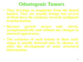
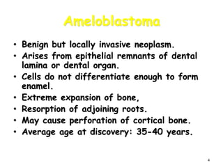
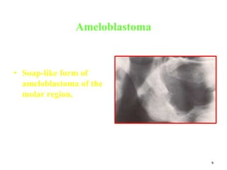
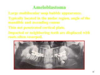
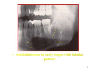
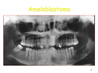
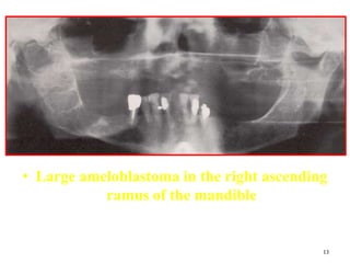
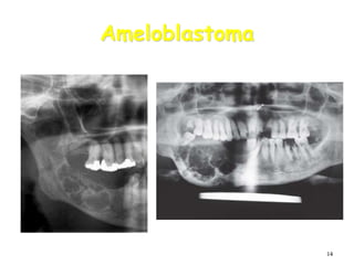
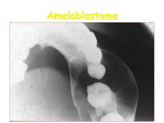
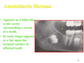
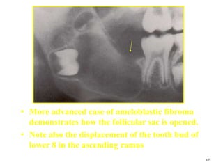
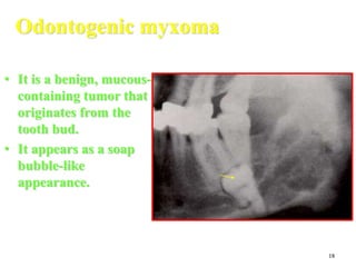
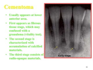
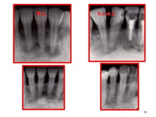
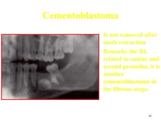
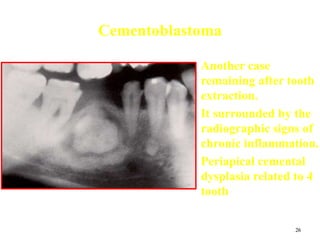
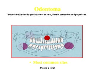
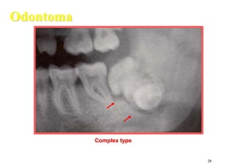
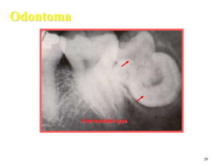
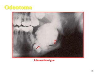
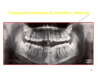
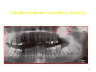
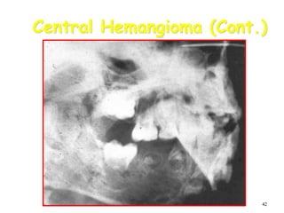
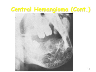
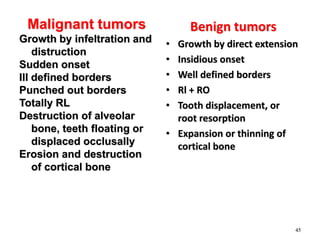
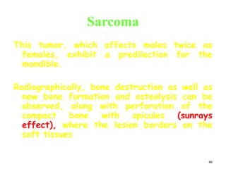
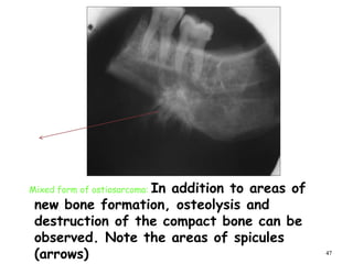
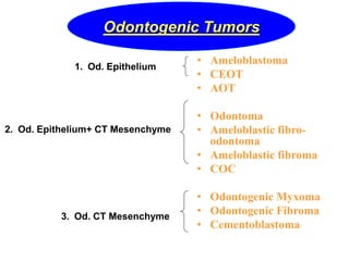
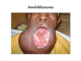
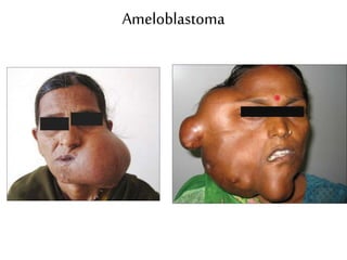
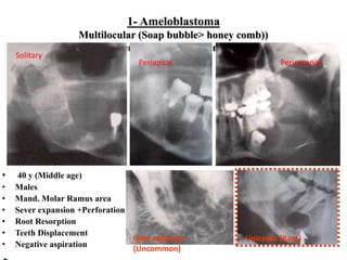
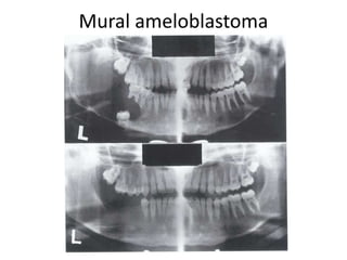
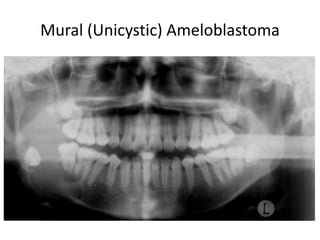
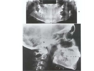
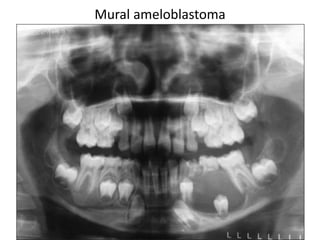
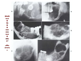
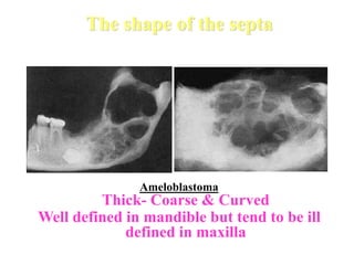
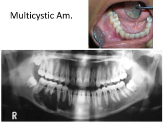
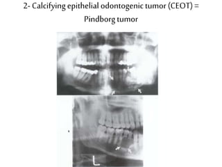
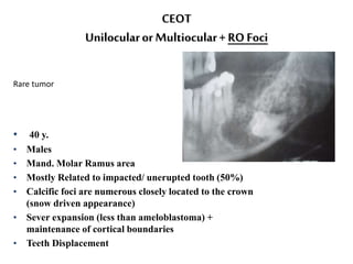
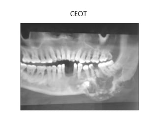
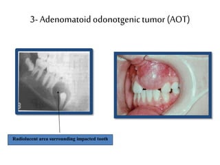
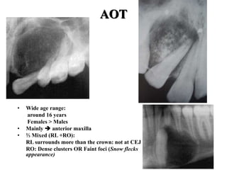
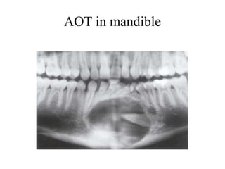
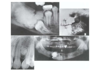
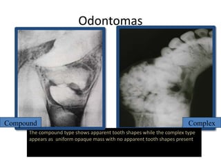
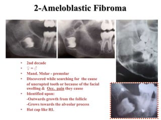
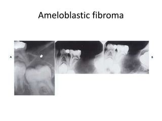
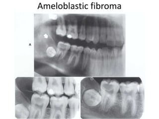
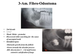
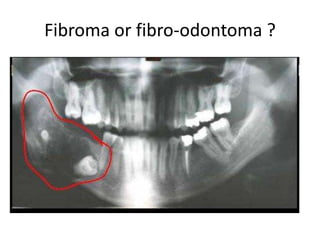
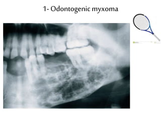
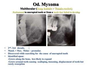
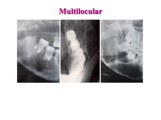
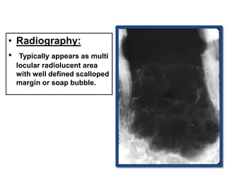
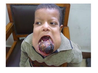
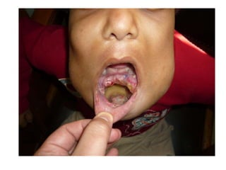
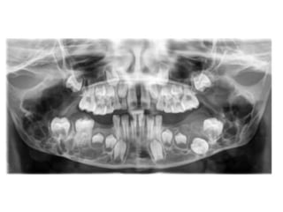
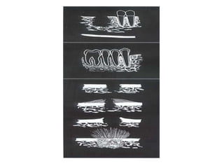
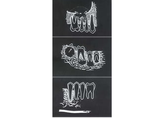
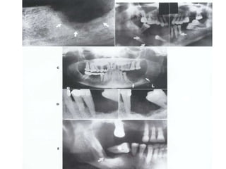
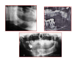
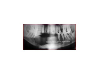
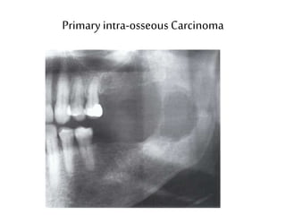
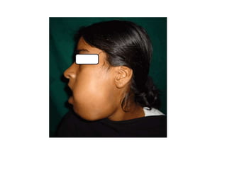
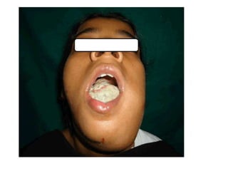
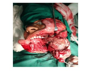
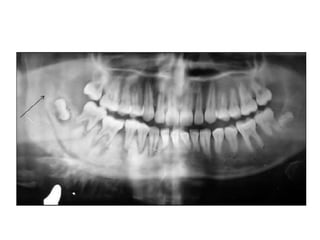
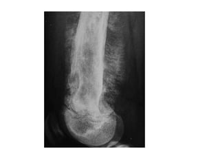
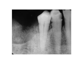
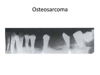
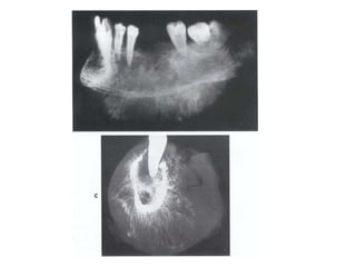
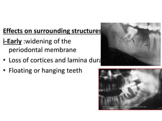
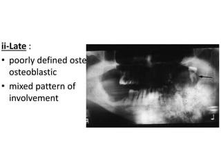
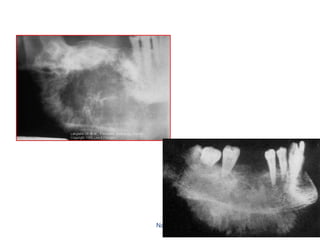
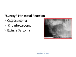
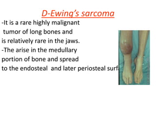
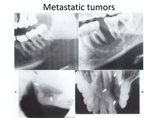
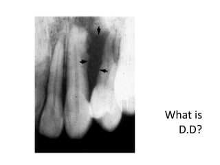
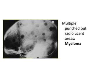
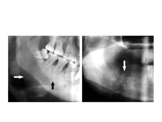
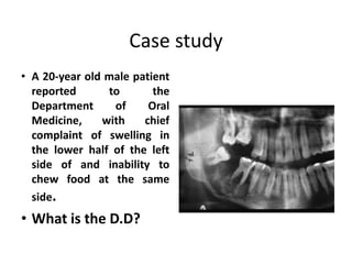
Practical Oral
Radiology 2
Ahmed A.Abdelazim
Total: 5 marks
1- Benign tumors
2-Malignant tumors
3
Odontogenic Tumors
• They develops as neoplasias from the dental
lamina. They are usually benign but several
of them have the tendency towards malignant
transformation.
• Because growth occurs only slowly,
asymptomatically and without any changes in
mucosal appearance,
• The existence of such lesions in their early
stages is usually detected only by chance, or
after the development of some structural
deformation.
4
Ameloblastoma
• Benign but locally invasive neoplasm.
• Arises from epithelial remnants of dental
lamina or dental organ.
• Cells do not differentiate enough to form
enamel.
• Extreme expansion of bone,
• Resorption of adjoining roots.
• May cause perforation of cortical bone.
• Average age at discovery: 35-40 years.
5
Most common sites of ameloblastoma
80%
20%
6
Ameloblastoma (Cont.)
• Occasionally develops in the wall of
dentigerous cyst (mural Ameloblatoma).
• 80% in mandible. ¾ of these in molar-
ramus area.
• Pain and paresthesia not common.
• Extremely high recurrence rate.
7
Ameloblastoma (Cont.)
• Most often a well-corticated multilocular
radiolucency.
• “Honey-comb”, “soap-bubble” or “tennis-racket”
appearance.
• May be a well-corticated unilocular lesion
resembling a cyst.
Honeycomb-like small
ameloblastoma at early stage with
evidence of root resorption.
8
Ameloblastoma
• Ameloblastoma at the
angle of the mandible.
• Expansive form with
oval RL traversed by
few very thin septa
9
Ameloblastoma
• Soap-like form of
ameloblastoma of the
molar region.
10
Ameloblastoma
Large multilocular soap bubble appearance.
Typically located in the molar region, angle of the
mandible and ascending ramus
Thin not penetrated cortical plate.
Impacted or neighboring teeth are displaced with
roots often resorped.
11
• Ameloblastoma in early stages with lobular
pattern
12
Ameloblastoma
13
• Large ameloblastoma in the right ascending
ramus of the mandible
14
Ameloblastoma
15
Ameloblastoma
16
Ameloblastic fibroma
• Appears as a follecular
cystic cavity
surrounding a crown
of a tooth.
• In early stages appears
as a hat upon the
occlusal surface of
affected tooth
17
• More advanced case of ameloblastic fibroma
demonstrates how the follicular sac is opened.
• Note also the displacement of the tooth bud of
lower 8 in the ascending ramus
18
Odontogenic myxoma
• It is a benign, mucous-
containing tumor that
originates from the
tooth bud.
• It appears as a soap
bubble-like
appearance.
19
Cementoma
• Usually appears at lower
anterior area.
• First appears as fibrous
tissue stage, which may
confused with a
granuloma (vitality test).
• The second stage is
characterized with
accumulation of calcified
materials.
• The third stage consists of
radio-opaque materials.
Early stage
20
R.L R.L+R.O
21
Periapical cemental
dysplasia
22
Periapical Cemental Dysplasia
23
Cementoblastoma
( True Cementoma )
• Slow growing
neoplasm composed
of cementum.
• Usually solitary
lesion seen as a
growth on root of
tooth.
• Most common in
mandible, premolar
or 1st molar (80%).
24
Cementoblastoma
• Appears as a well
defined RO area
with a thin RL band
around it
• May cause external
root resorption
25
Cementoblastoma
• It not removed after
tooth extraction
• Remarks the RL
related to canine and
second premolar, it is
another
cementoblastoma in
the fibrous stage.
26
Cementoblastoma
• Another case
remaining after tooth
extraction.
• It surrounded by the
radiographic signs of
chronic inflammation.
• Periapical cemental
dysplasia related to 4
tooth
Ossama El-Shall
Odontoma
• Most common sites
Tumor characterized by production of enamel, dentin, cementum and pulp tissue
28
Odontoma
Complex type
29
Odontoma
Intermediate type
30
Odontoma
Intermediate type
31
Odontoma
Compound type
32
Compound odontoma in maxillary tubrosity
33
Complex odontoma in maxillary tubrosity
34
Compound Composite Odontoma
• Composed of enamel
and dentin.
• Enamel and dentin
are laid down in an
orderly fashion so
that the mass has
some similarity to
normal teeth.
• Appears like a bunch
of small teeth.
35
Compound Composite Odontoma
36
Central Osteoma anterior to remaining roots of lower 7
37
Peripheral osteoma located in maxillary sinus
38
Peripheral osteoma in right angle of the mandible
It may confused with calcified lymph noads
39
Osteoma
40
Central Hemangioma
• Tumor characterized by
proliferation of blood
vessels.
• Central hemangiomas of
jaws uncommon.
• 50% occur in children
and teens.
• More common in females
and mandible.
• Well-defined or ill-
defined, unilocular or
multilocular radiolucency.
5/4/2016 Ossama El-Shall 41
Central Hemangioma (Cont.)
• May cause expansion of bone and
resorption of teeth.
• Early treatment is desirable in order
to avoid profuse bleeding due to
accidental trauma. Aspiration prior to
surgical procedure is advised.
42
Central Hemangioma (Cont.)
43
Central Hemangioma (Cont.)
44
Malignant tumors
• Sarcoma
• Carcinoma.
• Metastasis.
Benign tumors
• Growth by direct extension
• Insidious onset
• Well defined borders
• Rl + RO
• Tooth displacement, or
root resorption
• Expansion or thinning of
cortical bone
45
Malignant tumors
Growth by infeltration and
distruction
Sudden onset
Ill defined borders
Punched out borders
Totally RL
Destruction of alveolar
bone, teeth floating or
displaced occlusally
Erosion and destruction
of cortical bone
46
Sarcoma
This tumor, which affects males twice as
females, exhibit a predilection for the
mandible.
Radiographically, bone destruction as well as
new bone formation and osteolysis can be
observed, along with perforation of the
compact bone with spicules (sunrays
effect), where the lesion borders on the
soft tissues
47
Mixed form of ostiosarcoma: In addition to areas of
new bone formation, osteolysis and
destruction of the compact bone can be
observed. Note the areas of spicules
(arrows)
1-Benign
Tumors
• Ameloblastoma
• CEOT
• AOT
• Odontoma
• Ameloblastic fibro-
odontoma
• Ameloblastic fibroma
• COC
• Odontogenic Myxoma
• Odontogenic Fibroma
• Cementoblastoma
1. Od. Epithelium
2. Od. Epithelium+ CT Mesenchyme
3. Od. CT Mesenchyme
Odontogenic Tumors
Ameloblastoma
Ameloblastoma
1- Ameloblastoma
Multilocular (Soap bubble> honey comb))
origin (dental lamina and dental organ)
• 40 y (Middle age)
• Males
• Mand. Molar Ramus area
• Sever expansion +Perforation
• Root Resorption
• Teeth Displacement
• Negative aspiration
Unicystic (Rare)Inter radicular
(Uncommon)
Solitary
Periapical Pericoronal
Mural ameloblastoma
Mural (Unicystic) Ameloblastoma
Mural ameloblastoma
The shape of the septa
Ameloblastoma
Thick- Coarse & Curved
Well defined in mandible but tend to be ill
defined in maxilla
Multicystic Am.
2- Calcifyingepithelial odontogenictumor (CEOT)=
Pindborgtumor
CEOT
Unilocularor Multiocular+ RO Foci
• 40 y.
• Males
• Mand. Molar Ramus area
• Mostly Related to impacted/ unerupted tooth (50%)
• Calcific foci are numerous closely located to the crown
(snow driven appearance)
• Sever expansion (less than ameloblastoma) +
maintenance of cortical boundaries
• Teeth Displacement
Rare tumor
CEOT
3- Adenomatoidodonotgenictumor (AOT)
Radiolucent area surrounding impacted tooth
AOT
• Wide age range:
around 16 years
Females > Males
• Mainly  anterior maxilla
• ⅔ Mixed (RL +RO):
RL surrounds more than the crown: not at CEJ
RO: Dense clusters OR Faint foci (Snow flecks
appearance)
AOT in mandible
2- MixedOdontogenicTumors
1- Odontoma
2- Ameloblastic fibroma
3- Ameloblastic fibro-odontoma
1- Odontoma
Odontomas are developmental malformation
( hamartoma) of dental tissue, it is not neoplasm
Very important - very common – children
Two main Types
Compound = normal arrangement of dental tissues
Complex = abnormal mass of Calcification
1- Odontoma
Complex odontomas
Compound odontomas
Odontoma
• 2nd decade (young age  )
• Complex: ♀ Compound ♀=♂
Mand. Molar Max. Ant.
• Maturtion:RL…Mixed…..RO
• Surrounded by RL rim
• Discovered while searching for the cause of
unerupted permanent or retained deciduous
• Easily identified upon Shape & Density
• It’s the most common odontogenic tumor
Odontomas
The compound type shows apparent tooth shapes while the complex type
appears as uniform opaque mass with no apparent tooth shapes present
Compound Complex
2-Ameloblastic Fibroma
• 2nd decade
• ♀ = ♂
• Mand. Molar - premolar
• Discovered while searching for the cause
of unerupted tooth or because of the facial
swelling & Occ. pain they cause
• Identified upon:
-Outwards growth from the follicle
-Grows towards the alveolar process
• Hat cap like RL
Ameloblastic fibroma
3-Am. Fibro-Odontoma
• 2nd decade
• ♀ = ♂
• Mand. Molar - premolar
• Discovered while searching for the cause
of unerupted tooth
• Identified upon:
-Outwards growth from the follicle
-Grows towards the alveolar process
-RO: discrete foci 1 – 2 if small lesion
extensive calcification if large
40 y, ♂, Not as an outward growth
RL
Fibroma or fibro-odontoma ?
3- MesenchymalTumors
1-Odontogenic Myxomas
2-Benign Cementoblastoma
3-Central Odontogenic Fibroma
1- Odontogenicmyxoma
Od. Myxoma
Multilocular (Soap bubble > Tennis-racket)
Pericoronal to unerupted tooth or from a tooth that failed to develop
• 2nd- 3rd decade, ♀
• Mand. > Max. Molar – premolar.
• Discovered while searching for the cause of unerupted tooth
• Identified upon:
-Grows along the bone, lees likely to expand
-Grows around teeth causing scalloping, loosening, displacement of teeth but
rarely resorption
Multilocular
• Radiography:
• Typically appears as multi
locular radiolucent area
with well defined scalloped
margin or soap bubble.
A lateral radiograph of a surgical
specimen of a myxoma
An occlusal view shows
buccal expansion
2- Cementoblatoma
• ♂ >♀
• No race predilection
• Wide age range
• Vital teeth, Painful
• Mand. Premolars & 1st molars
• Fused with the roots
• Roots resorbed or obscured
Cementoma or
Periapical cemental dysplasia ?
Periapical cemental dysplasia
Tori - Exostosis - Enostosis
Known from clinical examination by:
•Their location,
•Lobulated shape,
• Adherent normally appearing
mucosa
•Asymptomatic
•Accidentally discovered
•Intra-bony
Osteoma
• ♂ >♀, 40 y & above
• Asymptomatic until interferes with function
• Overlying mucosa is normal and freely mobile.
• Mand. > Max. & Paranasal sinuses
frontoethmoidal
• Well-defined, RO (Compact),
Internal RL core (Cancellous)
A panoramic radiograph shows an osteoma
in the right mandibular angle region
Osteoma
Cherubism
2- Malignant
Tumors
Well defined borders
106
Clinical photograph shows
leukoplakia that transformed to
gingival cancer
Intraoral panoramic view shows
diffuse bone destruction
113
114
Primary intra-osseous Carcinoma
Osteosarcoma
124
Effects on surrounding structures:
i-Early :widening of the
periodontal membrane
• Loss of cortices and lamina dura.
• Floating or hanging teeth
125
ii-Late :
• poorly defined osteolytic,
osteoblastic
• mixed pattern of
involvement
Naglaa S. El Kilani
126
Naglaa S. El Kilani
127
“Sunray” Periosteal Reaction
• Osteosarcoma
• Chondrosarcoma
• Ewing’s Sarcoma
D-Ewing’s sarcoma
-It is a rare highly malignant
tumor of long bones and
is relatively rare in the jaws.
-The arise in the medullary
portion of bone and spread
to the endosteal and later periosteal surfaces.
Metastatic tumors
Metastatic tumors
Metastatic tumors
Multiple Myeloma
Naglaa S. El Kilani
135
Naglaa S. El Kilani
136
Punched Out” Skill Lesions
• Multiple Myeloma
• Langerhans Cell Histiocytosis
• Metastatic Carcinoma
• Neuroblastoma
Describe? D.D?
• Solitary ill
defined
radiolucent
area related to
lower right
molars and
causing
invasion of the
IAC.
What is the view? Describe? D.D?
• Inflammatory
1. Chronic osteomyelitis
2. Osteoradionecrosis
• Neoplastic
1. Squamous cell
carcinoma
2. Metastatic tumors to
the jaws
3. Osteosarcoma and
chondrosarcoma
Describe? D.D?
• What is the D.D? Solitary irregular periapical
radiolucent area related to
upper left lateral and
causing extensive
interproximal bone loss of
the adjacent tooth.
D.D:
• Chronic alveolar abscess
• Chronic osteomyelitis
• Osteoradionecrosis
• Squamous cell carcinoma
• Metastatic tumors to the jaws
• Osteosarcoma and
chondrosarcoma
• Fibrous dysplasia (early stage)
What is
D.D?
Multiple
punched out
radiolucent
areas:
Myeloma
Case study
• A 20-year old male patient
reported to the
Department of Oral
Medicine, with chief
complaint of swelling in
the lower half of the left
side of and inability to
chew food at the same
side.
• What is the D.D?

Practical oral radiology 2 2016