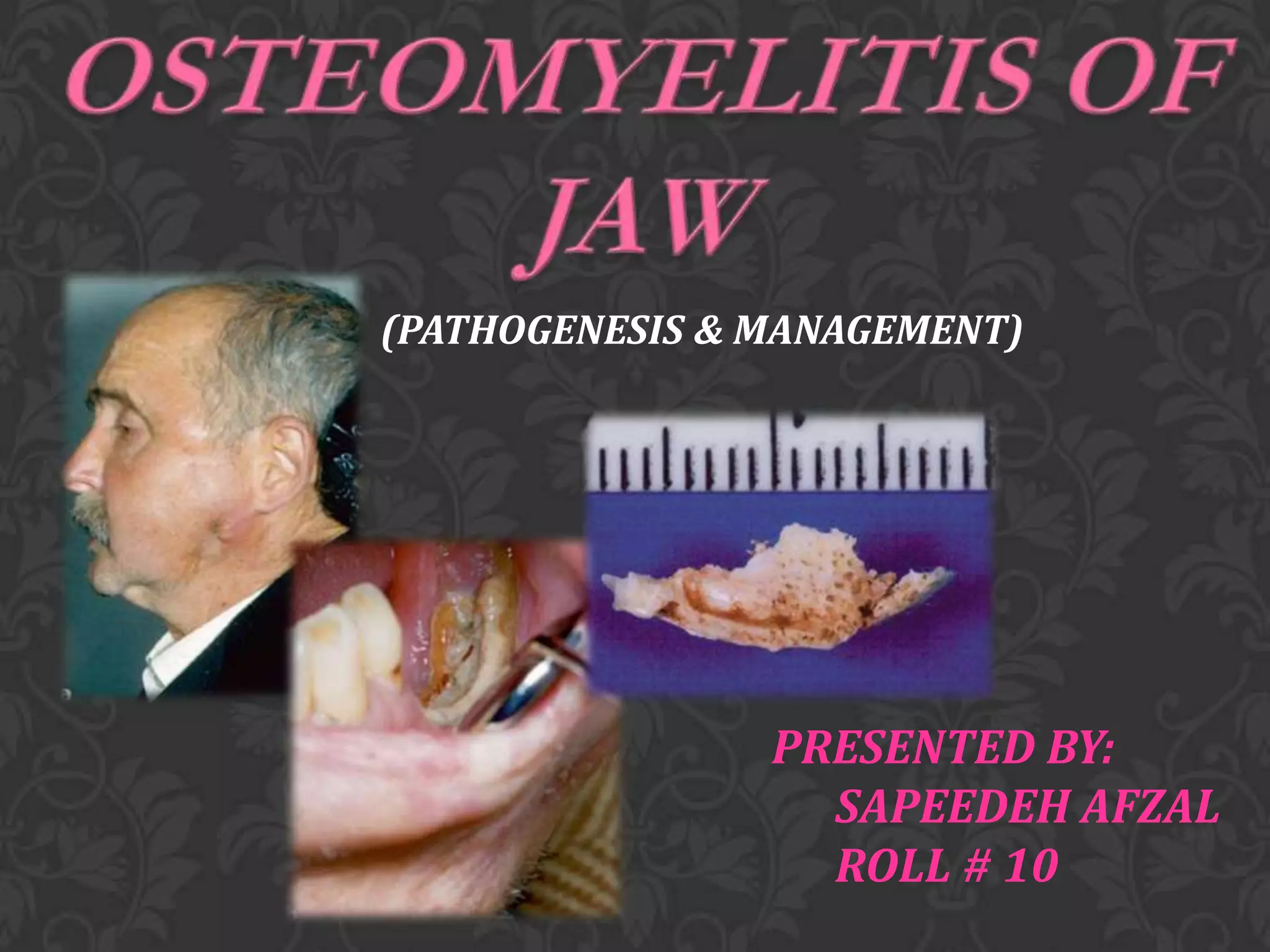
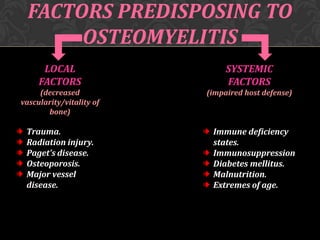
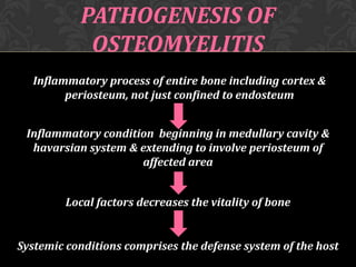
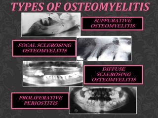
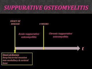
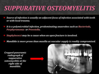
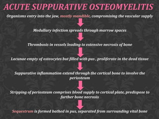
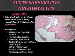
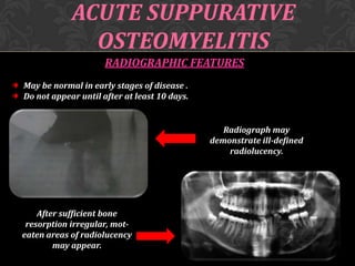
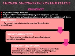
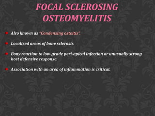
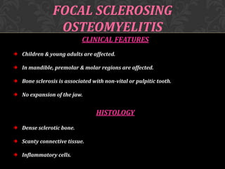
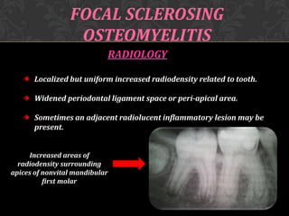
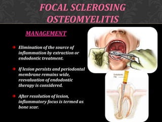
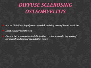
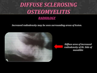
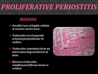
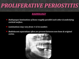
This document discusses osteomyelitis, including its pathogenesis and management. It defines osteomyelitis as an infection of the bone marrow and describes how local and systemic predisposing factors can lead to decreased bone vitality and impaired host defense. The main types of osteomyelitis covered are suppurative, focal sclerosing, diffuse sclerosing, and proliferative perositis. For each type, the document discusses pathogenesis, clinical features, histology, radiology, and management. Key points include how acute suppurative osteomyelitis can progress to chronic form if inadequately treated, and how eliminating infection sources is important but bone changes may persist radiographically for some types.
Odontogenic keratocyst (OKC) is the cyst arising from the cell rests of dental lamina. It can occur anywhere in the jaw, but commonly seen in the posterior part of the mandible. Radiographically, most OKCs are unilocular when presented at the periapex and can be mistaken for radicular or lateral periodontal cyst.
One of the most painful but easy-to-treat dental emergencies is a dry socket.
• Dry socket symptoms are experienced after a tooth extraction.
• This condition requires follow-up care by the doctor who performed the surgery, an oral surgeon or a dentist who is familiar with how to treat it.
For more information, contact :-
Dr Sachdeva's Dental Aesthetic And Implant Institute,
I 101, Ashok Vihar Phase 1, Delhi- 110052
Contact us at
• Phone : +919818894041,01142464041
• Our Websites:
• www.sachdevadentalcare.com
• www.dentalclinicindelhi.com
• www.dentalimplantindia.co.in
• www.dentalcoursesdelhi.com
• www.facialaestheticsdelhi.com
#drysocket #management #thirdmolarextraction #extractioncomplications
Fibro-osseous lesions of the jaws
Fibrous dysplasia
Cemento-osseous dysplasia
Focal cemento-osseous dysplasia
Periapical cemento-osseous dysplasia
Florid cemento-osseous dysplasia
Ossifying fibroma
Juvenile aggressive ossifying fibroma
Cherubism
Fibro-osseous lesions (FOL) are characterized by replacement of normal bone architecture by collagen fibers and fibroblasts containing calcified tissue.
They include a wide variety of lesions of developmental, dysplastic and neoplastic origins with clinical and radiographic presentation and behavior.
Because of the histological similarities between diverse diseases, proper diagnosis requires correlation of history, clinical and radiographic findings.Fibrous Dysplasia
2. Reactive (dysplastic lesions arising in the tooth-bearing area (presumably of periodontal origin).
a. Periapical cemento-osseous dysplasia
b. Focal cemento-osseous dysplasia
c. Florid cemento-osseous dysplasia
3. Fibro-osseous neoplasms (widely designated as cementifying fibroma, ossifying fibroma or cemento-ossifying fibroma.Bone dysplasias
a. Fibrous dyspla i. Monostoticii. Polyostotic
iii. Polyostotic with endocrinopathy (McCune-Albright)
iv Osteofibrous dysplasia
b. Osteitis deformansc. Pagetoid heritable bone dysplasias of childhood
d. Segmental odontomaxillary dysplasia
2. Cemento-osseous dysplasias
a. Focal cemento-osseous dysplasia b. Florid cemento-osseous dysplasia
3.Inflammatory/reactive processes
a. Focal sclerosing osteomyelitisb. Diffuse sclerosing osteomyelitis
c. Proliferative periostitis
4. Metabolic Disease: hyperparathyroidism
5. Neoplastic lesions (Ossifying fibromas)
a. Ossifying fibromab. Hyperparathyroidism jaw lesion syndrome
c. Juvenile ossifying fibroma i. Trabecular typeii. Psammomatoid type
d. Gigantiform cementomas
an overview of muscle pain disorder which regularly create some discomfort for patient to live a normal life as well as to the doctor regarding diagnosis of the problem.
Benign, locally aggressive tumor of odontogenic epithelium, Previously called adamantinoma, Second most common odontogenic tumor after odontoma, Mandible is most common site, Usually asymptomatic and can be found incidentally on routine dental examinations
osteomyelitis of jaw bones / dental implant courses by Indian dental academy Indian dental academy
The Indian Dental Academy is the Leader in continuing dental education , training dentists in all aspects of dentistry and
offering a wide range of dental certified courses in different formats.for more details please visit
www.indiandentalacademy.com
Factory Supply Best Quality Pmk Oil CAS 28578–16–7 PMK Powder in Stockrebeccabio
Factory Supply Best Quality Pmk Oil CAS 28578–16–7 PMK Powder in Stock
Telegram: bmksupplier
signal: +85264872720
threema: TUD4A6YC
You can contact me on Telegram or Threema
Communicate promptly and reply
Free of customs clearance, Double Clearance 100% pass delivery to USA, Canada, Spain, Germany, Netherland, Poland, Italy, Sweden, UK, Czech Republic, Australia, Mexico, Russia, Ukraine, Kazakhstan.Door to door service
Hot Selling Organic intermediates
Lung Cancer: Artificial Intelligence, Synergetics, Complex System Analysis, S...Oleg Kshivets
RESULTS: Overall life span (LS) was 2252.1±1742.5 days and cumulative 5-year survival (5YS) reached 73.2%, 10 years – 64.8%, 20 years – 42.5%. 513 LCP lived more than 5 years (LS=3124.6±1525.6 days), 148 LCP – more than 10 years (LS=5054.4±1504.1 days).199 LCP died because of LC (LS=562.7±374.5 days). 5YS of LCP after bi/lobectomies was significantly superior in comparison with LCP after pneumonectomies (78.1% vs.63.7%, P=0.00001 by log-rank test). AT significantly improved 5YS (66.3% vs. 34.8%) (P=0.00000 by log-rank test) only for LCP with N1-2. Cox modeling displayed that 5YS of LCP significantly depended on: phase transition (PT) early-invasive LC in terms of synergetics, PT N0—N12, cell ratio factors (ratio between cancer cells- CC and blood cells subpopulations), G1-3, histology, glucose, AT, blood cell circuit, prothrombin index, heparin tolerance, recalcification time (P=0.000-0.038). Neural networks, genetic algorithm selection and bootstrap simulation revealed relationships between 5YS and PT early-invasive LC (rank=1), PT N0—N12 (rank=2), thrombocytes/CC (3), erythrocytes/CC (4), eosinophils/CC (5), healthy cells/CC (6), lymphocytes/CC (7), segmented neutrophils/CC (8), stick neutrophils/CC (9), monocytes/CC (10); leucocytes/CC (11). Correct prediction of 5YS was 100% by neural networks computing (area under ROC curve=1.0; error=0.0).
CONCLUSIONS: 5YS of LCP after radical procedures significantly depended on: 1) PT early-invasive cancer; 2) PT N0--N12; 3) cell ratio factors; 4) blood cell circuit; 5) biochemical factors; 6) hemostasis system; 7) AT; 8) LC characteristics; 9) LC cell dynamics; 10) surgery type: lobectomy/pneumonectomy; 11) anthropometric data. Optimal diagnosis and treatment strategies for LC are: 1) screening and early detection of LC; 2) availability of experienced thoracic surgeons because of complexity of radical procedures; 3) aggressive en block surgery and adequate lymph node dissection for completeness; 4) precise prediction; 5) adjuvant chemoimmunoradiotherapy for LCP with unfavorable prognosis.
Ozempic: Preoperative Management of Patients on GLP-1 Receptor Agonists Saeid Safari
Preoperative Management of Patients on GLP-1 Receptor Agonists like Ozempic and Semiglutide
ASA GUIDELINE
NYSORA Guideline
2 Case Reports of Gastric Ultrasound
Pulmonary Thromboembolism - etilogy, types, medical- Surgical and nursing man...VarunMahajani
Disruption of blood supply to lung alveoli due to blockage of one or more pulmonary blood vessels is called as Pulmonary thromboembolism. In this presentation we will discuss its causes, types and its management in depth.
New Directions in Targeted Therapeutic Approaches for Older Adults With Mantl...i3 Health
i3 Health is pleased to make the speaker slides from this activity available for use as a non-accredited self-study or teaching resource.
This slide deck presented by Dr. Kami Maddocks, Professor-Clinical in the Division of Hematology and
Associate Division Director for Ambulatory Operations
The Ohio State University Comprehensive Cancer Center, will provide insight into new directions in targeted therapeutic approaches for older adults with mantle cell lymphoma.
STATEMENT OF NEED
Mantle cell lymphoma (MCL) is a rare, aggressive B-cell non-Hodgkin lymphoma (NHL) accounting for 5% to 7% of all lymphomas. Its prognosis ranges from indolent disease that does not require treatment for years to very aggressive disease, which is associated with poor survival (Silkenstedt et al, 2021). Typically, MCL is diagnosed at advanced stage and in older patients who cannot tolerate intensive therapy (NCCN, 2022). Although recent advances have slightly increased remission rates, recurrence and relapse remain very common, leading to a median overall survival between 3 and 6 years (LLS, 2021). Though there are several effective options, progress is still needed towards establishing an accepted frontline approach for MCL (Castellino et al, 2022). Treatment selection and management of MCL are complicated by the heterogeneity of prognosis, advanced age and comorbidities of patients, and lack of an established standard approach for treatment, making it vital that clinicians be familiar with the latest research and advances in this area. In this activity chaired by Michael Wang, MD, Professor in the Department of Lymphoma & Myeloma at MD Anderson Cancer Center, expert faculty will discuss prognostic factors informing treatment, the promising results of recent trials in new therapeutic approaches, and the implications of treatment resistance in therapeutic selection for MCL.
Target Audience
Hematology/oncology fellows, attending faculty, and other health care professionals involved in the treatment of patients with mantle cell lymphoma (MCL).
Learning Objectives
1.) Identify clinical and biological prognostic factors that can guide treatment decision making for older adults with MCL
2.) Evaluate emerging data on targeted therapeutic approaches for treatment-naive and relapsed/refractory MCL and their applicability to older adults
3.) Assess mechanisms of resistance to targeted therapies for MCL and their implications for treatment selection
Anti ulcer drugs and their Advance pharmacology ||
Anti-ulcer drugs are medications used to prevent and treat ulcers in the stomach and upper part of the small intestine (duodenal ulcers). These ulcers are often caused by an imbalance between stomach acid and the mucosal lining, which protects the stomach lining.
||Scope: Overview of various classes of anti-ulcer drugs, their mechanisms of action, indications, side effects, and clinical considerations.
Couples presenting to the infertility clinic- Do they really have infertility...Sujoy Dasgupta
Dr Sujoy Dasgupta presented the study on "Couples presenting to the infertility clinic- Do they really have infertility? – The unexplored stories of non-consummation" in the 13th Congress of the Asia Pacific Initiative on Reproduction (ASPIRE 2024) at Manila on 24 May, 2024.
Tom Selleck Health: A Comprehensive Look at the Iconic Actor’s Wellness Journeygreendigital
Tom Selleck, an enduring figure in Hollywood. has captivated audiences for decades with his rugged charm, iconic moustache. and memorable roles in television and film. From his breakout role as Thomas Magnum in Magnum P.I. to his current portrayal of Frank Reagan in Blue Bloods. Selleck's career has spanned over 50 years. But beyond his professional achievements. fans have often been curious about Tom Selleck Health. especially as he has aged in the public eye.
Follow us on: Pinterest
Introduction
Many have been interested in Tom Selleck health. not only because of his enduring presence on screen but also because of the challenges. and lifestyle choices he has faced and made over the years. This article delves into the various aspects of Tom Selleck health. exploring his fitness regimen, diet, mental health. and the challenges he has encountered as he ages. We'll look at how he maintains his well-being. the health issues he has faced, and his approach to ageing .
Early Life and Career
Childhood and Athletic Beginnings
Tom Selleck was born on January 29, 1945, in Detroit, Michigan, and grew up in Sherman Oaks, California. From an early age, he was involved in sports, particularly basketball. which played a significant role in his physical development. His athletic pursuits continued into college. where he attended the University of Southern California (USC) on a basketball scholarship. This early involvement in sports laid a strong foundation for his physical health and disciplined lifestyle.
Transition to Acting
Selleck's transition from an athlete to an actor came with its physical demands. His first significant role in "Magnum P.I." required him to perform various stunts and maintain a fit appearance. This role, which he played from 1980 to 1988. necessitated a rigorous fitness routine to meet the show's demands. setting the stage for his long-term commitment to health and wellness.
Fitness Regimen
Workout Routine
Tom Selleck health and fitness regimen has evolved. adapting to his changing roles and age. During his "Magnum, P.I." days. Selleck's workouts were intense and focused on building and maintaining muscle mass. His routine included weightlifting, cardiovascular exercises. and specific training for the stunts he performed on the show.
Selleck adjusted his fitness routine as he aged to suit his body's needs. Today, his workouts focus on maintaining flexibility, strength, and cardiovascular health. He incorporates low-impact exercises such as swimming, walking, and light weightlifting. This balanced approach helps him stay fit without putting undue strain on his joints and muscles.
Importance of Flexibility and Mobility
In recent years, Selleck has emphasized the importance of flexibility and mobility in his fitness regimen. Understanding the natural decline in muscle mass and joint flexibility with age. he includes stretching and yoga in his routine. These practices help prevent injuries, improve posture, and maintain mobilit
- Video recording of this lecture in English language: https://youtu.be/lK81BzxMqdo
- Video recording of this lecture in Arabic language: https://youtu.be/Ve4P0COk9OI
- Link to download the book free: https://nephrotube.blogspot.com/p/nephrotube-nephrology-books.html
- Link to NephroTube website: www.NephroTube.com
- Link to NephroTube social media accounts: https://nephrotube.blogspot.com/p/join-nephrotube-on-social-media.html
2. R
O
A
D
M
A
P
WHAT IS OSTEOMYELITIS
FACTORS PREDISPOSING TO OSTEOMYELITIS
PATHOGENESIS OF OSTEOMYELITIS
TYPES OF OSTEOMYELITIS
PATHOGENESIS
CLINICAL FEATURES
HISTOLOGY
RADIOLOGY
MANAGEMENT
3. OSTEOMYELITIS
WHAT’S IN THE NAME?
The word “osteomyelitis” originates from the ancient
Greek words osteon (bone) and muelinos (marrow)
and literally means infection of medullary portion of the bone.
WHAT IS IT?
It is an acute & chronic inflammatory process in the medullary
spaces or cortical surfaces of bone that extends away from the
initial site of involvement.
5. PATHOGENESIS OF
OSTEOMYELITIS
Inflammatory process of entire bone including cortex &
periosteum, not just confined to endosteum
Inflammatory condition beginning in medullary cavity &
havarsian system & extending to involve periosteum of
affected area
Local factors decreases the vitality of bone
Systemic conditions comprises the defense system of the host
7. SUPPURATIVE OSTEOMYELITIS
ONSET OF
DISEASE
4 WEEKS
Acute suppurative
osteomyelitis
Chronic suppurative
osteomyelitis
t
Onset of disease
Deep bacterial invasion
into medullary & cortical
bone
8. SUPPURATIVE OSTEOMYELITIS
Source of infection is usually an adjacent focus of infection associated with teeth
or with local trauma.
It is a polymicrobial infection, predominating anaerobes such as Bacteriods,
Porphyromonas or Provetella.
Staphylococci may be a cause when an open fracture is involved.
Mandible is more prone than maxilla as vascular supply is readily compromised.
Cropped panoramic
radiograph of
suppurative
osteomyelitis at the
right side of
mandible.
9. ACUTE SUPPURATIVE OSTEOMYELITIS
Organisms entry into the jaw, mostly mandible, compromising the vascular supply
Medullary infection spreads through marrow spaces
Thrombosis in vessels leading to extensive necrosis of bone
Lacunae empty of osteocytes but filled with pus , proliferate in the dead tissue
Suppurative inflammation extend through the cortical bone to involve the
periosteum
Stripping of periosteum comprises blood supply to cortical plate, predispose to
further bone necrosis
Sequestrum is formed bathed in pus, separated from surrounding vital bone
10. ACUTE SUPPURATIVE
OSTEOMYELITIS
CLINICAL FEATURES
EARLY :
Severe throbbing, deep- seated pain.
Swelling due to inflammatory
edema.
Gingiva appears red, swollen &
tender.
LATE :
Distension of periosteum with pus.
FINAL:
Subperiosteal bone formation cause
swelling to become firm.
11. ACUTE SUPPURATIVE
OSTEOMYELITIS
HISTOLOGY
Submitted material for biopsy
predominantly consists of
necrotic bone & is diagnosed as
sequestrum
Bone shows:
Loss of osteocytes from
lacunae.
Peripheral resorption.
Bacterial colonization.
Acute inflammatory infiltrate
consisting of
polymorphonuclear
leukocytes in haversian
canals & peripheral bone.
12. ACUTE SUPPURATIVE
OSTEOMYELITIS
RADIOGRAPHIC FEATURES
May be normal in early stages of disease .
Do not appear until after at least 10 days.
Radiograph may
demonstrate ill-defined
radiolucency.
After sufficient bone
resorption irregular, moteaten areas of radiolucency
may appear.
13. ACUTE SUPPURATIVE
OSTEOMYELITIS
MANAGEMENT
ESSENTIAL MEASURES
ADJUNCTIVE TREATMENT
• Bacterial sampling & culture.
• Sequestrectomy.
• Emperical antibiotic treatment.
• Decortication (if necessary)
• Drainage.
• Analgesics.
• Specific antibiotics based on culture
& sensitivity.
• Debridement.
• Remove source of infection, if
possible.
• Hyperbaric oxygen.
• Resection & reconstruction for
extensive bone destruction.
14. ACUTE SUPPURATIVE
OSTEOMYELITIS
COMPLICATIONS
Rare but include:
Pathological fracture
Extensive bone destruction.
Chronic osteomyelitis
Inadequate treatment.
Cellulitis
Septicemia
Spread of virulent bacteria.
Immuno-compromised patient.
15. CHRONIC SUPPURATIVE OSTEOMYELITIS
• Inadequate treatment of acute osteomyelitis
• Periodontal diseases
• Pulpal infections
• Extraction wounds
• Infected fractures
Infection in the medulllary spaces spread and form granulation
tissue
Granulation tissue forms dense scar to wall off the infected area
Encircled dead space acts as a reserviour for bacteria &
antibiotics have great difficulty reaching the site
18. CHRONIC SUPPURATIVE
OSTEOMYELITIS
RADIOLOGY
Patchy, ragged & ill defined radiolucency.
Often contains radiopaque sequestra.
• Sequestra lying close to the
peripheral sclerosis & lower
border.
• New bone formation is
evident below lower border.
19. CHRONIC SUPPURATIVE OSTEOMYELITIS
MANAGEMENT
Difficult to manage medically.
Surgical intervention is mandatory, depends on spread of process.
Antibiotics are same as in acute condition but are given through IV in high doses.
SMALL LESIONS
Curretage, removal of necrotic bone and decortication
are sufficient.
EXTENSIVE OSTEOMYELITIS
Decortication combined with transplantation of
cancellous bone chips.
PERSISTANT OSTEOMYELITIS
Resection of diseased bone followed by immediate
reconstruction with an autologous graft is required.
Weakened jawbones must be immobilized.
20. FOCAL SCLEROSING
OSTEOMYELITIS
Also known as “Condensing osteitis”.
Localized areas of bone sclerosis.
Bony reaction to low-grade peri-apical infection or unusually strong
host defensive response.
Association with an area of inflammation is critical.
21. FOCAL SCLEROSING
OSTEOMYELITIS
CLINICAL FEATURES
Children & young adults are affected.
In mandible, premolar & molar regions are affected.
Bone sclerosis is associated with non-vital or pulpitic tooth.
No expansion of the jaw.
HISTOLOGY
Dense sclerotic bone.
Scanty connective tissue.
Inflammatory cells.
22. FOCAL SCLEROSING
OSTEOMYELITIS
RADIOLOGY
Localized but uniform increased radiodensity related to tooth.
Widened periodontal ligament space or peri-apical area.
Sometimes an adjacent radiolucent inflammatory lesion may be
present.
Increased areas of
radiodensity surrounding
apices of nonvital mandibular
first molar
23. FOCAL SCLEROSING
OSTEOMYELITIS
MANAGEMENT
Elimination of the source of
inflammation by extraction or
endodontic treatment.
If lesion persists and periodontal
membrane remains wide,
reevaluation of endodontic
therapy is considered.
After resolution of lesion,
inflammatory focus is termed as
bone scar.
24. DIFFUSE SCLEROSING
OSTEOMYELITIS
It is an ill-defined, highly controversial, evolving area of dental medicine.
Exact etiology is unknown.
Chronic intraosseous bacterial infection creates a smoldering mass of
chronically inflammed granulation tissue.
25. DIFFUSE SCLEROSING
OSTEOMYELITIS
CLINICAL FEATURES
Arises exclusively in adult-hood with no sex pre-dominance.
Primarily occurs in mandible.
No pain.
No swelling.
HISTOLOGY
Bone sclerosis and remodling.
Scanty marrow spaces.
Necrotic bone separates from vital bone &
become surrounded by granulation tissue.
Secondary bacterial colonization often is
visible.
28. PROLIFERATIVE PERIOSTITIS
Also known as “ Periostitis ossificans” & “Garee’s osteomyelitis”.
It represents a periosteal reaction to the presence of inflammation.
Affected periosteum forms several rows of reactive vital bone that parallel each
other & expand surface of altered bone.
PATHOGENESIS
The spread of low-grade, chronic apical inflammation through cortical
bone
Periosteal reaction occurs
Stimulates proliferative reaction of periosteum
29. PROLIFERATIVE PERIOSTITIS
CLINICAL FEATURES
Affected patients are primarily children
& young adults.
Incidence is mean age of 13 years.
No sex predominance is noted.
Most cases arise in the premolar &
molar area of mandible.
Hyperplasia is located most commonly
along lower border of mandible.
Most cases are uni-focal, multiple
quadrants may be affected.
30. PROLIFERATIVE PERIOSTITIS
HISTOLOGY
Parallel rows of highly cellular
& reactive woven bone .
Trabeculae are frequently
oriented perpendicular to
surface.
Trabeculae sometimes form an
interconnecting meshwork of
bone.
Between trabeculae,
uninflammed fibrous tissue is
evident.
31. PROLIFERATIVE PERIOSTITIS
RADIOLOGY
Radiopaque laminations of bone roughly parallel each other & underlying
cortical surface.
Laminations may vary from 1-12 in number.
Radiolucent separations often are present between new bone & original
cortex.
33. QUESTIONS
1.WHY IS MANDIBLE MORE PRONE TO
OSTEOMYELITIS THAN MAXILLA?
2.WHY RADIOGRAPHIC FEATURES IN ACUTE
SUPPURATIVE OSTEOMYELITIS APPEAR AFTER 1014 DAYS OF DISEASE?