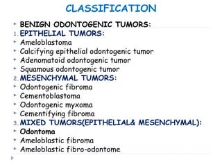
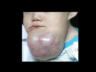
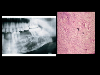
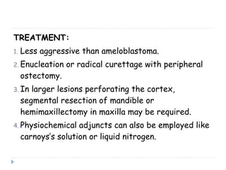
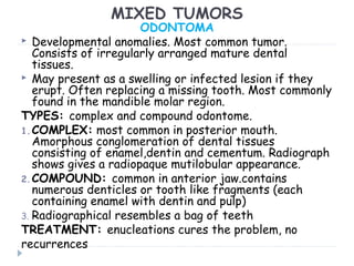
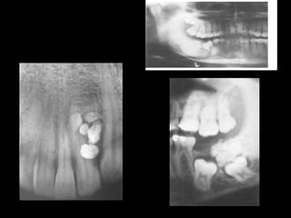
BENIGN TUMORS OF OROFACIAL
REGION
TYPES OF BENIGN TUMORS
1. ODONTOGENIC:
 These are derived from tooth forming elements.
 The more primitive the dental tissue found, the
more aggressive the tumor and vice versa
 Only found in the jaws
 Locally aggressive and rarely malignant
2. NON ODONTOGENIC :
 They are not derived from dental tissues
 They can be found in other regions of the body.
CLASSIFICATION
 BENIGN ODONTOGENIC TUMORS:
1.EPITHELIAL TUMORS:
 Ameloblastoma
 Calcifying epithelial odontogenic tumor
 Adenomatoid odontogenic tumor
 Squamous odontogenic tumor
2.MESENCHYMAL TUMORS:
 Odontogenic fibroma
 Cementoblastoma
 Odontogenic myxoma
 Cementifying fibroma
3.MIXED TUMORS(EPITHELIAL& MESENCHYMAL):
 Odontoma
 Ameloblastic fibroma
 Ameloblastic fibro-odontome
 NONODONTOGENIC TUMORS:
1. FIBRO-OSSEOUS TUMORS
 Ossifying fibroma
 Juvenile ossifying fibroma
2. LANGERHANS CELL DISEASE
 Chronic localized
 Chronic disseminated
 Acute disseminated
3. GIANT CELL LESIONS:
 Central giant cell granuloma
 Giant cell tumor
 Hyperparathyroidism
 Cherubism
 Aneurysmal bone cyst
4. NEUROGENIC TUMORS
 Schwannoma
 Neurofibroma
5. OSTEOID OSTEOMA AND
OSTEOBLASTOMA
6. OSTEOMA
CHRONDROMA
7. DESMOPLASTIC FIBROMA
AMELOBLASTOMA
 Benign but locally aggressive tumor of the jaw
 The name of the tumor derives from the fact the
diagnostic cells resemble ameloblast
 Histologically these cells are columnar , basally
staining arranged in palisaded along the basement
membrane.
 They are believe to express amelogenin, a
precursor of enamel
 They are divided into three distinct types:
1. Solid
2. Cystic
3. peripheral
PATHOGENESIS OF AMELOBLASTOMA
1. Over expression of antiapoptotic gene Bcl2
2. TNF alpha
3. MMPs and transforming growth factors
4. Low proliferation rate ki67
5. No p53 mutation
SOLID AMELOBLASTOMA
 Various histological patterns including follicular,
plexiform, and granular cell variants….does not have
affect treatment or prognosis
SITE: most common in mandible esp angle ,ramus
area. Rare in maxilla, but infiltration is much faster as
marrow spaces and cavities offer less resistance
OCCURRENCE: third to fifth decade of life. Male
to female ratio is approx equal
CLINICAL APPEARANCE:
1. slow growing, expansile lesion.
2. locally aggressive with buccal and lingual
cortical expansion,root resorption is seen
 INVESTIGATIONS:
1. Radiographic evaluation, OPG shows multilocular
radiolucency with root resorption and
displacement of teeth
2. CT scan in case of larger lesions and esp maxillary
ameloblastoma
3. Aspiration of the swelling will reveal nothing in
case of solid ameloblastoma
4. Incisional biospy, shows typical pallisaded
columnar cells resembling ameloblasts.
 TREATMENT:
1. For solid lesions, enucleation has a high recurrence rate 60-
80%. Cells are present beyond the radiographical margin.
2. Surgical resection should be with a 1 cm safe radiological
margin and soft tissue clearance if supraperiosteal.
3. IAN is sacrificed
4. Maxillary resections often end in maxillectomy with wide
margin clearance from the sinus, nasal and infratemporal
regions
5. After surgical resections or curettage , liquid nitrogen
cryotherapy plays a good role
 RECONSTRUCTION:
1. For the mandible, segmental defects of 5 cm can
be reconstructed with bone grafts most commonly
iliac crest.
2. For more larger defects free osseofasiocutaneous
flaps may be used.. DCIA and free fibular flap.
3. Reconstruction plates are used as adjuncts to
grafts or as an interim treatment modality
4. For maxilla, depending upon the defect, local,
regional flaps can be used but prosthetic options
serve the best purpose
CYSTIC AMELOBLASTOMA
 Unicystic ameloblastoma, less aggressive variant.
 Most common in younger population.
 Mostly located in posterior mandible and anterior
maxilla
 All other features are the same
 Enucleation alone may be insufficient, the cavity
should be treated with peripheral ostectomy, or
liquid nitrogen or both.
PERIPHERAL AMELOBLASTOMA
 Occurs in gingiva with no bony involvement
 Painless, sessile growth, firm and exophytic
 Complete excision solves the problem
CALCIFYING EPITHELIAL ODONTOGENIC
TUMOR
 Also called Pindborg tumor after he described it in
1955. derived from stratum intermedium
 Wide age range from 13- 80 yrs, no gender
predeliction.
SITE: mandibular premolar area.
CLINICAL BEHAVIOUR: most are asymptomatic,
slow growing in nature. In maxilla may cause, nasal
obstruction, proptosis or epistaxis
INVESTIGATIONS:
1. On radiographs, they appear as mixed radiolucent
radiopaque, unilocular or mutilocular lesions
2. Biopsy will reveal sheets of epithelial cells in a stroma
filled with hyaline like homogenous material and
concentric calcification rings called liesegang rings
TREATMENT:
 Less aggressive than ameloblastoma, however
malignant variant (odontogenic carcinoma) has
been identified.
 Wide excision with 5- 10 mm margin, which in
mandible may result in marginal or segmental
resection followed by reconstruction.
 Recurrence rate is 14-20%
ADENOMATOID ODONTOGENIC
TUMOR
 Occurs only in jaws. Resemblance to structures
present in enamel formation
 OCCURRENCE: most common in females; age of
occurrence is 5 -30 yrs.
 SITE: anterior maxilla, impacted teeth
 RADIOGRAPHIC APPEARANCE: pear shaped
radiolucency with speckled opaque foci indicating
calcification. Divergence of roots is seen
 TREATMENT: simple enucleation seems all that
is necessary.
SQUAMOUS ODONTOGENIC TUMOR
 Rare tumor originates from the rest of malassez
in the periodontium.
 Occurrence in both mandible(post) and maxilla
(ant)
 Presents with pain and tooth mobility
 Radiographic appearance is a semilunar
radiolucency associated with erupting teeth.
 Treatment is conservative excision
MESENCHYMAL ODONTOGENIC
TUMORS
CEMENTOBLASTOMA
 Rare tumor , most common in second and third
decade of life, lower molar region.
 Intimately associated with the root of tooth.
Tooth remains vital and symptoms of low grade
pain and cortical expansion are present.
RADIOGRAPH: radiopaque lesion attached to and
surrounding the root of a tooth. Must be
distinguished from focal sclerosing osteomyelitis,
odontoma and hypercementosis.
HISTOLOGY: Difficult to distinguish cementum
and bone. It is cemental variant of osteoblastoma
TREATMENT: surgical removal of tooth and lesion.
Peripheral ostectomy is recommended
ODONTOGENIC MYXOMA
 15-20% of odontogenic tumors. Second most
common tumor
OCCURRENCE: second through fourth decade of
life. More common in females. Two thirds occur in
mandible and one third in maxilla.
CLINICAL APPEARANCE: presents as a swelling
locally aggressive.
RADIOGRAPH: similar to ameloblastoma,
multilocular radiolucency
HISTOLOGY: loose mesenchymal tissue . Derived
from dental pulp or primitive dental papilla. Bland
histological appearance.
TREATMENT:
1. Less aggressive than ameloblastoma.
2. Enucleation or radical curettage with peripheral
ostectomy.
3. In larger lesions perforating the cortex,
segmental resection of mandible or
hemimaxillectomy in maxilla may be required.
4. Physiochemical adjuncts can also be employed like
carnoys’s solution or liquid nitrogen.
MIXED TUMORS
ODONTOMA
 Developmental anomalies. Most common tumor.
Consists of irregularly arranged mature dental
tissues.
 May present as a swelling or infected lesion if they
erupt. Often replacing a missing tooth. Most commonly
found in the mandible molar region.
TYPES: complex and compound odontome.
1.COMPLEX: most common in posterior mouth.
Amorphous conglomeration of dental tissues
consisting of enamel,dentin and cementum. Radiograph
shows gives a radiopaque mutilobular appearance.
2.COMPOUND: common in anterior jaw.contains
numerous denticles or tooth like fragments (each
containing enamel with dentin and pulp)
3. Radiographical resembles a bag of teeth
TREATMENT: enucleations cures the problem, no
recurrences
AMELOBLASTIC FIBROMA
OCCURRENCE: young people second and third
decade of life. No gender predilection. Most
common site is mandibular bicuspids area.
RADIORAPHIC VIEW: unilocular/ multilocular well
defined radiolucency. Teeth may be displaced but
rarely resorbed.
HISTOLOGY: islands of odontogenic epithel;ium in
myxomatous stroma
TREATMENT: encapsulated lesion so enucleation is
done. Malignant variant is ameloblastic
fibrosarcoma.
AMELOBLASTIC FIBRO-ODONTOMA
 Combination of ameloblastic fibroma with an
odontoma.(complex or compound).
 Radiographically a mixed radiopaque/ radiolucent
lesion
 Treatment is enucleation
NON ODONTOGENIC TUMORS
 FIBRO OSSEOUS LESIONS
1. Fibrous dysplasia
2. Ossifying fibroma
3. Cemento-osseous dysplasia
OSSIFYING FIBROMA:
replacement of normal bone by fibrous tissue and
variable amount of newly formed bone and cementum
like structures
CLINICAL FEATURES:
1. Painless, slowly growing swelling
2. Third and fourth decade of life
3. Females are more commonly affected
4. Mandible(premolar –molar ) most commonly affected
5. Juvenile OF is a variant, occurring in children and
involving the craniofacial complex
 RADIOLOGICAL FEATURES:
1. Well defined radiolucency with internal
calcifications.
2. The borders may be sclerotic
3. Root displacement and resorption is seen
 HISTOLOGY:
Fibrous stroma with bony trabaculae and cementum
like spherules
 TREATMENT:
1. Complete surgical excision
2. Stripping of the lesion is easy
3. Root that are resorbed are removed
FIBROUS DYSPLASIA
 Monostotic
 Polyostotic (Mc Cune Albright Syndrome)
 Usually in females
 In second and third decade of life
 Related to hormones
 Maxilla more favored site
 Ground glass appearance on radiograph
 The lesion’s margins are not well demarcated
 Similar histological picture as ossifying fibroma
 Recontouring after puberty
LANGERHANS CELL DISEASE
Formerly known as histiocytosis X and three
clinically distinct diseases
1. Eosinophilic granuloma
2. Hand- Schuller –Christian disease
3. Letterer-Siwe disease
PATHOGENESIS:
characterized by proliferation of Langerhans cells
and eosinophils along with other inflammatory cells.
 Langerhan cells are dendritic cells found in
mucosa, epidermis, lymph nodes that process
and present antigens to T lymphocytes.
 Overwelming allergenic response or viral
etiology
CLINICAL PRESENTATION
Affects children and young adults. 3 forms exists
1. Chronic localized or eosinophilic
granuloma….refers to solitary or multiple lesions
bone lesions only
2. Chronic disseminated disease or Hand- Schuller-
Christian disease …clinical triad of lytic bone
lesions, exophthalmos and diabetes inspidus
3. Acute Disseminated Langerhans or Letterer-
Siwe …affects infants, affecting skin, bones and
internal organs esp lungs and liver.
4. Bone lesions involve the skulls, mandible, ribs
and vertebrae. Jaw lesions produce pain,
mobility
RADIOGRAPHIC APPEARANCE:
 Well defined punched out radiolucency
 floating teeth may be seen
 Involved teeth are vital
HISTOLOGY:
They are diagnosed with immunohistochemical
studies with S-100 and CD1a antigen.
Langerhan cell contains cytoplasmic structures
called Birbeck granules
TREATMENT:
1. Accessible bone lesions are treated with curettage
and 5mm marginal resection
2. Less accessible lesions are treated with radiotherapy
3. Intralesional steroids are also employed in resolving
the lesions
GIANT CELL LESIONS
 CENTRAL GIANT CELL GRANULOMA
 GIANT CELL TUMOR
 HYPERPARATHYROIDISM(BROWN’S TUMOR)
 CHERUBISM
 ANEURYSMAL BONE CYST
GIANT CELL LESIONS
 CENTRAL GIANT CELL GRANULOMA:
Benign proliferation of fibroblasts and
multinucleated giant cells
PATHOGENESIS:
Proliferation of fibroblasts….cytokines release…
of monocytes…transform into multinucleated giant
Cells.. Osteoclasts begin to destroy bone
OCCURENCE:
1. Found in young children and young adults, before
30 yrs
2. Females most commonly affected
3. Lesions occur anterior to 1st
molar and cross the
midline
CLINICAL APPEARANCE
 Non aggressive , slowly expanding lesion
 Aggressive lesion , cortical expansion, root resorption
RADIOGRAPHICAL APPEARANCE:
Unilocular/ multilocular radiolucency
HISTOLOGY:
Few to many giant cells in a background of
Fibroblasts
TREATMENT:
1. Surgical curettage with 15-20 % recurrence
2. Intralesional steroids(weekly injections of
triamcinolone for 6 wks)
3. Subcutaneous injection of calcitonin (giant cell have
calcitonin receptors)…inhibits of osteoclastogenesis
4. Alpha-interferon is also given (will suppress the
angiogenic component of the lesion)
HYPERPARATHYROIDISM
 Overproduction of parathyroid hormone.
1. Primary hyperparathyroidism…uncontrolled
production due to an adenoma, hyperplasia
2. Secondary hyperparathyroidism..in response to
hypocalcemia due to chronic renal failure
PATHOGENESIS:
Excess PTH.. Stimulates osteoclast mediated bone
resorption…focal bone lesion…brown’s tumor due to
clinical appearance…erythrocyte extravasation
Radiologically and histologically indistinguishable
from GCG
HOW TO APPROACH SUCH PATIENTS
 Pay attention to clinical signs/ symptoms
 Groans, moans, stones and bones
 Serum calcium and phosphate, PTH, alkaline
phosphatase are done
 Raised PTH, raised serum Ca and raised ALP..
Primary hyperparathyroidism
 Raised PTH, Low serum Ca, Normal ALP, Renal
functions abnormal…secondary
hyperparathyroidism
 Normal PTH.. Giant cell granuloma
 TREATMENT:
 Treat the underlying cause.. Regression of lesion
CHERUBISM
 Autosomal dominant hereditary disorder.
 Genetic defect is in chromosome 4p16
CLINICAL APPEARANCE:
 Children btw 2 and 5 yrs
 Begins as painless bilateral symmetric expansion
of the jaws
 Mand angle, ramus, retromolar area, max
tuberosity
 Max involvement may raise the orbital floor,
globes may be displaced upwards, resembling
cherubes in renaissance paintings
RADIOGRAPHIC APPEARANCE:
 Multilocular radiolucency with thin cortices
 Premature exfoliation of primary teeth
TREATMENT:
Lesions enlarge uptil puberty, then regress
Later on, residual expansions can be contoured
OSTEOID OSTEOMA OSTEOBLASTOMA
 Less than 2cm
 Femur, tibia and phalanges
 Nocturnal pain relieved by
aspirin
 Radiographically, mixed
radiolucent-radiopaque
pattern
 Histologically irregular
trabeculae of osteoid and
immature bone
 Treatment.. Conservative
excision
 Greater than 2 cm
 Vertebrae and bones
of cranium, mandible
 Pain is not nocturnal
and not relieved by
aspirin
 Same as osteoid
osteoma
 Same
 same
OSTEOMA
 Benign tumors of mature cortical and cancellous
bone
 Two types: peripheral osteoma;endosteal osteoma
 May arise in paranasal sinuses, skull and facial
bones
 Periosteal osteoma may present as slow growing
lesions
 Endosteal osteoma are asymptomatic.
 Solitary lesions or multiple in no for eg Gardeners
syndrome(multiple osteomas, intestinal polyps,
fibromas skin, odontomes)
Benign tumors

Benign tumors

  • 1.
    BENIGN TUMORS OFOROFACIAL REGION
  • 2.
    TYPES OF BENIGNTUMORS 1. ODONTOGENIC:  These are derived from tooth forming elements.  The more primitive the dental tissue found, the more aggressive the tumor and vice versa  Only found in the jaws  Locally aggressive and rarely malignant 2. NON ODONTOGENIC :  They are not derived from dental tissues  They can be found in other regions of the body.
  • 3.
    CLASSIFICATION  BENIGN ODONTOGENICTUMORS: 1.EPITHELIAL TUMORS:  Ameloblastoma  Calcifying epithelial odontogenic tumor  Adenomatoid odontogenic tumor  Squamous odontogenic tumor 2.MESENCHYMAL TUMORS:  Odontogenic fibroma  Cementoblastoma  Odontogenic myxoma  Cementifying fibroma 3.MIXED TUMORS(EPITHELIAL& MESENCHYMAL):  Odontoma  Ameloblastic fibroma  Ameloblastic fibro-odontome
  • 4.
     NONODONTOGENIC TUMORS: 1.FIBRO-OSSEOUS TUMORS  Ossifying fibroma  Juvenile ossifying fibroma 2. LANGERHANS CELL DISEASE  Chronic localized  Chronic disseminated  Acute disseminated 3. GIANT CELL LESIONS:  Central giant cell granuloma  Giant cell tumor  Hyperparathyroidism  Cherubism  Aneurysmal bone cyst 4. NEUROGENIC TUMORS  Schwannoma  Neurofibroma 5. OSTEOID OSTEOMA AND OSTEOBLASTOMA 6. OSTEOMA CHRONDROMA 7. DESMOPLASTIC FIBROMA
  • 5.
    AMELOBLASTOMA  Benign butlocally aggressive tumor of the jaw  The name of the tumor derives from the fact the diagnostic cells resemble ameloblast  Histologically these cells are columnar , basally staining arranged in palisaded along the basement membrane.  They are believe to express amelogenin, a precursor of enamel  They are divided into three distinct types: 1. Solid 2. Cystic 3. peripheral
  • 7.
    PATHOGENESIS OF AMELOBLASTOMA 1.Over expression of antiapoptotic gene Bcl2 2. TNF alpha 3. MMPs and transforming growth factors 4. Low proliferation rate ki67 5. No p53 mutation
  • 8.
    SOLID AMELOBLASTOMA  Varioushistological patterns including follicular, plexiform, and granular cell variants….does not have affect treatment or prognosis SITE: most common in mandible esp angle ,ramus area. Rare in maxilla, but infiltration is much faster as marrow spaces and cavities offer less resistance OCCURRENCE: third to fifth decade of life. Male to female ratio is approx equal CLINICAL APPEARANCE: 1. slow growing, expansile lesion. 2. locally aggressive with buccal and lingual cortical expansion,root resorption is seen
  • 10.
     INVESTIGATIONS: 1. Radiographicevaluation, OPG shows multilocular radiolucency with root resorption and displacement of teeth 2. CT scan in case of larger lesions and esp maxillary ameloblastoma 3. Aspiration of the swelling will reveal nothing in case of solid ameloblastoma 4. Incisional biospy, shows typical pallisaded columnar cells resembling ameloblasts.
  • 12.
     TREATMENT: 1. Forsolid lesions, enucleation has a high recurrence rate 60- 80%. Cells are present beyond the radiographical margin. 2. Surgical resection should be with a 1 cm safe radiological margin and soft tissue clearance if supraperiosteal. 3. IAN is sacrificed 4. Maxillary resections often end in maxillectomy with wide margin clearance from the sinus, nasal and infratemporal regions 5. After surgical resections or curettage , liquid nitrogen cryotherapy plays a good role
  • 13.
     RECONSTRUCTION: 1. Forthe mandible, segmental defects of 5 cm can be reconstructed with bone grafts most commonly iliac crest. 2. For more larger defects free osseofasiocutaneous flaps may be used.. DCIA and free fibular flap. 3. Reconstruction plates are used as adjuncts to grafts or as an interim treatment modality 4. For maxilla, depending upon the defect, local, regional flaps can be used but prosthetic options serve the best purpose
  • 16.
    CYSTIC AMELOBLASTOMA  Unicysticameloblastoma, less aggressive variant.  Most common in younger population.  Mostly located in posterior mandible and anterior maxilla  All other features are the same  Enucleation alone may be insufficient, the cavity should be treated with peripheral ostectomy, or liquid nitrogen or both.
  • 17.
    PERIPHERAL AMELOBLASTOMA  Occursin gingiva with no bony involvement  Painless, sessile growth, firm and exophytic  Complete excision solves the problem
  • 18.
    CALCIFYING EPITHELIAL ODONTOGENIC TUMOR Also called Pindborg tumor after he described it in 1955. derived from stratum intermedium  Wide age range from 13- 80 yrs, no gender predeliction. SITE: mandibular premolar area. CLINICAL BEHAVIOUR: most are asymptomatic, slow growing in nature. In maxilla may cause, nasal obstruction, proptosis or epistaxis INVESTIGATIONS: 1. On radiographs, they appear as mixed radiolucent radiopaque, unilocular or mutilocular lesions 2. Biopsy will reveal sheets of epithelial cells in a stroma filled with hyaline like homogenous material and concentric calcification rings called liesegang rings
  • 20.
    TREATMENT:  Less aggressivethan ameloblastoma, however malignant variant (odontogenic carcinoma) has been identified.  Wide excision with 5- 10 mm margin, which in mandible may result in marginal or segmental resection followed by reconstruction.  Recurrence rate is 14-20%
  • 21.
    ADENOMATOID ODONTOGENIC TUMOR  Occursonly in jaws. Resemblance to structures present in enamel formation  OCCURRENCE: most common in females; age of occurrence is 5 -30 yrs.  SITE: anterior maxilla, impacted teeth  RADIOGRAPHIC APPEARANCE: pear shaped radiolucency with speckled opaque foci indicating calcification. Divergence of roots is seen  TREATMENT: simple enucleation seems all that is necessary.
  • 22.
    SQUAMOUS ODONTOGENIC TUMOR Rare tumor originates from the rest of malassez in the periodontium.  Occurrence in both mandible(post) and maxilla (ant)  Presents with pain and tooth mobility  Radiographic appearance is a semilunar radiolucency associated with erupting teeth.  Treatment is conservative excision
  • 23.
    MESENCHYMAL ODONTOGENIC TUMORS CEMENTOBLASTOMA  Raretumor , most common in second and third decade of life, lower molar region.  Intimately associated with the root of tooth. Tooth remains vital and symptoms of low grade pain and cortical expansion are present. RADIOGRAPH: radiopaque lesion attached to and surrounding the root of a tooth. Must be distinguished from focal sclerosing osteomyelitis, odontoma and hypercementosis. HISTOLOGY: Difficult to distinguish cementum and bone. It is cemental variant of osteoblastoma TREATMENT: surgical removal of tooth and lesion. Peripheral ostectomy is recommended
  • 25.
    ODONTOGENIC MYXOMA  15-20%of odontogenic tumors. Second most common tumor OCCURRENCE: second through fourth decade of life. More common in females. Two thirds occur in mandible and one third in maxilla. CLINICAL APPEARANCE: presents as a swelling locally aggressive. RADIOGRAPH: similar to ameloblastoma, multilocular radiolucency HISTOLOGY: loose mesenchymal tissue . Derived from dental pulp or primitive dental papilla. Bland histological appearance.
  • 27.
    TREATMENT: 1. Less aggressivethan ameloblastoma. 2. Enucleation or radical curettage with peripheral ostectomy. 3. In larger lesions perforating the cortex, segmental resection of mandible or hemimaxillectomy in maxilla may be required. 4. Physiochemical adjuncts can also be employed like carnoys’s solution or liquid nitrogen.
  • 28.
    MIXED TUMORS ODONTOMA  Developmentalanomalies. Most common tumor. Consists of irregularly arranged mature dental tissues.  May present as a swelling or infected lesion if they erupt. Often replacing a missing tooth. Most commonly found in the mandible molar region. TYPES: complex and compound odontome. 1.COMPLEX: most common in posterior mouth. Amorphous conglomeration of dental tissues consisting of enamel,dentin and cementum. Radiograph shows gives a radiopaque mutilobular appearance. 2.COMPOUND: common in anterior jaw.contains numerous denticles or tooth like fragments (each containing enamel with dentin and pulp) 3. Radiographical resembles a bag of teeth TREATMENT: enucleations cures the problem, no recurrences
  • 30.
    AMELOBLASTIC FIBROMA OCCURRENCE: youngpeople second and third decade of life. No gender predilection. Most common site is mandibular bicuspids area. RADIORAPHIC VIEW: unilocular/ multilocular well defined radiolucency. Teeth may be displaced but rarely resorbed. HISTOLOGY: islands of odontogenic epithel;ium in myxomatous stroma TREATMENT: encapsulated lesion so enucleation is done. Malignant variant is ameloblastic fibrosarcoma.
  • 32.
    AMELOBLASTIC FIBRO-ODONTOMA  Combinationof ameloblastic fibroma with an odontoma.(complex or compound).  Radiographically a mixed radiopaque/ radiolucent lesion  Treatment is enucleation
  • 33.
    NON ODONTOGENIC TUMORS FIBRO OSSEOUS LESIONS 1. Fibrous dysplasia 2. Ossifying fibroma 3. Cemento-osseous dysplasia OSSIFYING FIBROMA: replacement of normal bone by fibrous tissue and variable amount of newly formed bone and cementum like structures CLINICAL FEATURES: 1. Painless, slowly growing swelling 2. Third and fourth decade of life 3. Females are more commonly affected 4. Mandible(premolar –molar ) most commonly affected 5. Juvenile OF is a variant, occurring in children and involving the craniofacial complex
  • 34.
     RADIOLOGICAL FEATURES: 1.Well defined radiolucency with internal calcifications. 2. The borders may be sclerotic 3. Root displacement and resorption is seen  HISTOLOGY: Fibrous stroma with bony trabaculae and cementum like spherules  TREATMENT: 1. Complete surgical excision 2. Stripping of the lesion is easy 3. Root that are resorbed are removed
  • 36.
    FIBROUS DYSPLASIA  Monostotic Polyostotic (Mc Cune Albright Syndrome)  Usually in females  In second and third decade of life  Related to hormones  Maxilla more favored site  Ground glass appearance on radiograph  The lesion’s margins are not well demarcated  Similar histological picture as ossifying fibroma  Recontouring after puberty
  • 37.
    LANGERHANS CELL DISEASE Formerlyknown as histiocytosis X and three clinically distinct diseases 1. Eosinophilic granuloma 2. Hand- Schuller –Christian disease 3. Letterer-Siwe disease PATHOGENESIS: characterized by proliferation of Langerhans cells and eosinophils along with other inflammatory cells.  Langerhan cells are dendritic cells found in mucosa, epidermis, lymph nodes that process and present antigens to T lymphocytes.  Overwelming allergenic response or viral etiology
  • 38.
    CLINICAL PRESENTATION Affects childrenand young adults. 3 forms exists 1. Chronic localized or eosinophilic granuloma….refers to solitary or multiple lesions bone lesions only 2. Chronic disseminated disease or Hand- Schuller- Christian disease …clinical triad of lytic bone lesions, exophthalmos and diabetes inspidus 3. Acute Disseminated Langerhans or Letterer- Siwe …affects infants, affecting skin, bones and internal organs esp lungs and liver. 4. Bone lesions involve the skulls, mandible, ribs and vertebrae. Jaw lesions produce pain, mobility
  • 39.
    RADIOGRAPHIC APPEARANCE:  Welldefined punched out radiolucency  floating teeth may be seen  Involved teeth are vital HISTOLOGY: They are diagnosed with immunohistochemical studies with S-100 and CD1a antigen. Langerhan cell contains cytoplasmic structures called Birbeck granules TREATMENT: 1. Accessible bone lesions are treated with curettage and 5mm marginal resection 2. Less accessible lesions are treated with radiotherapy 3. Intralesional steroids are also employed in resolving the lesions
  • 41.
    GIANT CELL LESIONS CENTRAL GIANT CELL GRANULOMA  GIANT CELL TUMOR  HYPERPARATHYROIDISM(BROWN’S TUMOR)  CHERUBISM  ANEURYSMAL BONE CYST
  • 42.
    GIANT CELL LESIONS CENTRAL GIANT CELL GRANULOMA: Benign proliferation of fibroblasts and multinucleated giant cells PATHOGENESIS: Proliferation of fibroblasts….cytokines release… of monocytes…transform into multinucleated giant Cells.. Osteoclasts begin to destroy bone OCCURENCE: 1. Found in young children and young adults, before 30 yrs 2. Females most commonly affected 3. Lesions occur anterior to 1st molar and cross the midline
  • 43.
    CLINICAL APPEARANCE  Nonaggressive , slowly expanding lesion  Aggressive lesion , cortical expansion, root resorption RADIOGRAPHICAL APPEARANCE: Unilocular/ multilocular radiolucency HISTOLOGY: Few to many giant cells in a background of Fibroblasts TREATMENT: 1. Surgical curettage with 15-20 % recurrence 2. Intralesional steroids(weekly injections of triamcinolone for 6 wks) 3. Subcutaneous injection of calcitonin (giant cell have calcitonin receptors)…inhibits of osteoclastogenesis 4. Alpha-interferon is also given (will suppress the angiogenic component of the lesion)
  • 45.
    HYPERPARATHYROIDISM  Overproduction ofparathyroid hormone. 1. Primary hyperparathyroidism…uncontrolled production due to an adenoma, hyperplasia 2. Secondary hyperparathyroidism..in response to hypocalcemia due to chronic renal failure PATHOGENESIS: Excess PTH.. Stimulates osteoclast mediated bone resorption…focal bone lesion…brown’s tumor due to clinical appearance…erythrocyte extravasation Radiologically and histologically indistinguishable from GCG
  • 46.
    HOW TO APPROACHSUCH PATIENTS  Pay attention to clinical signs/ symptoms  Groans, moans, stones and bones  Serum calcium and phosphate, PTH, alkaline phosphatase are done  Raised PTH, raised serum Ca and raised ALP.. Primary hyperparathyroidism  Raised PTH, Low serum Ca, Normal ALP, Renal functions abnormal…secondary hyperparathyroidism  Normal PTH.. Giant cell granuloma  TREATMENT:  Treat the underlying cause.. Regression of lesion
  • 47.
    CHERUBISM  Autosomal dominanthereditary disorder.  Genetic defect is in chromosome 4p16 CLINICAL APPEARANCE:  Children btw 2 and 5 yrs  Begins as painless bilateral symmetric expansion of the jaws  Mand angle, ramus, retromolar area, max tuberosity  Max involvement may raise the orbital floor, globes may be displaced upwards, resembling cherubes in renaissance paintings
  • 49.
    RADIOGRAPHIC APPEARANCE:  Multilocularradiolucency with thin cortices  Premature exfoliation of primary teeth TREATMENT: Lesions enlarge uptil puberty, then regress Later on, residual expansions can be contoured
  • 51.
    OSTEOID OSTEOMA OSTEOBLASTOMA Less than 2cm  Femur, tibia and phalanges  Nocturnal pain relieved by aspirin  Radiographically, mixed radiolucent-radiopaque pattern  Histologically irregular trabeculae of osteoid and immature bone  Treatment.. Conservative excision  Greater than 2 cm  Vertebrae and bones of cranium, mandible  Pain is not nocturnal and not relieved by aspirin  Same as osteoid osteoma  Same  same
  • 53.
    OSTEOMA  Benign tumorsof mature cortical and cancellous bone  Two types: peripheral osteoma;endosteal osteoma  May arise in paranasal sinuses, skull and facial bones  Periosteal osteoma may present as slow growing lesions  Endosteal osteoma are asymptomatic.  Solitary lesions or multiple in no for eg Gardeners syndrome(multiple osteomas, intestinal polyps, fibromas skin, odontomes)