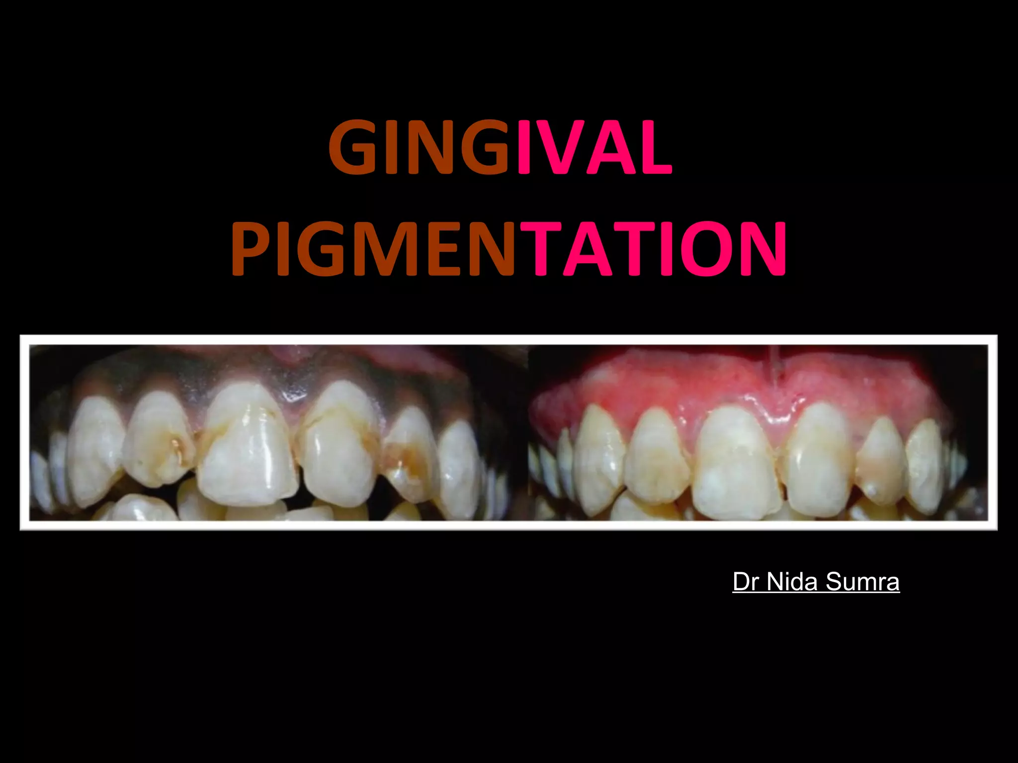
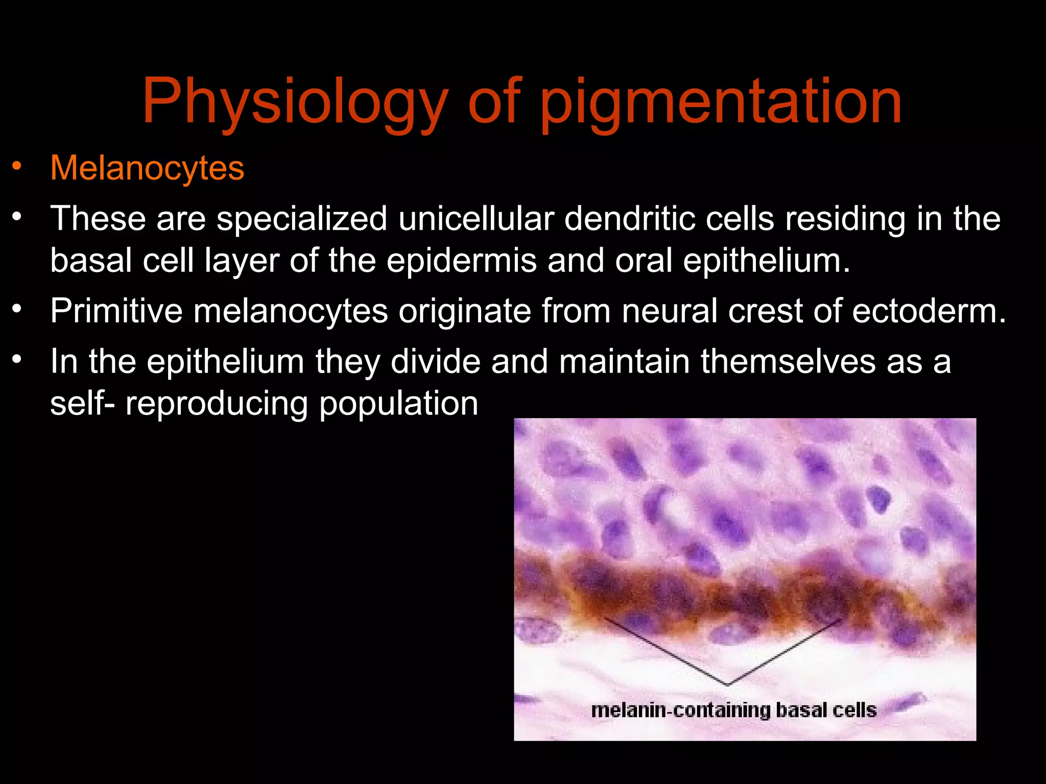
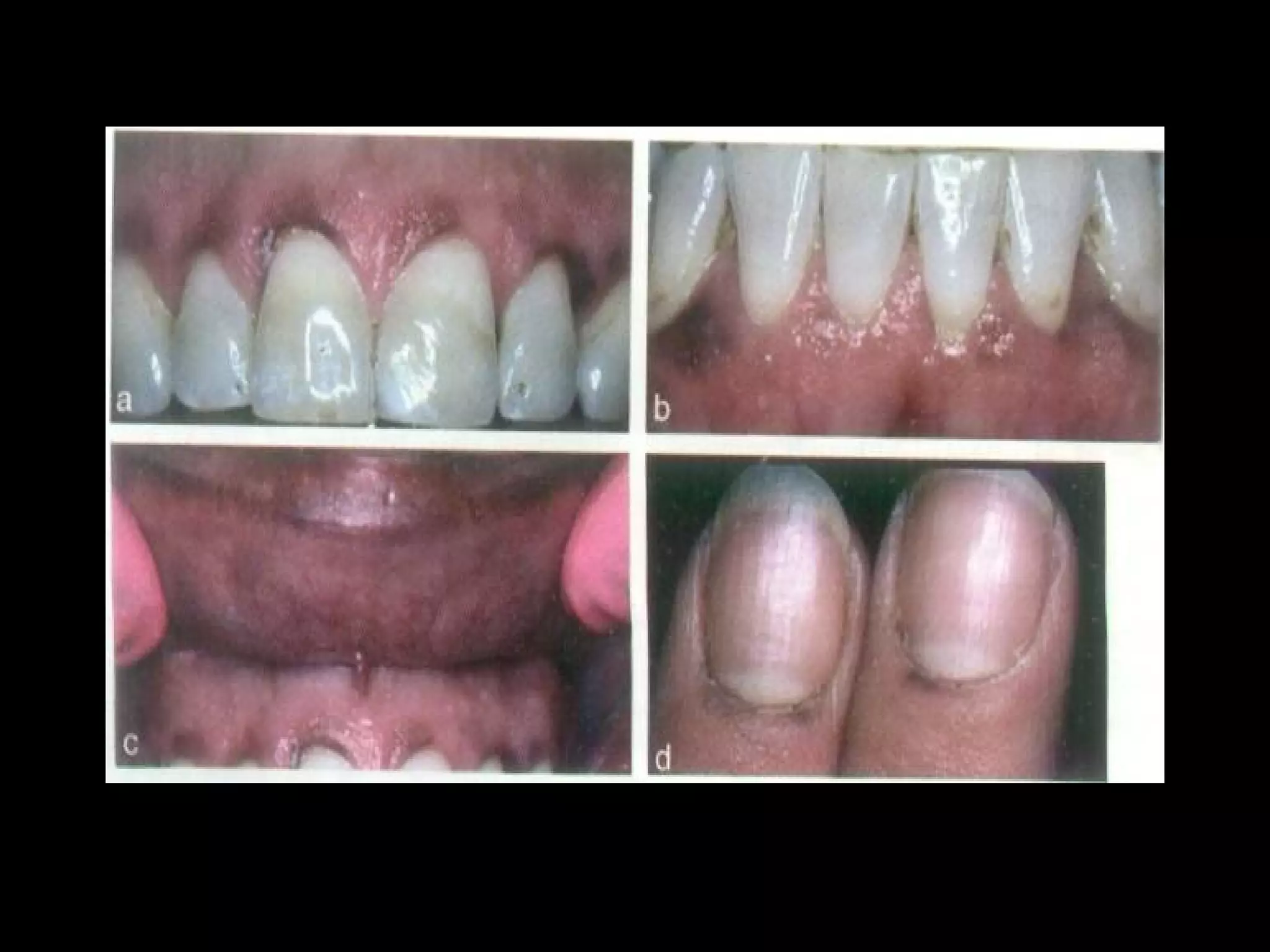
GINGIVAL
PIGMENTATION
Dr Nida Sumra
CONTENTS
• Introduction
• Historical aspects
• Physiology of pigmentation
• Normal racial variations
• Classification of oral pigmentation
• Pigmented lesions of the oral mucosa
• Methods of depigmentation
• Conclusion
• References
INTRODUCTION
Historical aspects
• 1912 - Bonnet and Laboef described physiologic
pigmentation of oral mucosa.
• 1914 - Brault and Monpellier and in 1923 - Lortat –
Jacob & Solente also reported the normal occurance of
pigmentation in the French people and fillipinos.
• 1924 - Reiche described the occurance of oral
pigmentation in Arabian, Chinese and East Indians.
• 1945 – Dummet C.O. reported pigmentation variations in
healthy oral tissues of more than 600 negros.
Physiology of pigmentation
• Melanocytes
• These are specialized unicellular dendritic cells residing in the
basal cell layer of the epidermis and oral epithelium.
• Primitive melanocytes originate from neural crest of ectoderm.
• In the epithelium they divide and maintain themselves as a
self- reproducing population
Structure
• Round cells with pale-
staining cytoplasm.
• Lack desmosomes and
tonofilaments but possess
long dendritic processes.
• Melanosomes.
• Melanocytes are cells capable of synthesizing
tyrosinase, which, when incorporated within specialized
organelles, the melanosomes, initiates events leading to
the synthesis and deposition of melanin.
• Routine hematoxylin
and eosin (H&E)
preparations
• Replicate throughout
their life, although at a
much slower rate
• According to the degree of maturation, melanosomes are
classified in 4 stages.
• Stage I. Membrane delinated vesicles containing
tyrosinase and a proteinateous matrix.
• Stage II. Oval organels with numerous membrane
filaments distinctive periodicity.
• Stage III. Less periodicity and melanin deposition.
• Stage IV. A dense uniform particle without any
distinguishable internal structure.
Melanin
Structure
• The exact structure of melanin has not been
determined.
• Melanin is defined as a biochrome of high
molecular weight formed by the enzymatic
oxidation of phenyl alanine.
• Insoluble polymer of tyrosine derivative formed
from tyrosine by the action of copper.
• The first step of the biosynthetic pathway for both eumelanins
and pheomelanins is catalysed by tyrosinase:
• Tyrosine → DOPA → dopaquinone
Dopaquinone can combine with cysteine - pheomelanins
• Dopaquinone + cysteine → benzothiazine intermediate →
pheomelanin
Also, dopaquinone can be converted to leucodopachrome -
eumelanins
• Dopaquinone → leucodopachrome → dopachrome →
quinone → eumelanin
Regulation of melanin synthesis
• Melanin synthesis can be controlled by three major
determinants:
Genes
Hormones
U-V Radiation
Endocrine control
• Melanocyte stimulating harmone (MSH)
• Adrenocorticotropic hormone (ACTH)
• Androgens
Normal racial variations
• Common characteristic of the darker races including
Caucasian brunettes.
• Most usual cases of brown oral pigmentation are ethnic.
• The pigmentation is usually symmetrically distributed
over the gingiva and palatal mucosa.
• Dumett examined 600 negroes of varying shades of
complexion to determine the relationship of oral to cutaneous
pigmentation and found that gingival pigmentation was directly
proportional to skin pigmentation
• Erica Amir et al (1991) studied the oral physiologic
pigmentation and found that it varied, according to the intensity
of the skin pigmentation.
• Patsakas et al (1991) conducted a study to determine the
distribution of melanin granules in different anatomical areas of
the gingiva and to relate the melanin granules to the degree of
gingival inflammation. The results showed positive correlation
between the density of melanin granules of the vestibular
epithelium and the severity of inflammation.
CLASSIFICATION OF ORAL
PIGMENTATION
• Endogenous pigmentation
• Exogenous pigmentation
PIGMENTED LESIONS OF
ORAL MUCOSA
Blue / purple vascular lesions
Brown melanotic lesions
Brown heme Associated lesions
Gray / black pigmentation
VASCULAR LESIONS.
Kaposi’s sarcoma
• Tumor of putative vascular origin
• Described by Morietz Kaposi (1872)
• Slow progressive growth.
• Diagnostic sign for AIDS
Clinical Appearance
• The oral mucosa is red, blue and purple and hard palate is
most favored site.
• Multifocal with numerous isolated and coalescing plaques.
Some will involve entire palate.
• Facial gingiva second most favored oral site
Management
Early plaque or macular lesions no treatment.
Nodular lesions are surgically excised.
Intra lesions injection of 1% sodium, tetradecyl
sulfate.
BROWN MELANOTIC LESIONS
NEVOCELLULAR AND BLUE NEVI
• Nevi are benign proliferation of melanocytes.
Nevo cellular nevi
• Arise from basal cell layer melanocyte early in life
• Called as junctional nevi when macular.
• Flat, brown and have a regular round or oval out line.
• Dome shaped navei
Blue nevus
• Not derived from basal
layer melanocytes
• Blue in color on the skin
• Biopsy necessary for
diagnostic confirmation.
Treatment
• Simple excision
• Dyer et al (1993) reported a case of pigmented naevus
of the gingiva which was a recurrent lesion. The
histological examination confirmed the presence of
junctional activity of melanocytes at the epithelio
mesenchymal interface.
MALIGNANT MELANOMA
Melanoma may arise from neoplastic transformation of either
melanocytes or nevus cells.
Clinical features
• Melanomas of oral mucosa less common than cutaneous.
• Oral lesions mainly in hard palate and gingiva followed by lips
and buccal mucosa.
• Mixture of color such as brown, black, blue and red
• Can be asymmetrical or irregular margins.
• Depending on radial and vertical growth sub classified
Nodular melanoma
– Superficial spreading melanoma
– Lentigo malignant melanoma
– Acral lentiguous melanoma
• Management
– Oral melanomas highly malignant and have high
mortality.
– Biopsy essential for diagnosis.
– Destruction of underlying bone or metastasis
indicate poor outcome.
– Excision with centimeter wide margins followed by
radical radiotherapy.
– Immunotherapy with interferon and gene therapy
is also under trial.
DRUG INDUCED MELANOSIS
• A variety of drugs can
induce oral mucosal
pigmentation.
• May be localized usually to
the hard palate or multifocal
throughout the mouth.
• Lesions are flat without any
evidence of nodularity or
swelling.
• Antimalarial therapy, tricyclic
antidepressants, Busulfan,
cyclophosphamide,
bleomycin and fluoracil
• Birek et al (1988) reported two cases of pigmentation of the
palatal mucosa due to quindine therapy both the clinical
appearances were those of melanin pigmentation and had
characteristic bluish black color.
• Siller et al (1994) reported oral mucosal pigmentation in two
patients taking the drug minocycline and demonstrated
minocycline deposition within teeth and bone by fluorescence
microscopy.
• Meyerson et al (1995) reported acquired isolated lingual
hyperpigmentation caused by minocycline in two women .They
had been receiving oral minocycline for approximately 4 weeks
to 4 months.
SMOKING ASSOCIATED MELANOSIS
Clinical features
• Diffuse macular melanosis
• Seen on the buccal mucosa,
lateral tongue, palate and
floor of the mouth.
• Lesions are brown, flat and
irregular.
• Hedin et al (1993) compared the frequency of oral
melanin pigmentation in a large number of former
smokers with that of non smokers. Results showed
that although clinically pigment free, the oral mucosa
may have elevated melanin content for a period of
time until the melanocyte activity declines.
• Brown et al (1991) found smoker’s melanosis to be
secondary to tobacco smoking as described by Hedin
and as such has an identifiable etiologic agent or
factor.
ENDOCRINOPATHIC
PIGMENTATION
• Addison’s disease
• Cushings syndrome
Bronzing of the skin and patchy
melanosis, of the gingiva, palate
and buccal mucosa are classical
signs
Oral melanotic pigmentation
accompanied by cutaneous
bronzing.
Serum steroid and ACTH
determination will aid in the
diagnosis
Treatment
• The pigment will disappear once
appropriate therapy for the
endocrine problem is initiated.
GENO DERMATOSES
• Peutz–Jeghers syndrome.
• Albright’s syndrome.
• Neurofibromatosis.
PEUTZ – JEGHERS
SYNDROME
CLINICAL FEATURES
Melanin pigmentation of the
lips and oral mucosa is
usually present from birth.
Buccal mucosa more
frequently involved
followed by gingiva and
hard palate.
Facial pigmentation tends to
fade later on, the mucosa
pigmentation persists.
ALBRIGHTS SYNDROME
• It is also called polyostotic fibrous dysplasia .
• Associated with skin lesions and endocrine
dysfunction
Clinical Features
• Bowing or thickening of long bones – unilateral /
bilateral
• Bone of face and skull are frequently involved.
• Skin lesions described as “Café au-lait” spots
NEUROFIBROMATOSIS
(Von- Reckling hausen’s Disease)
– Multiple neural tumors
Clinical Features
– “Café au-lait” spots.
– Oral lesions present as discrete nodules, which tend to be of
same colour as the oral mucosal,
– Palate, alveolar ridges, vestibules and tongue.
• Most sero positive patients present with diffuse,
multifocal macular, brown pigmentation of the buccal
mucosa
• Gingiva, palate and tongue may also be involved.
Histologically
• Characterized by basilar melanin pigment.
• Alangford et al (1989) reported six cases of oral
hyperpigmentation in HIV infected patients. The lesions
could be related to systemic clofazimine or ketoconazole
therapy.
HIV ORAL MELANOSIS
BROWN HEME ASSOCIATED
LESIONS
HEMOCHROMATOSIS
• Deposition of hemosiderin
pigment in multiple organs
and tissue
• Primary heritable disease or
secondary to variety of
disease
• Clinical features
– Mucosal lesions appear
brown to gray diffuse
macules.
– Seen on the palate and
gingiva.
GRAY BLACK
PIGMENTATIONS
AMALGAM TATTOO
• The lesions are macular and
bluish gray or even black.
• Buchner and Hansen (1990)
evaluated series of cases of
amalgam tattoo, which were
analyzed clinically and
histologically. The most
common location was the
gingiva and alveolar mucosa
followed by the buccal
mucosa. Histologically the
amalgam was present in the
tissues as discrete fine, dark
granules and as irregular solid
fragments.
GRAPHIC TATTOO
• Graphic tattoo occur on
the palate representing
traumatic implantation
from a lead pencil.
• The lesions are usually
macular, focal and gray
or black in colour.
Microscopically
• Graphite resembles
amalgam in tissue although
special stains can
segregate the two.
HEAVY METAL
INGESTION
• Ingestion of heavy
metals or metal salts can
be occupational hazard.
• In oral cavity found along
the free marginal gingiva
• Metallic line is gray or
black in colour.
• Bismuth, lead, mercury,
silver.
DEPIGMENTATION
Hanioka T et al. Association of melanin pigmentation in the gingiva of children with
parents who smoke. Pediatrics Aug 2005;116(2):186-90.
- Santosh Kumar et al
Oral Pigmentation Index –Dummett et al 1964
0: No clinical pigmentation (pink gingiva)
1: Mild clinical pigmentation (mild light brown
color)
2: Moderate clinical pigmentation (medium
brown or mixed pink and brown color)
3: Heavy clinical pigmentation (deep brown or
bluish black color)
METHODS OF DEPIGMENTATION
1. De-epithelization
• a. Scalpel technique
• b. Gingival abrasion technique using diamond bur
• c. Combination of the scalpel and bur
2. Gingivectomy
3. Free gingival autografting
4. Acellular dermal matrix allograft (ADMA)
5. Electrosurgery
6. Cryosurgery
• a. Using liquid nitrogren
• Using a gas expansion system
7. Chemical agents
• a. 90% phenol and 95% alcohol
• b. Ascorbic acid
8. Laser[
• a. Nd:YAG
• b. Semiconductor diode laser
• c. CO2 laser
• d. Argon laser
SURGICAL METHODS
Criteria for patient selection
• Skin shade not very dark toned, but gingiva is deeply
pigmented.
• Periodontal health not compromised or is pretreated.
• Adequate thickness of the periodontal tissues.
Scalpel
• The procedure essentially involves surgical removal of
the gingival epithelium along with a layer of the
underlying connective tissue under adequate local
anesthesia and allowing the denuded connective tissue
to heal by secondary intention.
Split thickness epithelial excision
technique
Gingival abrasion technique using
diamond bur
• Similar to the scalpel technique.
• Comparatively simple, safe and non-aggressive method
that can be easily performed and readily repeated, if
necessary, to eradicate any residual repigmentation.
• Extra care should be taken to control the speed and
pressure of the handpiece bur so as not to cause
unwanted abrasion or pitting of the tissue.
• Minimum pressure with feather light brushing strokes
with copious saline irrigation should be used without
holding the bur in one place to perceive excellent results
Gingivo abrasion technique
Gingivectomy
• Removing the gingival margin by gingivectomy or the entire
attached gingiva by the “push back” procedure may also be
used.
• However, these procedures are associated with alveolar bone
loss, prolonged healing by secondary intention and excessive
pain and discomfort caused by exposure and denudation of the
underlying bone.
• Oswaldo Bergamaschi et al (1993) treated five patients with
gingival pigmentation by gingivectomy to remove band like
melanin pigmentation. They concluded that gingival resective
procedure is not of permanent value, because pigmentation
tends to return to baseline values.
Free gingival graft
• Tamizi et al (1996) used free gingival auto graft treatment of
pigmentation and obtained successful results.
• Nicholas (1981) reported a case of amalgam tattoo of the
labial gingiva in the maxillary anterior region treated by
palatal gingival auto graft successfully.
• Esthetic result was not always satisfactory due to color
differences between the palatal tissues and the gingiva
• The presence of a demarcated line commonly observed
around the graft in the recipient site may itself pose an
esthetic problem.
Acellular dermal matrix allograft
• Novaes et al. reported the use of ADMA for elimination
of the gingival pigmentation.
• These studies demonstrated the efficacy of the ADMA,
with the advantages of reduced surgical time, decreased
postoperative complications, unlimited amount of graft
material and a predictable and satisfactory esthetic
result.
• However, it is expensive and requires clinical expertise.
Electrosurgery
• According to Oringer’s “Exploding cell theory, it is predicted
that electrical energy leads to the molecular disintegration of
melanin cells of the operated and surrounding sites.
• Thus, electrosurgery has a strong influence in retarding
migration of melanin cells.
• However, it requires more expertise.
• Prolonged or repeated application of current to the tissues
induces heat accumulation and undesired tissue destruction.
• Contact of current with the periosteum and vital teeth should
be avoided.
• Hence, it is to be used in light brushing strokes and the tip
has to be kept moving.
CRYOSURGICAL DEPIGMENTATION
• It is a method of tissue destruction by rapid freezing
• The cytoplasm of the cells freezes, leading to
denaturation of proteins and cell death. It does not
require use of local anesthesia or periodontal dressing,
is relatively painless and has shown excellent results
lasting for several years.
• The cryotherapy procedure requires a special container
for storage of liquid nitrogen.
• The depth of penetration is difficult to control and
prolonged freezing may cause excessive tissue
destruction.
• The Dip-stick method utilizes a small cotton bud/swab
dipped in LN, which can be applied on the pigmented
area and maintained in contact for around 20–30 s as
described by Tal et al.
• It requires a separate sitting after about 5 days, during
which the residual pigmentation, if any, may be removed.
• It is followed by considerable swelling and accompanied
by increased soft tissue destruction
• Spray technique is the most popular method using open
end cryoprobes i.e. spray tip cone or cylinder.
• In this method, the cryogen is sprayed directly on to the
lesion. The spray tip is held 1 cm away from the lesion
and a steady spray of liquid nitrogen is directed at the
centre of the lesion.
• In the cryoprobe technique, liquid nitrogen is circulated
so as to cool the tip of the cryoprobe, which is to be
applied on to the lesion.
• It will be not possible to observe immediate tissue
changes after application of the cryogen
CRYOSURGER
Y
Pre-op
Post-op
Liq. N2O
Chemical agents (chemoexfoliation)
• It is a treatment method that destroys the epidermis
and/or dermis using a chemical peeling agent.
• A variety of chemical peeling agents are available;
phenols, salicylic acid, glycolic acid, trichloracetic acid,
etc.
• These agents have been classified into four types: Very
superficial, superficial, medium depth and deep, based
on their penetration
• Phenol penetrates the subepithelial connective tissue
and causes necrosis or apoptosis of melanocytes. It
result in incapacity of melanocytes to normally
synthesize melanin.
• Phenol does not seem to determine melanocyte
destruction; rather, it compromises its activity.
• Hirschfeld reported a series with 20 cases that were
treated for melanin pigmentation with the phenol–alcohol
method.
• It requires the area to be air-dried before application The
phenol pellet is applied and maintained for 1 min and the
area needs to be rinsed with 99% alcohol.
ASCORBIC ACID
• Ascorbic acid significantly inhibits tyrosinase activity
LASERS
• Lasers combine the advantages of rapid healing of the
scalpel surgery and the minimal bleeding of electrosurgery.
• A one-step laser treatment is usually sufficient to eliminate
the pigmented gingiva and does not require a periodontal
dressing.
• Easy handling, short treatment line, hemostasis, sterilization
effects and excellent coagulation.
• The treated area should be left exposed in the mouth. Few
myofibroblasts present in the base of the wound cause
minimal contraction and scarring, with little restriction in
movement of the soft tissues.
• Emin Essen et al (2004) used CO2 laser for gingival
depigmentation. Ablation of the hyperpigmented gingiva was
accomplished with minimal carbonization and almost no
bleeding and concluded that CO2 laser appears to be safe
and effective procedure.
• Yousuf et al (2000) - a semiconductor diode laser and it
was effective in removing melanin. Post operative
reevaluation showed continuous healing process with the
proliferation of squamous epithelial cells.
• Ishikawa et al (2004) The Er. YAG laser -possesses
suitable characteristics for oral soft and hard tissue ablation.
It had been applied for effective elimination of granulation
tissue and gingival melanin pigmentation.
REPIGMENTATION
• Repigmentation is described as "spontanoues" and has
been attributed to the activity and migration of cells from
surrounding areas..
• Although the exact mechanism of repigmetnation is not
known, it is suggested that the melanocytes from normal
skin proliferate and migrate into the depigmented areas.
• Further research is needed on gingival repigmentation
to study factors affecting the rate and length of time
required for reappearance of the pigmented areas.
• It should be noted, however, that the simplicity of the
technique prevents permanent damage, unlike many
other periodontal treatments, hence deepithelialization
can be done repeatedly in the same areas for a number
of times.
CONCLUSION.
• Gingival melanin pigmentations can occur as a consequence
of local, systemic, environmental or genetic factors.
• To ensure treatment success, the potential causative or
aggravating agent of the pigmentation should be identified
and eliminated, if possible, before the surgical treatment.
• Various techniques are available with some advantages and
some drawbacks.
• However, choice of the technique should be dependent on
individual preference, clinical expertise and patient
affordability.
• More data is required on comparative techniques to ensure
the long-term predictability and success.
• Clinical Periodontology – Carranza 9th edition.
• Burkets oral medicine – Malcolm A. Lynch.
• Text book of oral pathology – IV ed. – Shafer.
• Split Mouth Gingival Depigmentation Using Blade and Diode Laser- A Case Report -
Kumara Ajeya et al Annals of Dental Research (2011) Vol 1 (1): 91-95
• Cryosurgical Treatment of Gingival Melanin Pigmentation — A 30-Month Follow-Up Case
Report-Shaeesta Khaleel Ahmed-Clin Adv Periodontics 2012;2:73-78.
• Complication of Cryosurgery Treatment of Gingival Melanin Pigmentation: A Case Report
Clinical Advances in Periodontics; Copyright 2013, Mustafa et al
• Oromucosal Pigmentation: an Updated Literary Review CLIFTON O. DUMMETT JOP
Volume 42 Number 11
• Melanin Pigmentation and Inflammation in Human Gingiva- A. Patsakas et al J.
Periodontol.November, 1981
• Treatment of severe physiologic gingival pigmentation with free gingival antograft
Mahmoud Tamizí -Quintessence Int 1996:27
• Effects of Ascorbic Acid on Gingival Melanin Pigmentation In Vitro and In Vivo Yasuko
Shimada et al J Periodontol 2009;80:317-323.
• Gingival Depigmentation: A Case Report.SSV Prasad et al- People’s Journal of Scientific
Research Vol.3(1), Jan 2010
• Management of Gingival Pigmentation with Diode Laser: Is It a Predictive Tool? Doshi et
al International Journal of Laser Dentistry, January-Apri l2012;2(1):29-32.
• Split mouth de-epithelization techniques for gingival depigmentation: A case series and
review of literature. Kathariya et al. Journal of Indian Society of Periodontology - Vol 15,
Issue 2, Apr-Jun 2011
Thank You

Gingival pigmentation

  • 1.
  • 2.
    CONTENTS • Introduction • Historicalaspects • Physiology of pigmentation • Normal racial variations • Classification of oral pigmentation • Pigmented lesions of the oral mucosa • Methods of depigmentation • Conclusion • References
  • 3.
  • 4.
    Historical aspects • 1912- Bonnet and Laboef described physiologic pigmentation of oral mucosa. • 1914 - Brault and Monpellier and in 1923 - Lortat – Jacob & Solente also reported the normal occurance of pigmentation in the French people and fillipinos. • 1924 - Reiche described the occurance of oral pigmentation in Arabian, Chinese and East Indians. • 1945 – Dummet C.O. reported pigmentation variations in healthy oral tissues of more than 600 negros.
  • 5.
    Physiology of pigmentation •Melanocytes • These are specialized unicellular dendritic cells residing in the basal cell layer of the epidermis and oral epithelium. • Primitive melanocytes originate from neural crest of ectoderm. • In the epithelium they divide and maintain themselves as a self- reproducing population
  • 6.
    Structure • Round cellswith pale- staining cytoplasm. • Lack desmosomes and tonofilaments but possess long dendritic processes. • Melanosomes.
  • 7.
    • Melanocytes arecells capable of synthesizing tyrosinase, which, when incorporated within specialized organelles, the melanosomes, initiates events leading to the synthesis and deposition of melanin.
  • 8.
    • Routine hematoxylin andeosin (H&E) preparations • Replicate throughout their life, although at a much slower rate
  • 9.
    • According tothe degree of maturation, melanosomes are classified in 4 stages. • Stage I. Membrane delinated vesicles containing tyrosinase and a proteinateous matrix. • Stage II. Oval organels with numerous membrane filaments distinctive periodicity. • Stage III. Less periodicity and melanin deposition. • Stage IV. A dense uniform particle without any distinguishable internal structure.
  • 10.
    Melanin Structure • The exactstructure of melanin has not been determined. • Melanin is defined as a biochrome of high molecular weight formed by the enzymatic oxidation of phenyl alanine. • Insoluble polymer of tyrosine derivative formed from tyrosine by the action of copper.
  • 11.
    • The firststep of the biosynthetic pathway for both eumelanins and pheomelanins is catalysed by tyrosinase: • Tyrosine → DOPA → dopaquinone Dopaquinone can combine with cysteine - pheomelanins • Dopaquinone + cysteine → benzothiazine intermediate → pheomelanin Also, dopaquinone can be converted to leucodopachrome - eumelanins • Dopaquinone → leucodopachrome → dopachrome → quinone → eumelanin
  • 13.
    Regulation of melaninsynthesis • Melanin synthesis can be controlled by three major determinants: Genes Hormones U-V Radiation
  • 14.
    Endocrine control • Melanocytestimulating harmone (MSH) • Adrenocorticotropic hormone (ACTH) • Androgens
  • 15.
    Normal racial variations •Common characteristic of the darker races including Caucasian brunettes. • Most usual cases of brown oral pigmentation are ethnic. • The pigmentation is usually symmetrically distributed over the gingiva and palatal mucosa.
  • 16.
    • Dumett examined600 negroes of varying shades of complexion to determine the relationship of oral to cutaneous pigmentation and found that gingival pigmentation was directly proportional to skin pigmentation • Erica Amir et al (1991) studied the oral physiologic pigmentation and found that it varied, according to the intensity of the skin pigmentation. • Patsakas et al (1991) conducted a study to determine the distribution of melanin granules in different anatomical areas of the gingiva and to relate the melanin granules to the degree of gingival inflammation. The results showed positive correlation between the density of melanin granules of the vestibular epithelium and the severity of inflammation.
  • 17.
    CLASSIFICATION OF ORAL PIGMENTATION •Endogenous pigmentation • Exogenous pigmentation
  • 18.
    PIGMENTED LESIONS OF ORALMUCOSA Blue / purple vascular lesions Brown melanotic lesions Brown heme Associated lesions Gray / black pigmentation
  • 19.
  • 20.
    Kaposi’s sarcoma • Tumorof putative vascular origin • Described by Morietz Kaposi (1872) • Slow progressive growth. • Diagnostic sign for AIDS Clinical Appearance • The oral mucosa is red, blue and purple and hard palate is most favored site. • Multifocal with numerous isolated and coalescing plaques. Some will involve entire palate. • Facial gingiva second most favored oral site
  • 21.
    Management Early plaque ormacular lesions no treatment. Nodular lesions are surgically excised. Intra lesions injection of 1% sodium, tetradecyl sulfate.
  • 22.
  • 23.
    NEVOCELLULAR AND BLUENEVI • Nevi are benign proliferation of melanocytes. Nevo cellular nevi • Arise from basal cell layer melanocyte early in life • Called as junctional nevi when macular. • Flat, brown and have a regular round or oval out line. • Dome shaped navei
  • 24.
    Blue nevus • Notderived from basal layer melanocytes • Blue in color on the skin • Biopsy necessary for diagnostic confirmation. Treatment • Simple excision
  • 25.
    • Dyer etal (1993) reported a case of pigmented naevus of the gingiva which was a recurrent lesion. The histological examination confirmed the presence of junctional activity of melanocytes at the epithelio mesenchymal interface.
  • 26.
    MALIGNANT MELANOMA Melanoma mayarise from neoplastic transformation of either melanocytes or nevus cells. Clinical features • Melanomas of oral mucosa less common than cutaneous. • Oral lesions mainly in hard palate and gingiva followed by lips and buccal mucosa. • Mixture of color such as brown, black, blue and red • Can be asymmetrical or irregular margins. • Depending on radial and vertical growth sub classified Nodular melanoma – Superficial spreading melanoma – Lentigo malignant melanoma – Acral lentiguous melanoma
  • 28.
    • Management – Oralmelanomas highly malignant and have high mortality. – Biopsy essential for diagnosis. – Destruction of underlying bone or metastasis indicate poor outcome. – Excision with centimeter wide margins followed by radical radiotherapy. – Immunotherapy with interferon and gene therapy is also under trial.
  • 29.
  • 30.
    • A varietyof drugs can induce oral mucosal pigmentation. • May be localized usually to the hard palate or multifocal throughout the mouth. • Lesions are flat without any evidence of nodularity or swelling. • Antimalarial therapy, tricyclic antidepressants, Busulfan, cyclophosphamide, bleomycin and fluoracil
  • 32.
    • Birek etal (1988) reported two cases of pigmentation of the palatal mucosa due to quindine therapy both the clinical appearances were those of melanin pigmentation and had characteristic bluish black color. • Siller et al (1994) reported oral mucosal pigmentation in two patients taking the drug minocycline and demonstrated minocycline deposition within teeth and bone by fluorescence microscopy. • Meyerson et al (1995) reported acquired isolated lingual hyperpigmentation caused by minocycline in two women .They had been receiving oral minocycline for approximately 4 weeks to 4 months.
  • 33.
    SMOKING ASSOCIATED MELANOSIS Clinicalfeatures • Diffuse macular melanosis • Seen on the buccal mucosa, lateral tongue, palate and floor of the mouth. • Lesions are brown, flat and irregular.
  • 34.
    • Hedin etal (1993) compared the frequency of oral melanin pigmentation in a large number of former smokers with that of non smokers. Results showed that although clinically pigment free, the oral mucosa may have elevated melanin content for a period of time until the melanocyte activity declines. • Brown et al (1991) found smoker’s melanosis to be secondary to tobacco smoking as described by Hedin and as such has an identifiable etiologic agent or factor.
  • 35.
  • 36.
    Bronzing of theskin and patchy melanosis, of the gingiva, palate and buccal mucosa are classical signs Oral melanotic pigmentation accompanied by cutaneous bronzing. Serum steroid and ACTH determination will aid in the diagnosis Treatment • The pigment will disappear once appropriate therapy for the endocrine problem is initiated.
  • 37.
    GENO DERMATOSES • Peutz–Jegherssyndrome. • Albright’s syndrome. • Neurofibromatosis.
  • 38.
    PEUTZ – JEGHERS SYNDROME CLINICALFEATURES Melanin pigmentation of the lips and oral mucosa is usually present from birth. Buccal mucosa more frequently involved followed by gingiva and hard palate. Facial pigmentation tends to fade later on, the mucosa pigmentation persists.
  • 39.
    ALBRIGHTS SYNDROME • Itis also called polyostotic fibrous dysplasia . • Associated with skin lesions and endocrine dysfunction Clinical Features • Bowing or thickening of long bones – unilateral / bilateral • Bone of face and skull are frequently involved. • Skin lesions described as “Café au-lait” spots
  • 40.
    NEUROFIBROMATOSIS (Von- Reckling hausen’sDisease) – Multiple neural tumors Clinical Features – “Café au-lait” spots. – Oral lesions present as discrete nodules, which tend to be of same colour as the oral mucosal, – Palate, alveolar ridges, vestibules and tongue.
  • 41.
    • Most seropositive patients present with diffuse, multifocal macular, brown pigmentation of the buccal mucosa • Gingiva, palate and tongue may also be involved. Histologically • Characterized by basilar melanin pigment. • Alangford et al (1989) reported six cases of oral hyperpigmentation in HIV infected patients. The lesions could be related to systemic clofazimine or ketoconazole therapy. HIV ORAL MELANOSIS
  • 42.
  • 43.
    HEMOCHROMATOSIS • Deposition ofhemosiderin pigment in multiple organs and tissue • Primary heritable disease or secondary to variety of disease • Clinical features – Mucosal lesions appear brown to gray diffuse macules. – Seen on the palate and gingiva.
  • 44.
  • 45.
    AMALGAM TATTOO • Thelesions are macular and bluish gray or even black. • Buchner and Hansen (1990) evaluated series of cases of amalgam tattoo, which were analyzed clinically and histologically. The most common location was the gingiva and alveolar mucosa followed by the buccal mucosa. Histologically the amalgam was present in the tissues as discrete fine, dark granules and as irregular solid fragments.
  • 46.
    GRAPHIC TATTOO • Graphictattoo occur on the palate representing traumatic implantation from a lead pencil. • The lesions are usually macular, focal and gray or black in colour. Microscopically • Graphite resembles amalgam in tissue although special stains can segregate the two.
  • 47.
    HEAVY METAL INGESTION • Ingestionof heavy metals or metal salts can be occupational hazard. • In oral cavity found along the free marginal gingiva • Metallic line is gray or black in colour. • Bismuth, lead, mercury, silver.
  • 48.
  • 49.
    Hanioka T etal. Association of melanin pigmentation in the gingiva of children with parents who smoke. Pediatrics Aug 2005;116(2):186-90.
  • 50.
  • 51.
    Oral Pigmentation Index–Dummett et al 1964 0: No clinical pigmentation (pink gingiva) 1: Mild clinical pigmentation (mild light brown color) 2: Moderate clinical pigmentation (medium brown or mixed pink and brown color) 3: Heavy clinical pigmentation (deep brown or bluish black color)
  • 52.
    METHODS OF DEPIGMENTATION 1.De-epithelization • a. Scalpel technique • b. Gingival abrasion technique using diamond bur • c. Combination of the scalpel and bur 2. Gingivectomy 3. Free gingival autografting 4. Acellular dermal matrix allograft (ADMA) 5. Electrosurgery
  • 53.
    6. Cryosurgery • a.Using liquid nitrogren • Using a gas expansion system 7. Chemical agents • a. 90% phenol and 95% alcohol • b. Ascorbic acid 8. Laser[ • a. Nd:YAG • b. Semiconductor diode laser • c. CO2 laser • d. Argon laser
  • 54.
    SURGICAL METHODS Criteria forpatient selection • Skin shade not very dark toned, but gingiva is deeply pigmented. • Periodontal health not compromised or is pretreated. • Adequate thickness of the periodontal tissues.
  • 55.
    Scalpel • The procedureessentially involves surgical removal of the gingival epithelium along with a layer of the underlying connective tissue under adequate local anesthesia and allowing the denuded connective tissue to heal by secondary intention.
  • 56.
    Split thickness epithelialexcision technique
  • 58.
    Gingival abrasion techniqueusing diamond bur • Similar to the scalpel technique. • Comparatively simple, safe and non-aggressive method that can be easily performed and readily repeated, if necessary, to eradicate any residual repigmentation. • Extra care should be taken to control the speed and pressure of the handpiece bur so as not to cause unwanted abrasion or pitting of the tissue. • Minimum pressure with feather light brushing strokes with copious saline irrigation should be used without holding the bur in one place to perceive excellent results
  • 59.
  • 61.
    Gingivectomy • Removing thegingival margin by gingivectomy or the entire attached gingiva by the “push back” procedure may also be used. • However, these procedures are associated with alveolar bone loss, prolonged healing by secondary intention and excessive pain and discomfort caused by exposure and denudation of the underlying bone. • Oswaldo Bergamaschi et al (1993) treated five patients with gingival pigmentation by gingivectomy to remove band like melanin pigmentation. They concluded that gingival resective procedure is not of permanent value, because pigmentation tends to return to baseline values.
  • 62.
    Free gingival graft •Tamizi et al (1996) used free gingival auto graft treatment of pigmentation and obtained successful results. • Nicholas (1981) reported a case of amalgam tattoo of the labial gingiva in the maxillary anterior region treated by palatal gingival auto graft successfully. • Esthetic result was not always satisfactory due to color differences between the palatal tissues and the gingiva • The presence of a demarcated line commonly observed around the graft in the recipient site may itself pose an esthetic problem.
  • 64.
    Acellular dermal matrixallograft • Novaes et al. reported the use of ADMA for elimination of the gingival pigmentation. • These studies demonstrated the efficacy of the ADMA, with the advantages of reduced surgical time, decreased postoperative complications, unlimited amount of graft material and a predictable and satisfactory esthetic result. • However, it is expensive and requires clinical expertise.
  • 66.
    Electrosurgery • According toOringer’s “Exploding cell theory, it is predicted that electrical energy leads to the molecular disintegration of melanin cells of the operated and surrounding sites. • Thus, electrosurgery has a strong influence in retarding migration of melanin cells. • However, it requires more expertise. • Prolonged or repeated application of current to the tissues induces heat accumulation and undesired tissue destruction. • Contact of current with the periosteum and vital teeth should be avoided. • Hence, it is to be used in light brushing strokes and the tip has to be kept moving.
  • 68.
    CRYOSURGICAL DEPIGMENTATION • Itis a method of tissue destruction by rapid freezing • The cytoplasm of the cells freezes, leading to denaturation of proteins and cell death. It does not require use of local anesthesia or periodontal dressing, is relatively painless and has shown excellent results lasting for several years. • The cryotherapy procedure requires a special container for storage of liquid nitrogen. • The depth of penetration is difficult to control and prolonged freezing may cause excessive tissue destruction.
  • 69.
    • The Dip-stickmethod utilizes a small cotton bud/swab dipped in LN, which can be applied on the pigmented area and maintained in contact for around 20–30 s as described by Tal et al. • It requires a separate sitting after about 5 days, during which the residual pigmentation, if any, may be removed. • It is followed by considerable swelling and accompanied by increased soft tissue destruction
  • 70.
    • Spray techniqueis the most popular method using open end cryoprobes i.e. spray tip cone or cylinder. • In this method, the cryogen is sprayed directly on to the lesion. The spray tip is held 1 cm away from the lesion and a steady spray of liquid nitrogen is directed at the centre of the lesion. • In the cryoprobe technique, liquid nitrogen is circulated so as to cool the tip of the cryoprobe, which is to be applied on to the lesion. • It will be not possible to observe immediate tissue changes after application of the cryogen
  • 71.
  • 74.
    Chemical agents (chemoexfoliation) •It is a treatment method that destroys the epidermis and/or dermis using a chemical peeling agent. • A variety of chemical peeling agents are available; phenols, salicylic acid, glycolic acid, trichloracetic acid, etc. • These agents have been classified into four types: Very superficial, superficial, medium depth and deep, based on their penetration
  • 75.
    • Phenol penetratesthe subepithelial connective tissue and causes necrosis or apoptosis of melanocytes. It result in incapacity of melanocytes to normally synthesize melanin. • Phenol does not seem to determine melanocyte destruction; rather, it compromises its activity. • Hirschfeld reported a series with 20 cases that were treated for melanin pigmentation with the phenol–alcohol method. • It requires the area to be air-dried before application The phenol pellet is applied and maintained for 1 min and the area needs to be rinsed with 99% alcohol.
  • 76.
    ASCORBIC ACID • Ascorbicacid significantly inhibits tyrosinase activity
  • 77.
    LASERS • Lasers combinethe advantages of rapid healing of the scalpel surgery and the minimal bleeding of electrosurgery. • A one-step laser treatment is usually sufficient to eliminate the pigmented gingiva and does not require a periodontal dressing. • Easy handling, short treatment line, hemostasis, sterilization effects and excellent coagulation. • The treated area should be left exposed in the mouth. Few myofibroblasts present in the base of the wound cause minimal contraction and scarring, with little restriction in movement of the soft tissues.
  • 79.
    • Emin Essenet al (2004) used CO2 laser for gingival depigmentation. Ablation of the hyperpigmented gingiva was accomplished with minimal carbonization and almost no bleeding and concluded that CO2 laser appears to be safe and effective procedure. • Yousuf et al (2000) - a semiconductor diode laser and it was effective in removing melanin. Post operative reevaluation showed continuous healing process with the proliferation of squamous epithelial cells. • Ishikawa et al (2004) The Er. YAG laser -possesses suitable characteristics for oral soft and hard tissue ablation. It had been applied for effective elimination of granulation tissue and gingival melanin pigmentation.
  • 82.
    REPIGMENTATION • Repigmentation isdescribed as "spontanoues" and has been attributed to the activity and migration of cells from surrounding areas.. • Although the exact mechanism of repigmetnation is not known, it is suggested that the melanocytes from normal skin proliferate and migrate into the depigmented areas. • Further research is needed on gingival repigmentation to study factors affecting the rate and length of time required for reappearance of the pigmented areas. • It should be noted, however, that the simplicity of the technique prevents permanent damage, unlike many other periodontal treatments, hence deepithelialization can be done repeatedly in the same areas for a number of times.
  • 83.
    CONCLUSION. • Gingival melaninpigmentations can occur as a consequence of local, systemic, environmental or genetic factors. • To ensure treatment success, the potential causative or aggravating agent of the pigmentation should be identified and eliminated, if possible, before the surgical treatment. • Various techniques are available with some advantages and some drawbacks. • However, choice of the technique should be dependent on individual preference, clinical expertise and patient affordability. • More data is required on comparative techniques to ensure the long-term predictability and success.
  • 84.
    • Clinical Periodontology– Carranza 9th edition. • Burkets oral medicine – Malcolm A. Lynch. • Text book of oral pathology – IV ed. – Shafer. • Split Mouth Gingival Depigmentation Using Blade and Diode Laser- A Case Report - Kumara Ajeya et al Annals of Dental Research (2011) Vol 1 (1): 91-95 • Cryosurgical Treatment of Gingival Melanin Pigmentation — A 30-Month Follow-Up Case Report-Shaeesta Khaleel Ahmed-Clin Adv Periodontics 2012;2:73-78. • Complication of Cryosurgery Treatment of Gingival Melanin Pigmentation: A Case Report Clinical Advances in Periodontics; Copyright 2013, Mustafa et al • Oromucosal Pigmentation: an Updated Literary Review CLIFTON O. DUMMETT JOP Volume 42 Number 11 • Melanin Pigmentation and Inflammation in Human Gingiva- A. Patsakas et al J. Periodontol.November, 1981 • Treatment of severe physiologic gingival pigmentation with free gingival antograft Mahmoud Tamizí -Quintessence Int 1996:27 • Effects of Ascorbic Acid on Gingival Melanin Pigmentation In Vitro and In Vivo Yasuko Shimada et al J Periodontol 2009;80:317-323. • Gingival Depigmentation: A Case Report.SSV Prasad et al- People’s Journal of Scientific Research Vol.3(1), Jan 2010 • Management of Gingival Pigmentation with Diode Laser: Is It a Predictive Tool? Doshi et al International Journal of Laser Dentistry, January-Apri l2012;2(1):29-32. • Split mouth de-epithelization techniques for gingival depigmentation: A case series and review of literature. Kathariya et al. Journal of Indian Society of Periodontology - Vol 15, Issue 2, Apr-Jun 2011
  • 85.