OCT provides high-resolution, cross-sectional images of the retina and anterior eye using low-coherence interferometry. It allows detection of morphological changes and measurement of retinal thickness, volume, and nerve fiber layer thickness. Newer variants such as ultra-high resolution OCT, Doppler OCT, and anterior segment OCT provide additional structural and functional information. OCT is a non-invasive imaging technique that has become an essential tool for diagnosing and managing retinal diseases.
Describes the basic of applanation tonometry, the factors affecting it and also how to perform the ideal tonometry. The slide are borrowed but it gives complete idea of mastering Applanation tonometry.
If the original owner of the slides has an objection i shall take down the ppt with due apologies.
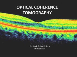
This presentation is mainly focused on the clinical diagnosis and interpretation of oct macula.This is presented on 4th year optometry as topic presentation.
Recent advances in ophthalmology - Dr. Parag Apteparag apte
A full presentation on recent advances in ophthalmology till today. There is not a single presentation on recent advances in ophthalmology in slide share till September 2017. I have tried my best to cover most of the topics so that the lecture gets over in one hour
OCT is a great technology,Many ophthalmologist find very difficult to understand it ,SO I have tired to simplify it as much as possible .Hope everyone can understand now onwards the basic about OCT .
Every feedback s most welcomed sothat i can improve further in coming days
Please email your feedback to me in the following address
yourgyanu@gmail.com
Review of the imaging modalities in Glaucoma. Structural loss precedes functional loss. Presentation includes a review of OCT, HRT and GDxVcc for posterior segment as well as AS-OCT and UBM for anterior segment.
optical coherence tomography is a new tool that makes retinal diagnosis easier. the above ppt includes a detailed and precise notes on OCT and its interpretation.
HOT NEW PRODUCT! BIG SALES FAST SHIPPING NOW FROM CHINA!! EU KU DB BK substit...GL Anaacs
Contact us if you are interested:
Email / Skype : kefaya1771@gmail.com
Threema: PXHY5PDH
New BATCH Ku !!! MUCH IN DEMAND FAST SALE EVERY BATCH HAPPY GOOD EFFECT BIG BATCH !
Contact me on Threema or skype to start big business!!
Hot-sale products:
NEW HOT EUTYLONE WHITE CRYSTAL!!
5cl-adba precursor (semi finished )
5cl-adba raw materials
ADBB precursor (semi finished )
ADBB raw materials
APVP powder
5fadb/4f-adb
Jwh018 / Jwh210
Eutylone crystal
Protonitazene (hydrochloride) CAS: 119276-01-6
Flubrotizolam CAS: 57801-95-3
Metonitazene CAS: 14680-51-4
Payment terms: Western Union,MoneyGram,Bitcoin or USDT.
Deliver Time: Usually 7-15days
Shipping method: FedEx, TNT, DHL,UPS etc.Our deliveries are 100% safe, fast, reliable and discreet.
Samples will be sent for your evaluation!If you are interested in, please contact me, let's talk details.
We specializes in exporting high quality Research chemical, medical intermediate, Pharmaceutical chemicals and so on. Products are exported to USA, Canada, France, Korea, Japan,Russia, Southeast Asia and other countries.
The prostate is an exocrine gland of the male mammalian reproductive system
It is a walnut-sized gland that forms part of the male reproductive system and is located in front of the rectum and just below the urinary bladder
Function is to store and secrete a clear, slightly alkaline fluid that constitutes 10-30% of the volume of the seminal fluid that along with the spermatozoa, constitutes semen
A healthy human prostate measures (4cm-vertical, by 3cm-horizontal, 2cm ant-post ).
It surrounds the urethra just below the urinary bladder. It has anterior, median, posterior and two lateral lobes
It’s work is regulated by androgens which are responsible for male sex characteristics
Generalised disease of the prostate due to hormonal derangement which leads to non malignant enlargement of the gland (increase in the number of epithelial cells and stromal tissue)to cause compression of the urethra leading to symptoms (LUTS
TEST BANK for Operations Management, 14th Edition by William J. Stevenson, Ve...kevinkariuki227
TEST BANK for Operations Management, 14th Edition by William J. Stevenson, Verified Chapters 1 - 19, Complete Newest Version.pdf
TEST BANK for Operations Management, 14th Edition by William J. Stevenson, Verified Chapters 1 - 19, Complete Newest Version.pdf
Explore natural remedies for syphilis treatment in Singapore. Discover alternative therapies, herbal remedies, and lifestyle changes that may complement conventional treatments. Learn about holistic approaches to managing syphilis symptoms and supporting overall health.
263778731218 Abortion Clinic /Pills In Harare ,sisternakatoto
263778731218 Abortion Clinic /Pills In Harare ,ABORTION WOMEN’S CLINIC +27730423979 IN women clinic we believe that every woman should be able to make choices in her pregnancy. Our job is to provide compassionate care, safety,affordable and confidential services. That’s why we have won the trust from all generations of women all over the world. we use non surgical method(Abortion pills) to terminate…Dr.LISA +27730423979women Clinic is committed to providing the highest quality of obstetrical and gynecological care to women of all ages. Our dedicated staff aim to treat each patient and her health concerns with compassion and respect.Our dedicated group ABORTION WOMEN’S CLINIC +27730423979 IN women clinic we believe that every woman should be able to make choices in her pregnancy. Our job is to provide compassionate care, safety,affordable and confidential services. That’s why we have won the trust from all generations of women all over the world. we use non surgical method(Abortion pills) to terminate…Dr.LISA +27730423979women Clinic is committed to providing the highest quality of obstetrical and gynecological care to women of all ages. Our dedicated staff aim to treat each patient and her health concerns with compassion and respect.Our dedicated group of receptionists, nurses, and physicians have worked together as a teamof receptionists, nurses, and physicians have worked together as a team wwww.lisywomensclinic.co.za/
Prix Galien International 2024 Forum ProgramLevi Shapiro
June 20, 2024, Prix Galien International and Jerusalem Ethics Forum in ROME. Detailed agenda including panels:
- ADVANCES IN CARDIOLOGY: A NEW PARADIGM IS COMING
- WOMEN’S HEALTH: FERTILITY PRESERVATION
- WHAT’S NEW IN THE TREATMENT OF INFECTIOUS,
ONCOLOGICAL AND INFLAMMATORY SKIN DISEASES?
- ARTIFICIAL INTELLIGENCE AND ETHICS
- GENE THERAPY
- BEYOND BORDERS: GLOBAL INITIATIVES FOR DEMOCRATIZING LIFE SCIENCE TECHNOLOGIES AND PROMOTING ACCESS TO HEALTHCARE
- ETHICAL CHALLENGES IN LIFE SCIENCES
- Prix Galien International Awards Ceremony
Couples presenting to the infertility clinic- Do they really have infertility...Sujoy Dasgupta
Dr Sujoy Dasgupta presented the study on "Couples presenting to the infertility clinic- Do they really have infertility? – The unexplored stories of non-consummation" in the 13th Congress of the Asia Pacific Initiative on Reproduction (ASPIRE 2024) at Manila on 24 May, 2024.
Report Back from SGO 2024: What’s the Latest in Cervical Cancer?bkling
Are you curious about what’s new in cervical cancer research or unsure what the findings mean? Join Dr. Emily Ko, a gynecologic oncologist at Penn Medicine, to learn about the latest updates from the Society of Gynecologic Oncology (SGO) 2024 Annual Meeting on Women’s Cancer. Dr. Ko will discuss what the research presented at the conference means for you and answer your questions about the new developments.
New Drug Discovery and Development .....NEHA GUPTA
The "New Drug Discovery and Development" process involves the identification, design, testing, and manufacturing of novel pharmaceutical compounds with the aim of introducing new and improved treatments for various medical conditions. This comprehensive endeavor encompasses various stages, including target identification, preclinical studies, clinical trials, regulatory approval, and post-market surveillance. It involves multidisciplinary collaboration among scientists, researchers, clinicians, regulatory experts, and pharmaceutical companies to bring innovative therapies to market and address unmet medical needs.
Lung Cancer: Artificial Intelligence, Synergetics, Complex System Analysis, S...Oleg Kshivets
RESULTS: Overall life span (LS) was 2252.1±1742.5 days and cumulative 5-year survival (5YS) reached 73.2%, 10 years – 64.8%, 20 years – 42.5%. 513 LCP lived more than 5 years (LS=3124.6±1525.6 days), 148 LCP – more than 10 years (LS=5054.4±1504.1 days).199 LCP died because of LC (LS=562.7±374.5 days). 5YS of LCP after bi/lobectomies was significantly superior in comparison with LCP after pneumonectomies (78.1% vs.63.7%, P=0.00001 by log-rank test). AT significantly improved 5YS (66.3% vs. 34.8%) (P=0.00000 by log-rank test) only for LCP with N1-2. Cox modeling displayed that 5YS of LCP significantly depended on: phase transition (PT) early-invasive LC in terms of synergetics, PT N0—N12, cell ratio factors (ratio between cancer cells- CC and blood cells subpopulations), G1-3, histology, glucose, AT, blood cell circuit, prothrombin index, heparin tolerance, recalcification time (P=0.000-0.038). Neural networks, genetic algorithm selection and bootstrap simulation revealed relationships between 5YS and PT early-invasive LC (rank=1), PT N0—N12 (rank=2), thrombocytes/CC (3), erythrocytes/CC (4), eosinophils/CC (5), healthy cells/CC (6), lymphocytes/CC (7), segmented neutrophils/CC (8), stick neutrophils/CC (9), monocytes/CC (10); leucocytes/CC (11). Correct prediction of 5YS was 100% by neural networks computing (area under ROC curve=1.0; error=0.0).
CONCLUSIONS: 5YS of LCP after radical procedures significantly depended on: 1) PT early-invasive cancer; 2) PT N0--N12; 3) cell ratio factors; 4) blood cell circuit; 5) biochemical factors; 6) hemostasis system; 7) AT; 8) LC characteristics; 9) LC cell dynamics; 10) surgery type: lobectomy/pneumonectomy; 11) anthropometric data. Optimal diagnosis and treatment strategies for LC are: 1) screening and early detection of LC; 2) availability of experienced thoracic surgeons because of complexity of radical procedures; 3) aggressive en block surgery and adequate lymph node dissection for completeness; 4) precise prediction; 5) adjuvant chemoimmunoradiotherapy for LCP with unfavorable prognosis.
Title: Sense of Taste
Presenter: Dr. Faiza, Assistant Professor of Physiology
Qualifications:
MBBS (Best Graduate, AIMC Lahore)
FCPS Physiology
ICMT, CHPE, DHPE (STMU)
MPH (GC University, Faisalabad)
MBA (Virtual University of Pakistan)
Learning Objectives:
Describe the structure and function of taste buds.
Describe the relationship between the taste threshold and taste index of common substances.
Explain the chemical basis and signal transduction of taste perception for each type of primary taste sensation.
Recognize different abnormalities of taste perception and their causes.
Key Topics:
Significance of Taste Sensation:
Differentiation between pleasant and harmful food
Influence on behavior
Selection of food based on metabolic needs
Receptors of Taste:
Taste buds on the tongue
Influence of sense of smell, texture of food, and pain stimulation (e.g., by pepper)
Primary and Secondary Taste Sensations:
Primary taste sensations: Sweet, Sour, Salty, Bitter, Umami
Chemical basis and signal transduction mechanisms for each taste
Taste Threshold and Index:
Taste threshold values for Sweet (sucrose), Salty (NaCl), Sour (HCl), and Bitter (Quinine)
Taste index relationship: Inversely proportional to taste threshold
Taste Blindness:
Inability to taste certain substances, particularly thiourea compounds
Example: Phenylthiocarbamide
Structure and Function of Taste Buds:
Composition: Epithelial cells, Sustentacular/Supporting cells, Taste cells, Basal cells
Features: Taste pores, Taste hairs/microvilli, and Taste nerve fibers
Location of Taste Buds:
Found in papillae of the tongue (Fungiform, Circumvallate, Foliate)
Also present on the palate, tonsillar pillars, epiglottis, and proximal esophagus
Mechanism of Taste Stimulation:
Interaction of taste substances with receptors on microvilli
Signal transduction pathways for Umami, Sweet, Bitter, Sour, and Salty tastes
Taste Sensitivity and Adaptation:
Decrease in sensitivity with age
Rapid adaptation of taste sensation
Role of Saliva in Taste:
Dissolution of tastants to reach receptors
Washing away the stimulus
Taste Preferences and Aversions:
Mechanisms behind taste preference and aversion
Influence of receptors and neural pathways
Impact of Sensory Nerve Damage:
Degeneration of taste buds if the sensory nerve fiber is cut
Abnormalities of Taste Detection:
Conditions: Ageusia, Hypogeusia, Dysgeusia (parageusia)
Causes: Nerve damage, neurological disorders, infections, poor oral hygiene, adverse drug effects, deficiencies, aging, tobacco use, altered neurotransmitter levels
Neurotransmitters and Taste Threshold:
Effects of serotonin (5-HT) and norepinephrine (NE) on taste sensitivity
Supertasters:
25% of the population with heightened sensitivity to taste, especially bitterness
Increased number of fungiform papillae
ARTIFICIAL INTELLIGENCE IN HEALTHCARE.pdfAnujkumaranit
Artificial intelligence (AI) refers to the simulation of human intelligence processes by machines, especially computer systems. It encompasses tasks such as learning, reasoning, problem-solving, perception, and language understanding. AI technologies are revolutionizing various fields, from healthcare to finance, by enabling machines to perform tasks that typically require human intelligence.
2. INTRODUCTION
• OCT is a diagnostic technology,provides a
cross sectional image of the anterior eye and
retina in-vivo with a high resolution, similar to
histological section.
• OCT allows assessment of retinal disease,
understanding of pathology and correlations
between structure and function.
3. It allows detection and measurement of:
• Morphological changes in retina
• Retinal thickness
• Retinal volume
• Retinal nerve fiber layer thickness (RNFL)
• Various parameters of the optic nerve head
(ONH)
4. PRINCIPLE
• Low coherence interferometry.
Michelson Interferometer
• A beam of light passes through
semitransparent mirror that splits the beam
into two.
• These two beams are then thrown on two
equidistant mirrors; reflected light from these
mirrors is then picked up and summed up by a
detector.
5. • The equidistant mirrors reflect the light wave in
same phase; however, if one of the mirrors is
moved by a distance less than the wavelength of
the incident light, the reflected lights from the
two mirrors will then possess a phase difference.
• This phase difference then produces an
interference pattern at the level of the detector.
6.
7.
8. • The resulting interference patterns are used to
reconstruct an axial A-scan, which represents
the scattering properties of the tissue along
the beam path.
• Moving the beam of light along the tissue in a
line results in a compilation of A-scans with
each A-scan having a different incidence point.
9. • From all these A-scans, a two-dimensional
crosssectional image of the target tissue can be
reconstructed and this is known as a B-scan.
• OCT operates like a fundus camera but resolves
like a USG machine.
USG OCT
• Source Sound waves Infrared light
• Resolution 150 μ 10 μ
• Patient contact Needed Non-invasive
11. • In TD OCT, image resolution and acquisition
speed are inversely related.
• Simultaneous increase in imaging speed and
resolution can be brought about by spectral
domain OCT (SD OCT).
13. Technique
• In the presence of clear media and cooperative
patients quality images can be taken even with a 3
mm pupil; otherwise dilatation is recommended.
• The patient is seated comfortably in front of the
OCT machine with chin positioned on the chin rest.
He is asked to fixate on the fixation target. The
internal fixation (green color light) target is the
commonly used fixation target. Those patients who
are unable to fixate with macula can focus with the
opposite eye on an external target. After Fixation
the operator selects the desired scan and aligns the
instrument so that fundus image and scan beam is
displayed on the screen.
14. Scanning protocols
• The OCT software offers a variety of retinal
scanning protocols including linear, circular,
radial and parallel line scans
• The duration of scan acquisition can be
shortened by using fast scan protocols, which
acquire three fast scans and averages them to
give the final interpretation.
15. • The alignment algorithm reduces artifacts caused by
axial movement of the eye during scan acquisition.
• The normalization algorithm allows comparison of
scans with varying signals. Apart from this the machine
has Gaussian smoothing, proportional evaluation and
profile algorithm scan for further refinement of the
scan image
• Application of gray scale image allows better
sensitivity for detection of minute difference in the
contrast.
• A normative database is also available for comparison
of peripapillary retinal nerve fiber layer as well as the
macular thickness in the latest software.
16. Specific scanning protocols
• Retina scanning protocols: Line scan, Raster lines,
Cross hair, Radial lines, X-line and Circle.
• Macular scanning protocols: Macular thickness scan,
Fast macular thickness, Raster lines and Single line
scan
• RNFL scanning protocols: RNFL thickness protocol
(3.4 mm), Fast RNFL thickness protocol (3.4 mm),
Proportional circle, Concentric 3 rings, RNFL
thickness (2.27Xdisc), RNFL map.
• ONH scanning protocols: Optical disc scan and Fast
Optical disc scan.
17.
18.
19.
20.
21.
22. NORMAL RETINAL SCAN
• Posterior hyaloid ,visible as very faint, fine and
slightly reflective line.
• Internal limiting membrane is clearly defined
in the OCT scans due to contrast between the
reflective retina and non-reflective vitreous.
• The nerve fiber layer is highly reflective and
more visible on the nasal side due to the
density of papillomacular bundle.
23. • The fovea shows a characteristic depression
on the macular scan.
• The plexiform layers with reflectivity that is
slightly greater than the reflectivity of the
nuclear layers.
• The outer retina is bounded by a highly
reflective band (70 microns thick) that
represents the retinal pigment epithelium.
24. • This band can be divided into three layers.
• The first is thin and hyperreflective
representing the junction of inner and outer
photoreceptors.
• The second of hyporeflective,outer segment
• The third one is the thickest and most
hyperreflective
25. • The Bruchs’ membrane and the
choriocapillaris are seen as a single less
reflective structure but in some scans the
choriocapillaris may be visible separate from
the RPE and the Bruchs’ membrane.
• The larger retinal vessels are located
indirectly by the shadow cone that they form
on the posterior layers
26. • The OCT image can be displayed on a gray
scale where more highly reflected light is
brighter than less highly reflected light.
• Alternatively, it can be displayed in color,
colors correspond to different degrees of
reflectivity.
• Highly reflective:bright colors(red and yellow)
• Low reflectivity:darker colors(black and blue).
• Intermediate reflectivity -green.
27.
28.
29. • First generation OCT became available in 1996
as Humphrey Optical Coherence Tomography
Scanner.
• Infrared light source with wavelength of 850
nm,with a resolution of 10 to 17 μm.
30. • Commercial 3rd generation OCT (StratusOCT, Carl
Zeiss Meditec, Dublin, CA) was introduced in
2002.
Light source - super luminescent diode (SLD)
Wavelength - 820 nm
STRATUS OCT:
Scanning speed - 400 A-scan/seconds (4х 1st
gen).
Axial resolution of 10 μm
Transverse resolution of 20 μm.
31. Various newer OCT systems are:
• Ultra-high resolution OCT
• Combined OCT/SLO
• Doppler-OCT
• High Speed UHR-OCT
• CAS OCT-Visante™ OCT
• Polarization sensitive OCT
• Combined FFA and en-face OCT
• Intraoperative OCT
32. ULTRA HIGH RESOLUTION OCT
• Axial resolution - 3 μm.
• Transverse resolution - 15-20 μm.
• Uses femto-second titanium sapphire laser that
generates light with bandwidth of 125 nm
centered at 815 nm.
• Time for image acquisition is 4.3 sec(1.3 sec with
Stratus OCT).
• Hence, UHR OCT images need correction for axial
motion.
33. • With OCT (Stratus OCT); GCL,ELM and
photoreceptor details are not well visualized.
• UHR OCT may be useful in evaluation of these
retinal layers.
1.Showing photoreceptor integrity in patients with
macular hole. Foveal photoreceptor degeneration
is represented by outer hyporeflective
disruptions of the junction between the inner
and outer segments of the photoreceptors
34. 2.Detection of milder forms of ERM and
vitreomacular traction.
3.Earlier detection of RNFL thinning and
treatment of glaucoma patients.
• UHR OCT can not only demonstrate focal RNFL
changes before appearance of field defect but
also detect progression of disease in an
established case.
35. HIGH SPEED UHR OCT
High Speed UHR-OCT uses SD OCT technology.
• It allows simultaneous ultra-high speed and
ultra-high resolution.
• Imaging speed is 100 times faster than time
domain UHR OCT.
• 40 times faster than the standard- resolution
OCT.
36. • It not only gives structural information but
also functional retinal blood flow similar to
Doppler ultrasound.
• It may reduce the need for FFA.
37. • Raster scan to obtain 3-dimensional (3-D)
images of ocular structure.
• Quantitative mapping of retina layers,
including measurements of the retinal
thickness, RNFL photoreceptor layer and other
intraretinal layers can be performed.
• Its applications would be similar to that
indicated for UHR-OCT.
38.
39.
40. COLOR DOPPLER OCT
• CD OCT is the technique that combines laser
Doppler velocimetry and OCT for imaging the
depth, diameter, flow rate and retinal
hemodynamic characteristics.
• Color coded velocity data are superimposed
on the conventional OCT image for CD OCT
display. The direction and magnitude of blood
flow are designated by red and blue color and
intensity respectively.
41. • As it can measure blood flow profile in a few
milliseconds it is able to show vascular
autoregulation and response to changes in
perfusion pressure, oxygen contents and
following laser photocoagulation.
• Presently only larger vessels near the optic
disc have been mostly studied.
42. CORNEA ANTERIOR SEGMENT OCT
• ASOCT with 1300 nm wavelength ASOCT was
first reported by Radhakrishnan.
• The CAS OCT image is a gray scale or false
color two-dimensional representation of
backscattered light intensity in a cross-
sectional plane.
• The scanning speed of the system is 4000 axial
scans per second
43. • CAS OCT with a wavelength of 1.3-μm
(present model) provides adequate resolution
of both the cornea and the AC angle.
• Detailed image of the cornea, iris root,angle
recess, anterior ciliary body, scleral spur,
and,in some eyes, the canal of Schlemm is
possible.
44. ADVANTAGE OVER OTHERS
• Being noncontact, the patient comfort, cooperation and
safety is increased (pediatric) and there is no mechanical
distortion of the tissue.
• Provides more accurate biometry of anterior segment than
Orbscan or Scheimpflug photography.
• Though confocal scanning microscopy gives higher
resolution than CAS OCT, it can scan only a small area of the
eye at a time.
• Unlike UBM, CAS OCT can perform measurements without
any need of anesthesia or coupling medium. CAS OCT can
also perform scanning of all 4 quadrants at a time.
45. OTHER USES
• Measure corneal thickness, flap and residual posterior
stromal bed following refractive surgery.
• Important landmarks such as the scleral spur are more
distinct in CAS OCT images.
• Traumatic angle recession can be easily picked up.
• Detecting gonioscopically occludable angle.
• Also being used for imaging ocular surface and iris
neoplasia.
46. LIMITATIONS
• Speed and depth of penetration.
• It cannot obtain clear images through opaque
media.
• Is obstructed by the eyelids making imaging of
the superior and inferior angles difficult.
• It provides limited visualization of the ciliary
body.
47. INTERPRETATION
EPIRETINAL MEMBRANE:
• ERMs can be classified as idiopathic or secondary.
• Idiopathic ERMs -fibroglial proliferation on the
inner surface of the retina,secondary to a break
in ILM,during posterior vitreous detachment.
• Secondary ERMs result from an already-existing
ocular pathology such as central or branch retinal
vein occlusion, diabetic retinopathy, uveitis,and
retinal breaks with or without detachment.
48. • ERMs are seen as a highly reflective layer on
the inner retinal surface.
• In most eyes, the membrane is globally
adherent to the retina but,in some cases, it
can be separated from the inner aspect of the
retina, which enhances its visibility by OCT.
49.
50.
51.
52. MACULAR HOLE
• Macular hole is partial or full thickness
dissolution of retinal tissue at the foveal region.
• It may occur following blunt trauma, long-
standing macular edema or as an idiopathic
condition.
• Pseudoholes are seen in dense sheet of ERM
with a central defect that overlies the foveal
center, giving the ophthalmoscopic appearance of
a true macular hole.
53. OCT STAGING OF MACULAR HOLE
• Stage 1
• Stage 1a: Foveolar detachment with yellow spot. OCT
shows a cystoid space occupying the inner part of the
foveal tissue.
• Stage 1b: Foveolar detachment with yellow halo. OCT
shows impending hole with extension of cystoid space
posteriorly, disrupting the outer retinal layers.
• Stage 2: Formation of minute eccentric holes. OCT
shows eccentric opening of the roof of the hole with
presence of an operculum (Figs 23.6 to 23.8).
54. • Stage 3: Full thickness macular hole with or
without operculum. OCT shows a central full
thickness macular hole with detached
posterior vitreous.
• Stage 4: Full thickness macular hole with
posterior vitreous detachment. OCT shows a
central full thickness macular hole with a cuff
of subretinal fluid and completely detached
posterior vitreous.
55.
56.
57.
58.
59.
60.
61. BERLINS EDEMA
• Acute retinal opacification (macula or elsewhere)
following closed globe injury.
• Mild cases resolve spontaneously without any
sequelae; severe cases result in permanent vision loss.
• Histopathological features:
• Disruption of photoreceptor outer segments
• Phagocytosis of retinal pigment epithelial cells (RPE)
• Intraretinal migration of RPE
• Multilayered, disorganized RPE
62. • OCT findings depend on the severity and the
duration of commotio retinae .
• Photoreceptor disruption is seen as optically
clear spaces in the area corresponding to the
photoreceptors.
63.
64.
65. RETINAL ARTERY OCCLUSIONS
• The clinical presentation depends on the type
of vessel that is occluded. It may be
asymptomatic or sudden rapid loss of vision or
no PL.
• The ophthalmoscopic include cotton wool
exudates, opacification of retina and a cherry
red spot.
66. OCT FINDINGS
• Diffuse thickening of the neurosensory retina.
• Increased reflectivity in the inner retinal
layers, decreased reflectivity of the
photoreceptor layers and the retinal pigment
epithelium secondary to the shadowing effect.
• Involvement of macula,of cystoid changes in
the macular area with loss of the macular
contour.
• In old cases decrease in macular thickness.
67.
68. RETINAL VENOUS OCCLUSIONS
• Retinal thickening and cystoid macular edema (CME):
Increase in retinal thickness is seen as loss of macular
contour on OCT. In the area of retinal edema,
presence of cystoid spaces with reduced reflectivity
depicts CME.
• Intraretinal and subretinal hemorrhages are seen as
focal areas with bright and high reflectivity with back
scattering. Area of shadowing appears as black
spaces in the RPE and choriocapillaris layer
69. • Cotton wool spots are seen as highly reflective
well demarcated areas with reduced
reflectivity from outer retinal layers.
• Optic disc edema is a common feature in
CRVO. OCT can demonstrate disc edema and
help in monitoring it in a quantitative manner.
70.
71. AGE RELATED MACULAR
DEGENERATION
• Age related macular degeneration is the most
common cause of blindness.
• Early grades of the disease more frequent
than the advanced grade.
• Early stage – drusen
• Advanced stage by geographic atrophy (non-
exudative or dry form)
choroidal neovascularization (exudative or
wet form).
72. • Geographic atrophy - large area of
irregular,well-defined chorioretinal atrophy
involving the macula.
• Exudative form-choroidal neovascular
membrane and its sequlae like serous
detachment, hemorrhagic
detachment,exudation and distortion of the
retinal photoreceptors.
73. • Drusen - hyper-reflective discrete protrusions
within the RPE complex.
• Active choroidal neovascular membrane -
multi-layered, highly hyper-reflective fusiform
or irregular mass with loss of retinal contour
in the overlying region.
• It may be located in the pre choriocapillaris
region or in front of the pigment epithelial
layer or in both these spaces.
74.
75.
76.
77. CENTRAL SEROUS
CHORIORETINOPATHY
• Central serous chorioretinopathy is a
noninflammatory, idiopathic serous
detachment of the macula with or without
associated RPE detachment.
• Spontaneous resolution occurs - majority.
Concurrent pigment epithelial detachments
persist for a longer time despite resolution of
the serous detachment.
78. • OCT to detect subtle serous detachments of
the macula, either in the first episode, during
recurrences or following treatment.
• Serous detachment - hypo-reflective, shallow
separation of the neurosensory retina from
the RPE.
A) Stage 1b macular hole and vitreofoveal
traction; (B and C) Evolving into stage 2 macular hole (full
thickness eccentric defect with operculum
Stage 2 macular hole
(full thickness eccentric defect with operculum
full thickness defect with
cystoid changes at the edge and pseudo-operculum
nd yellow pigment deposit at the base of macular
hol
central area of retinal elevation and subneurosensory
collection of moderate reflectivity material
suggestive of fibrin along with retinal pigment epithelium
detachment.