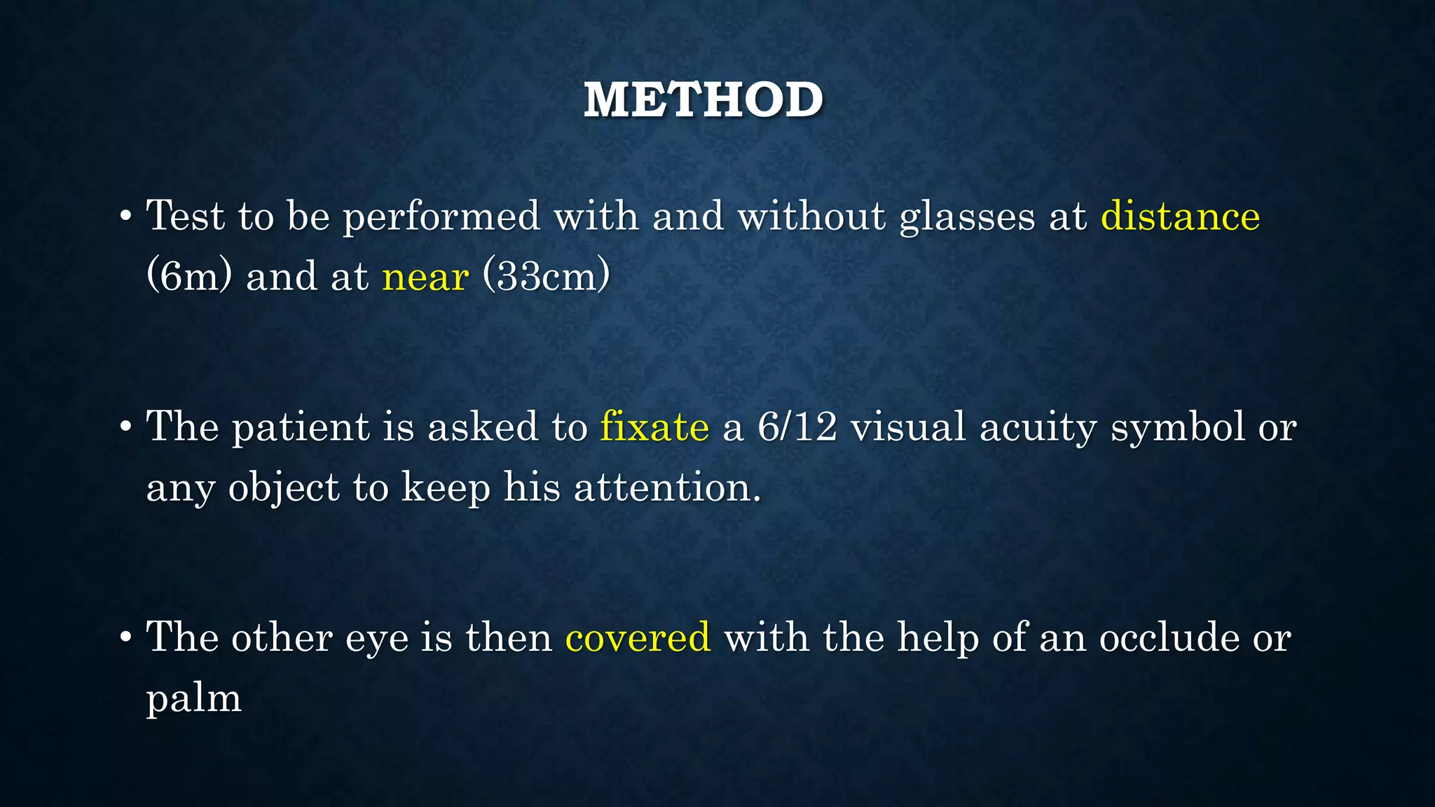
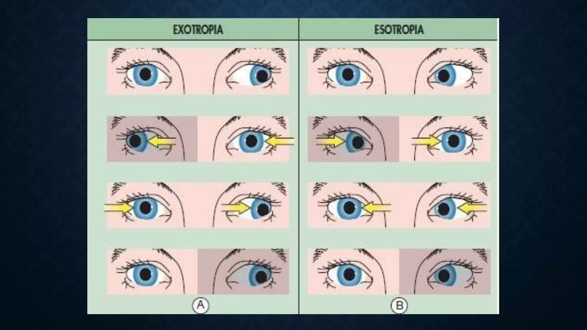
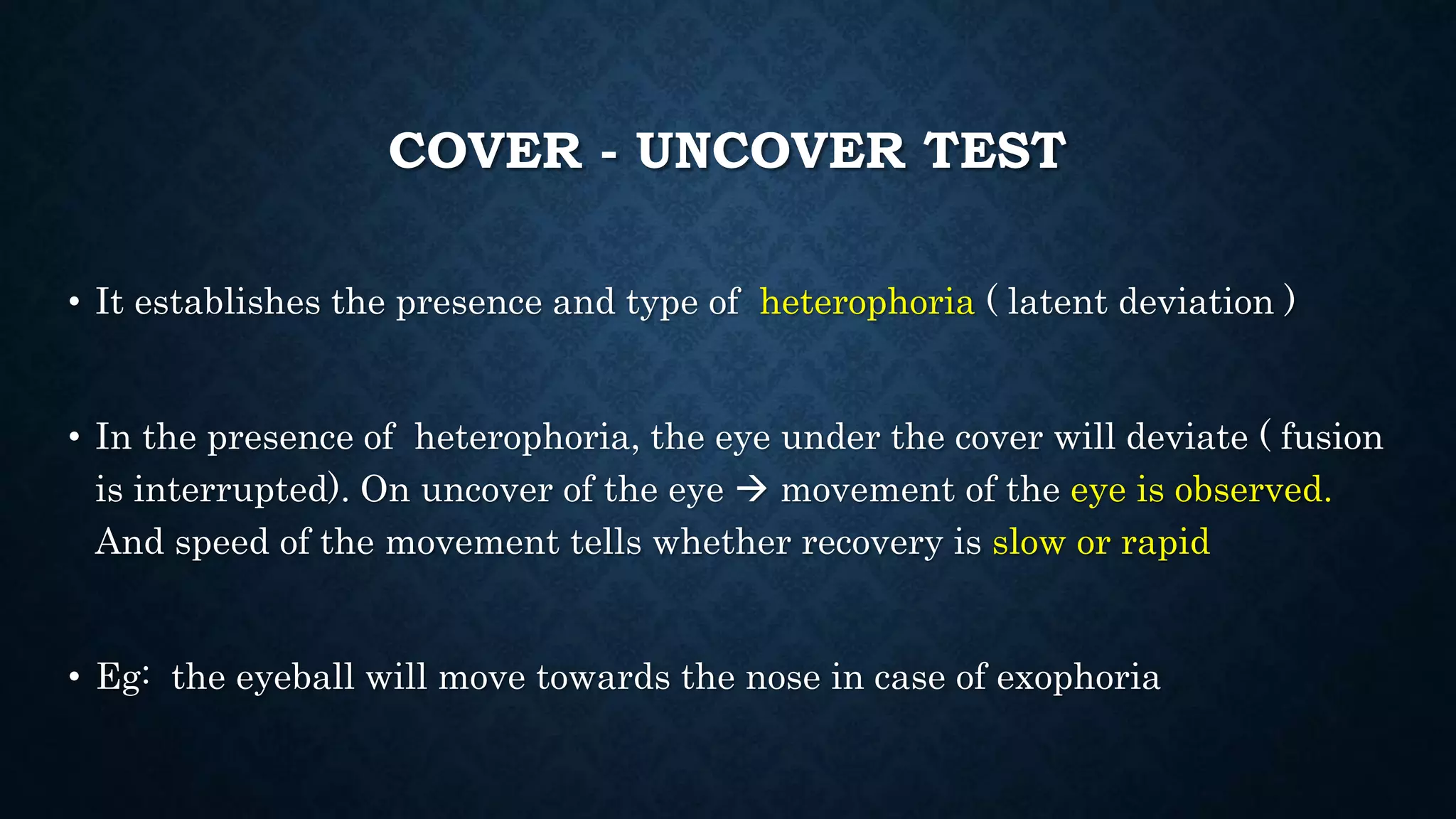
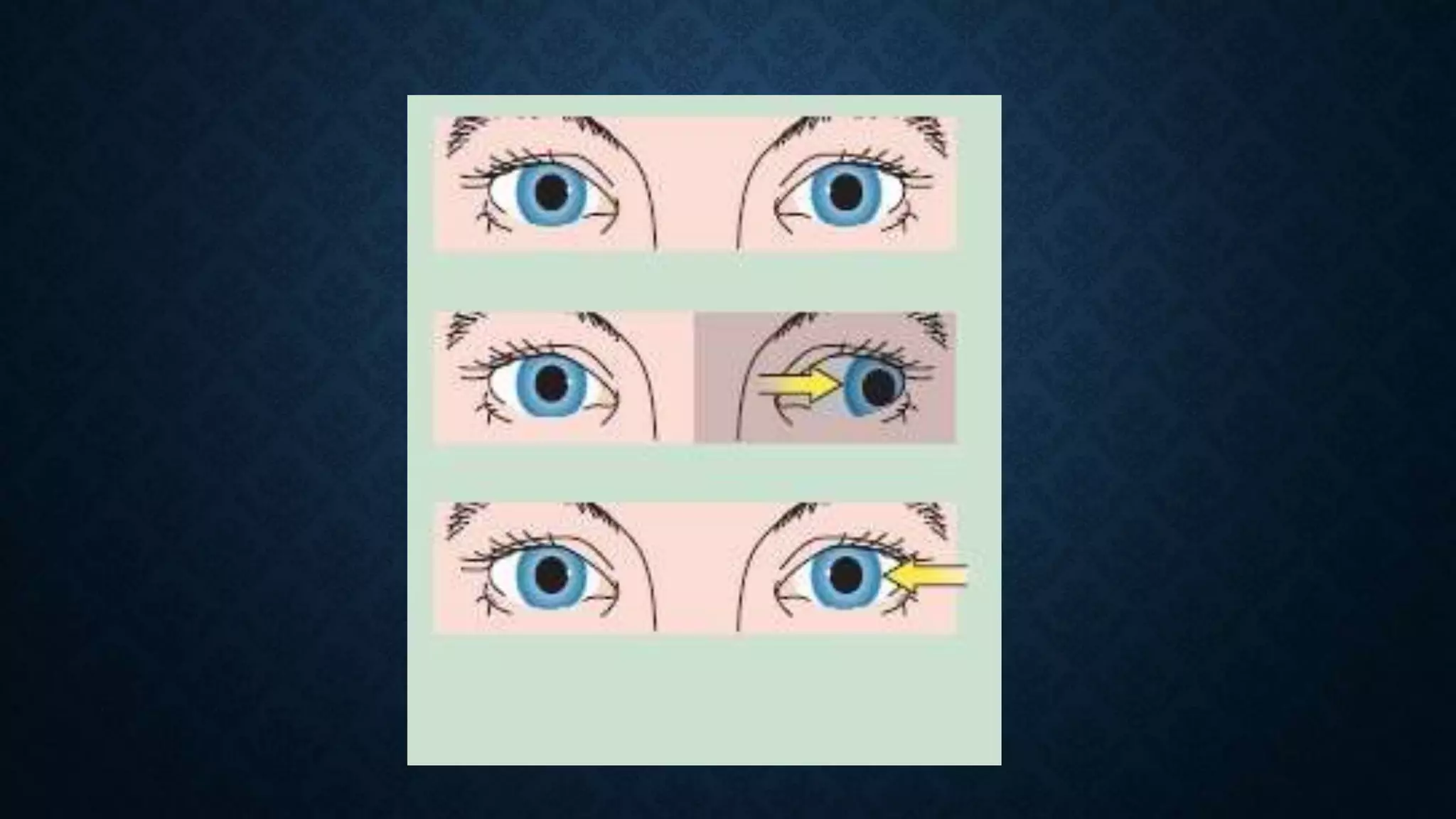
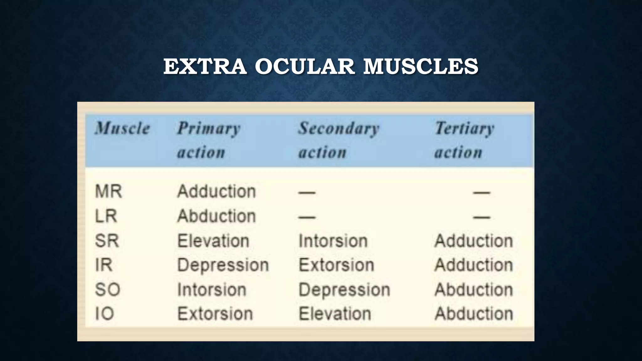
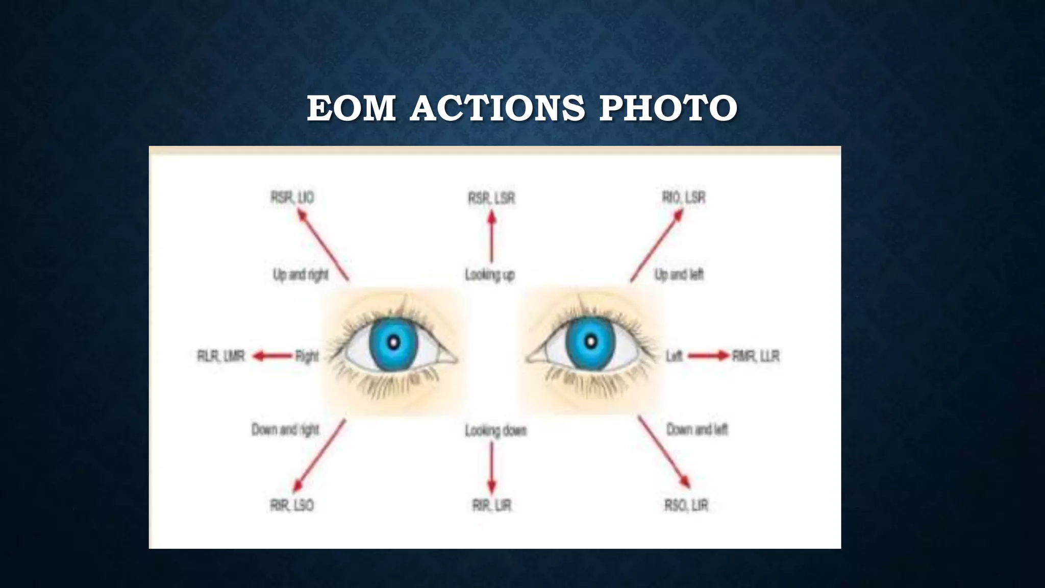
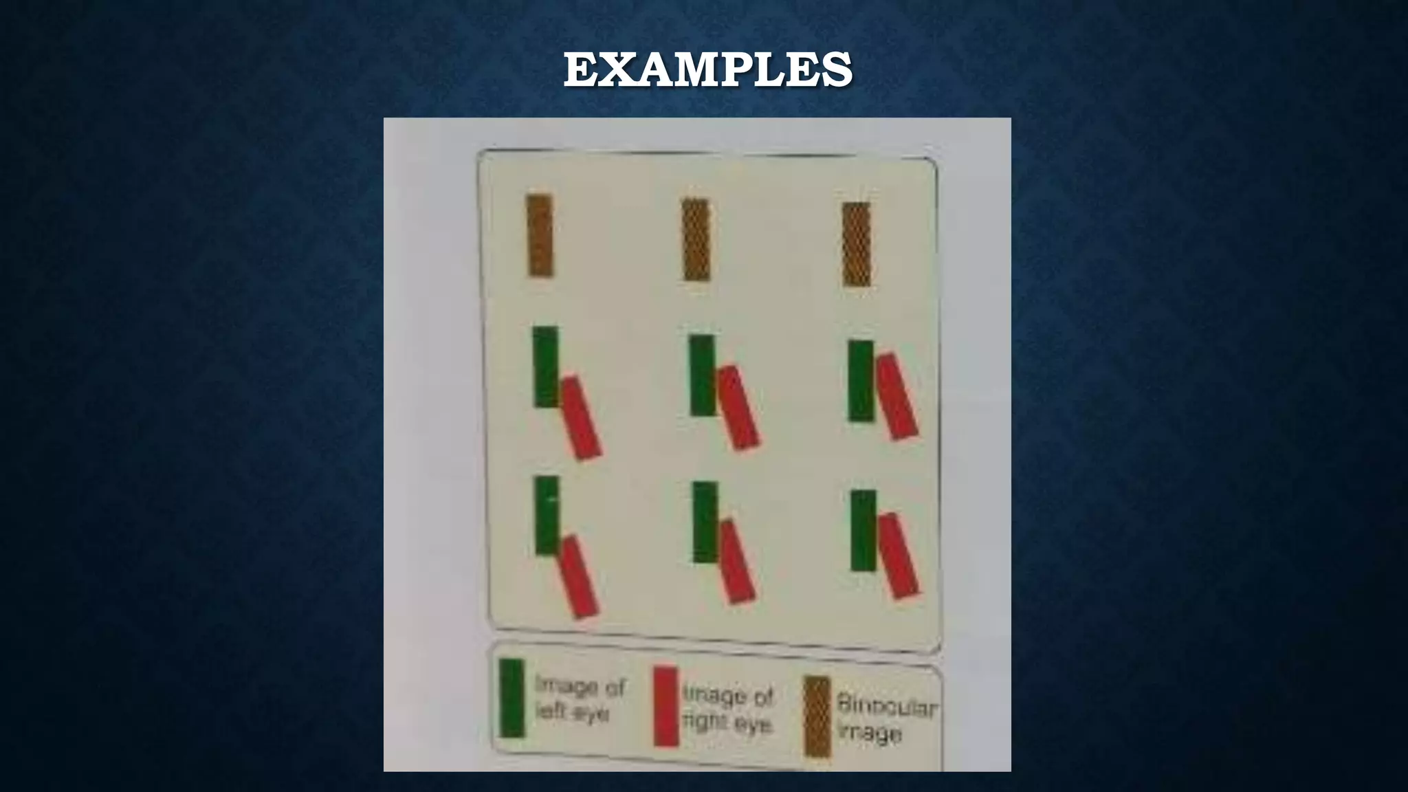
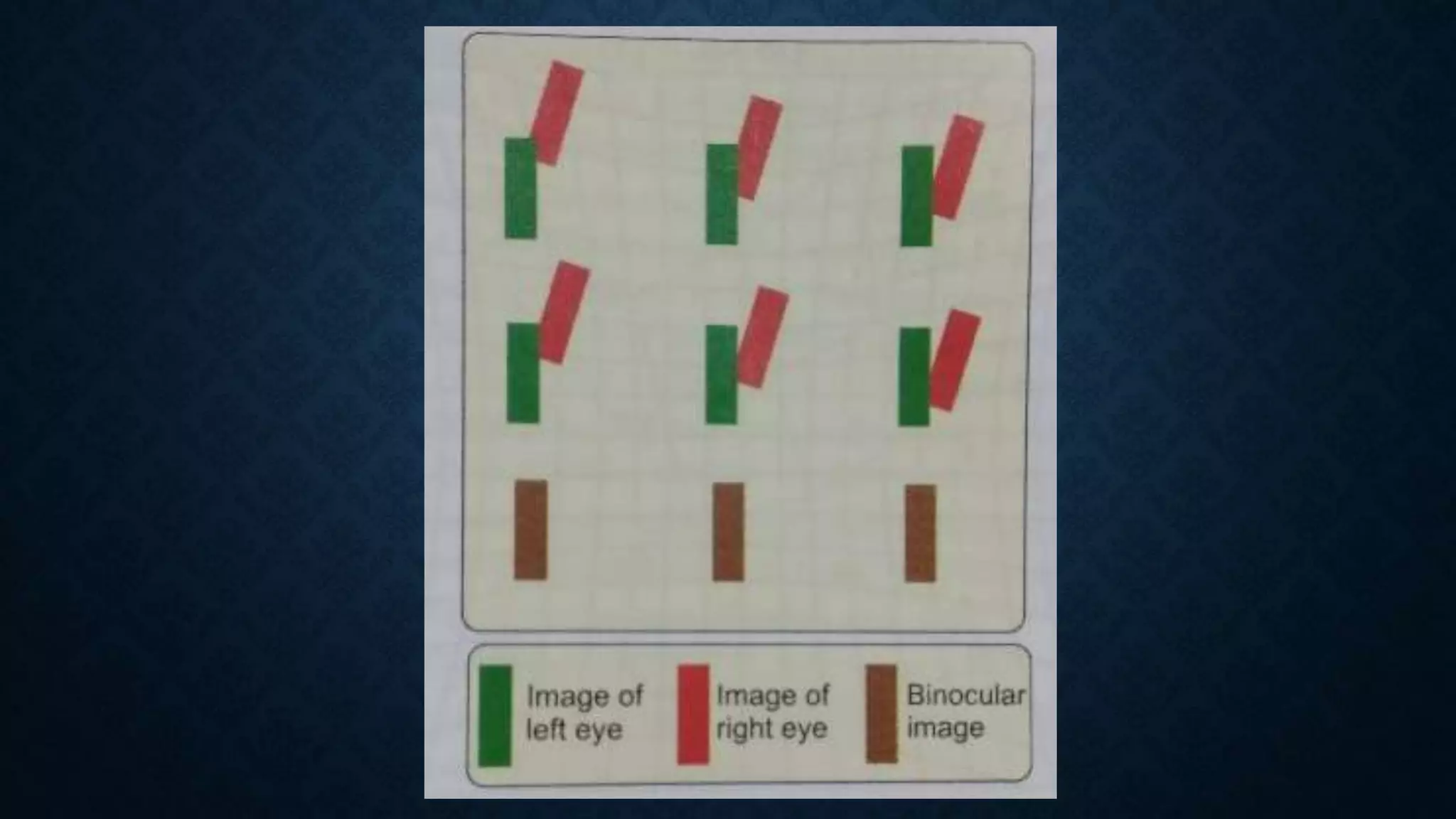
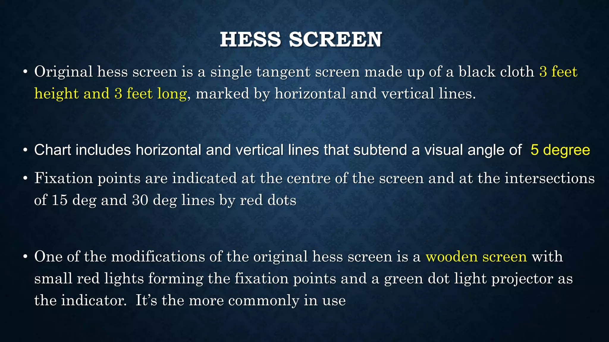
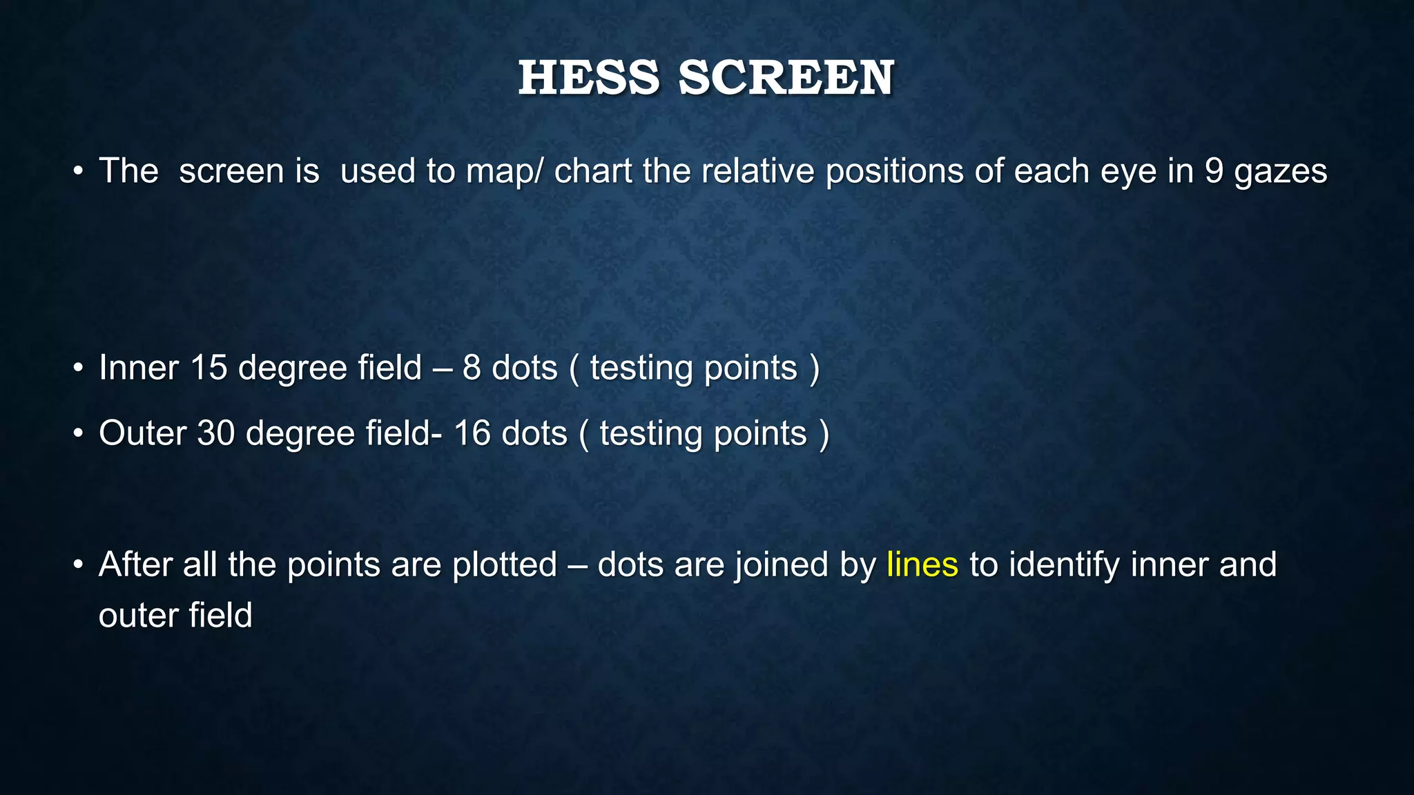
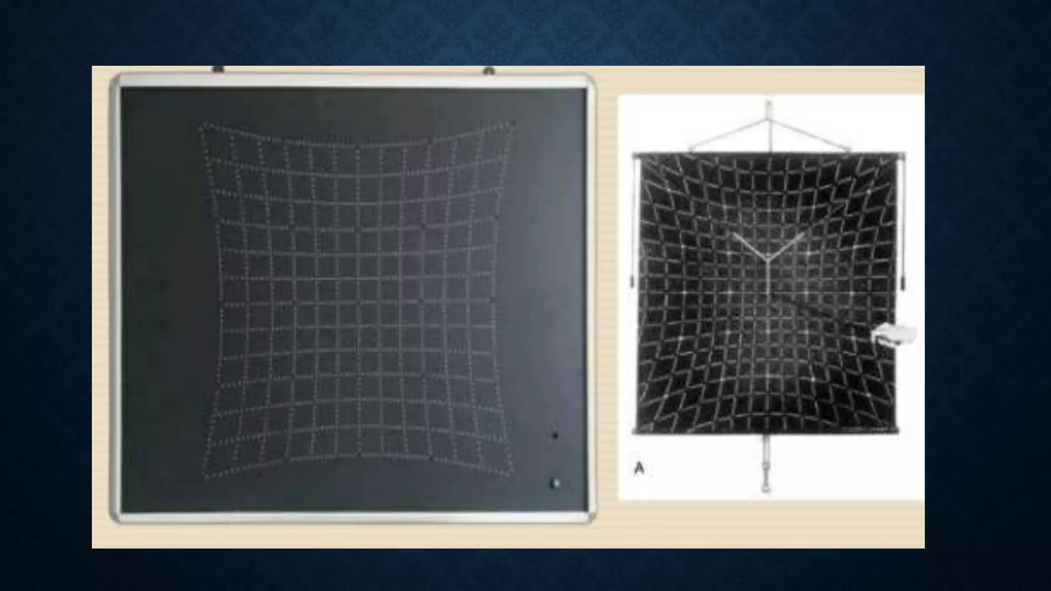
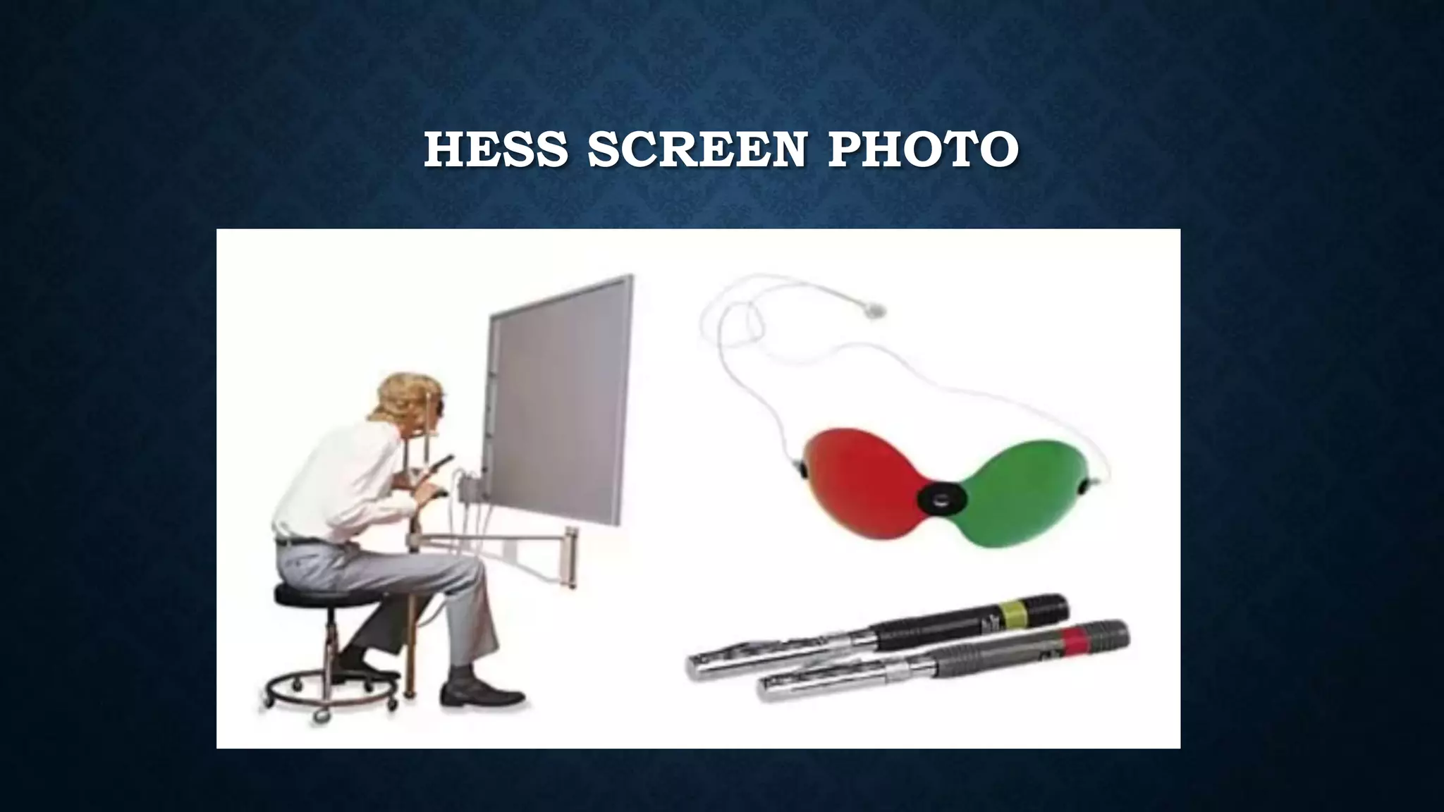
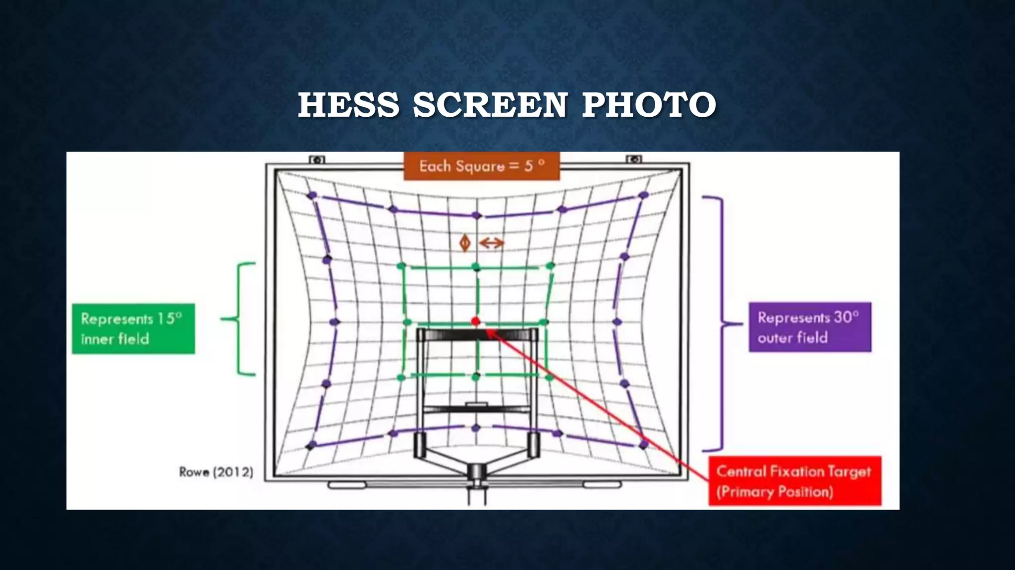
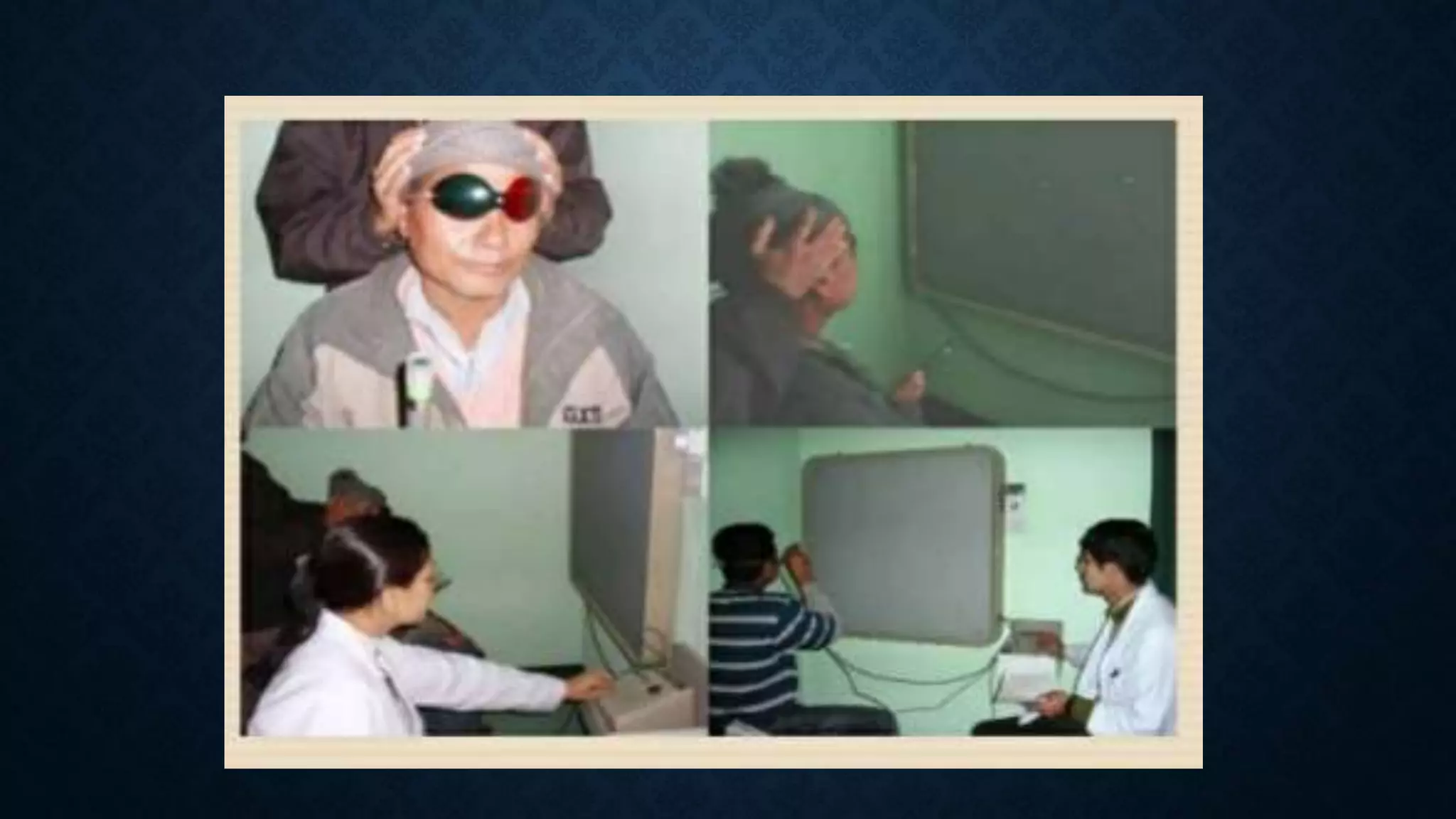
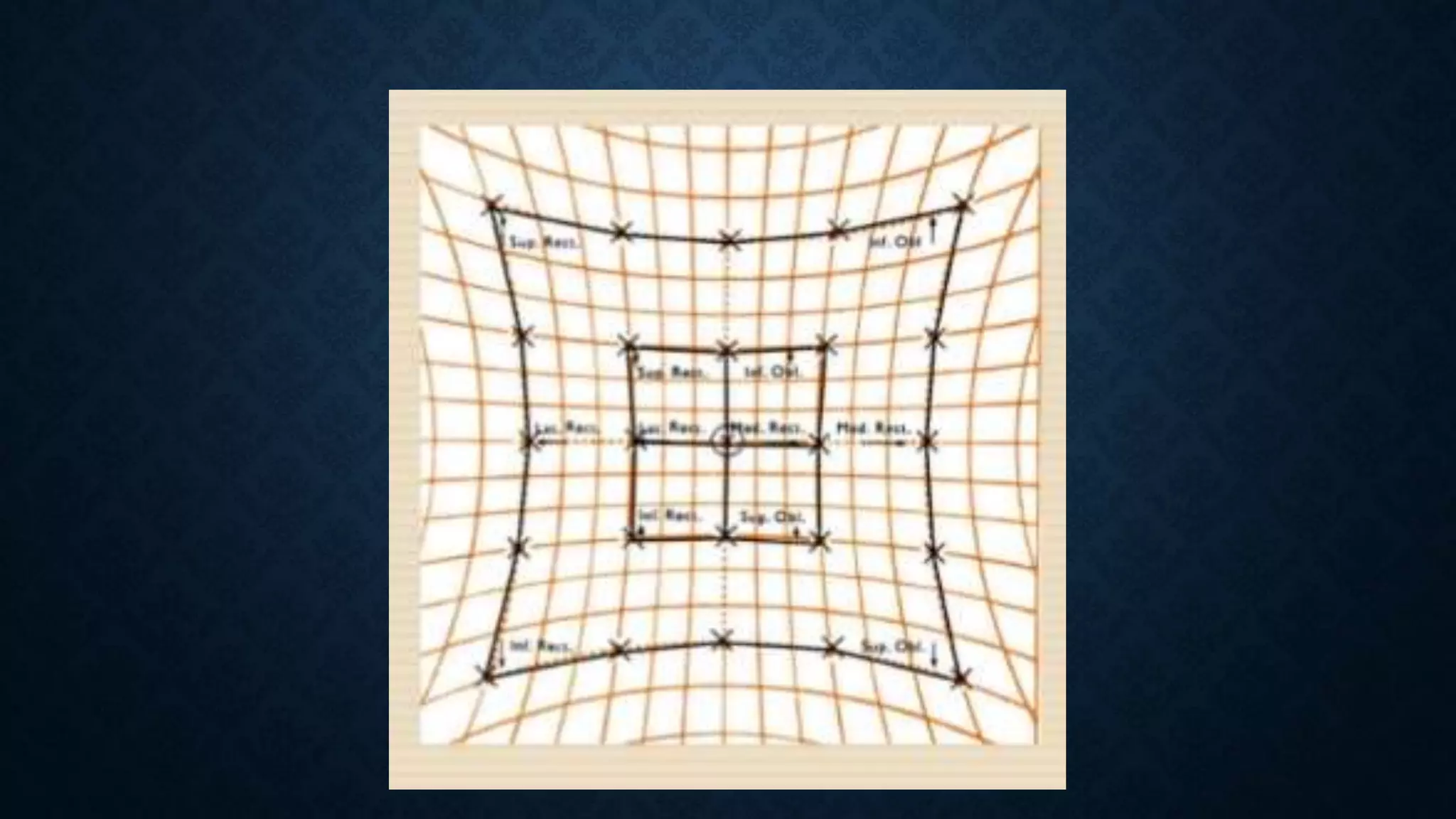
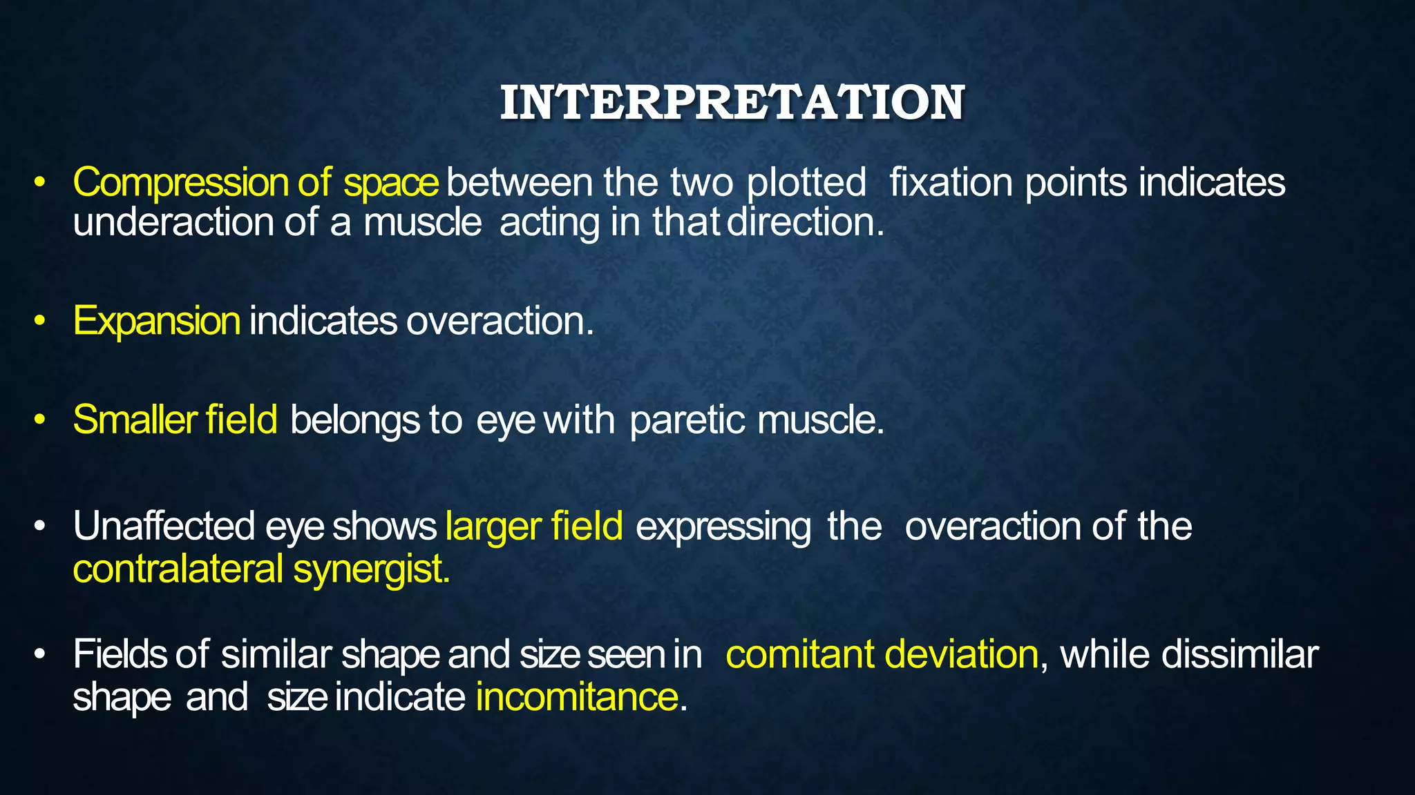
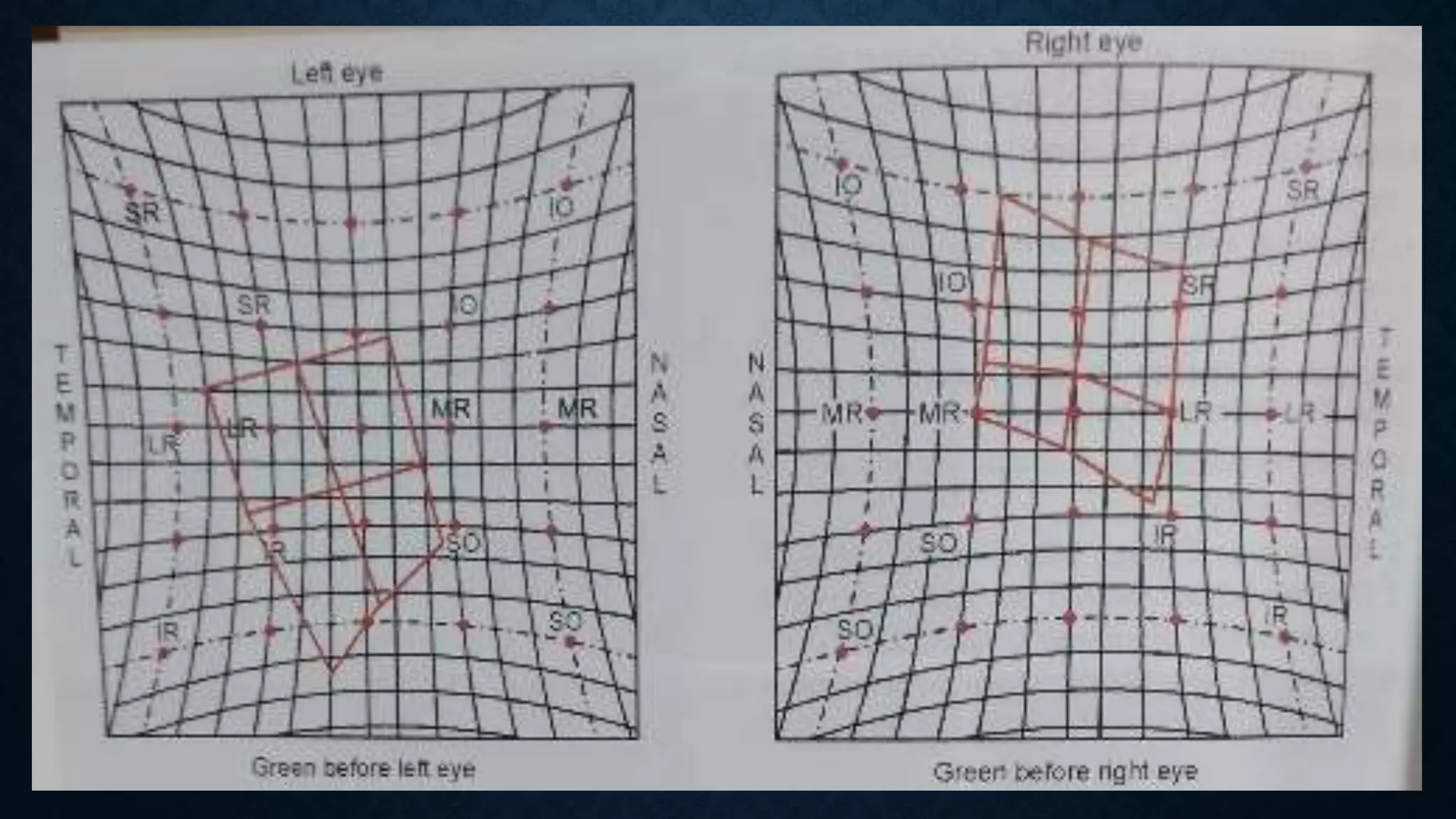
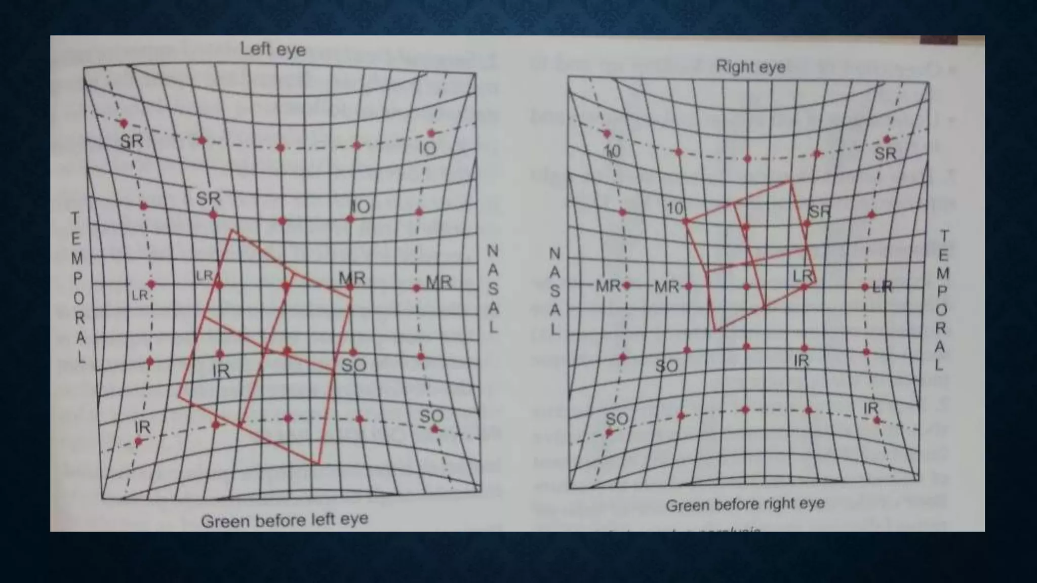
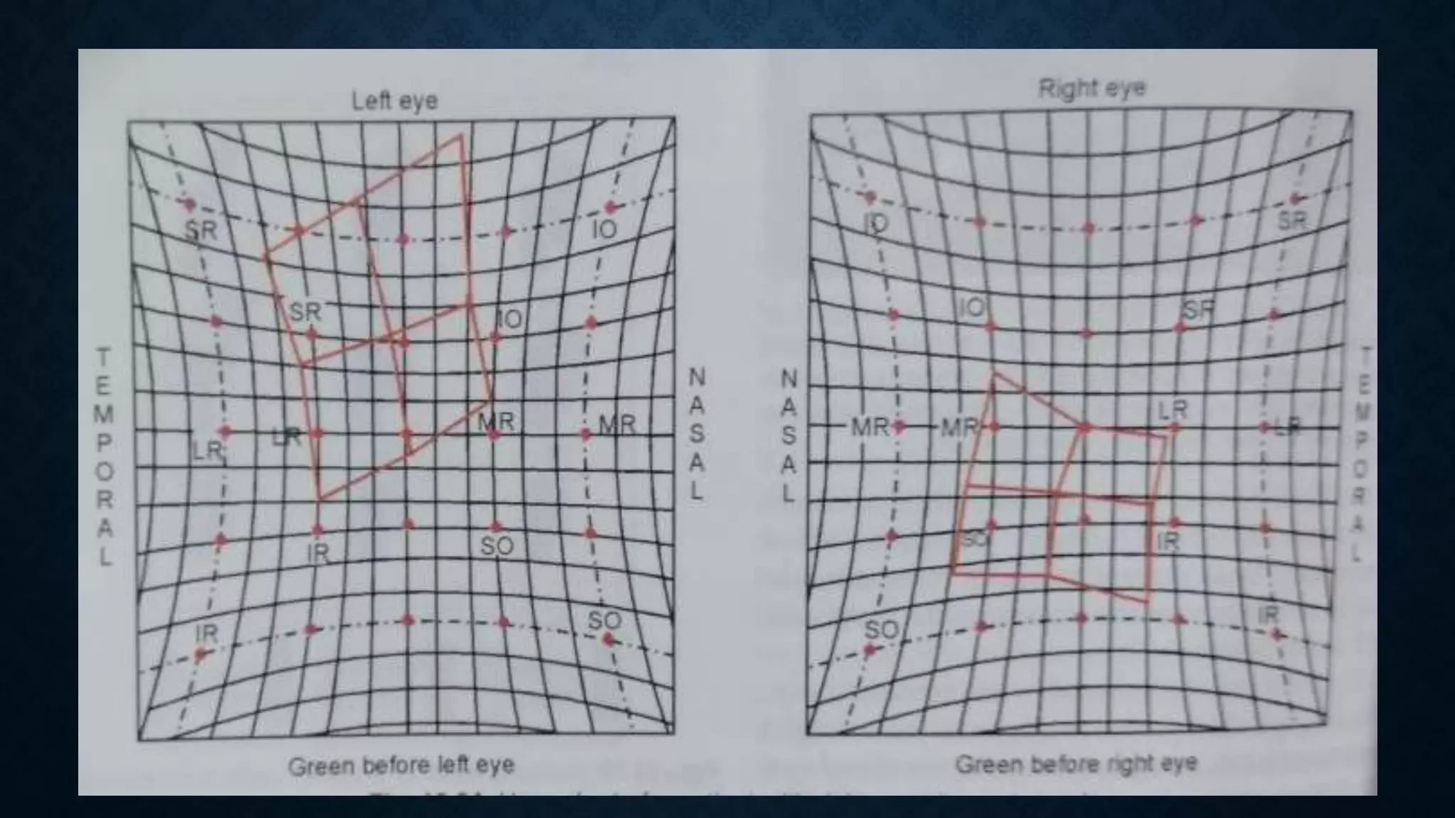
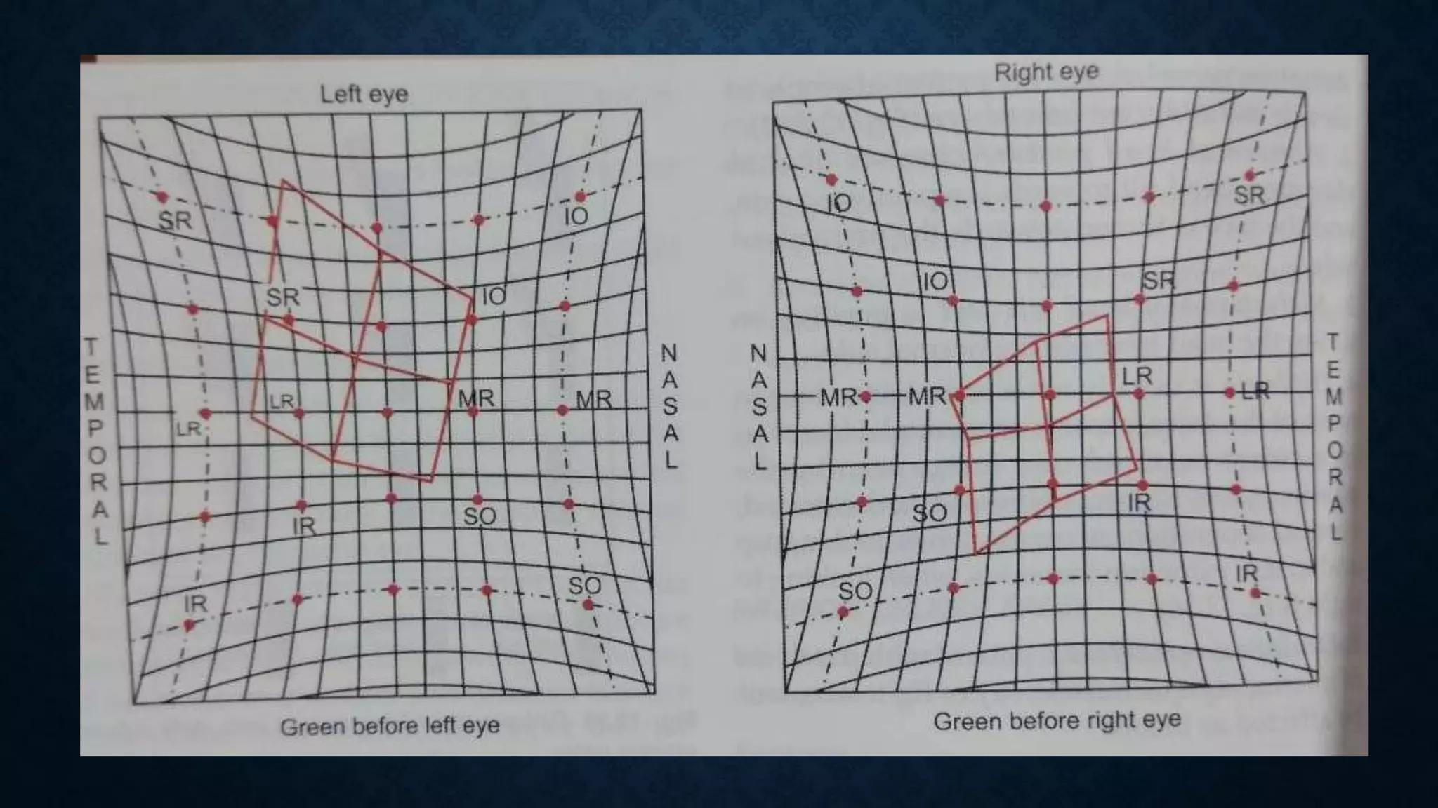
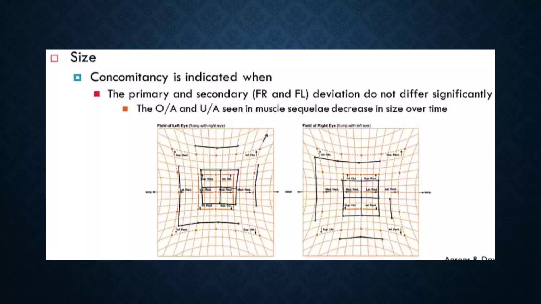
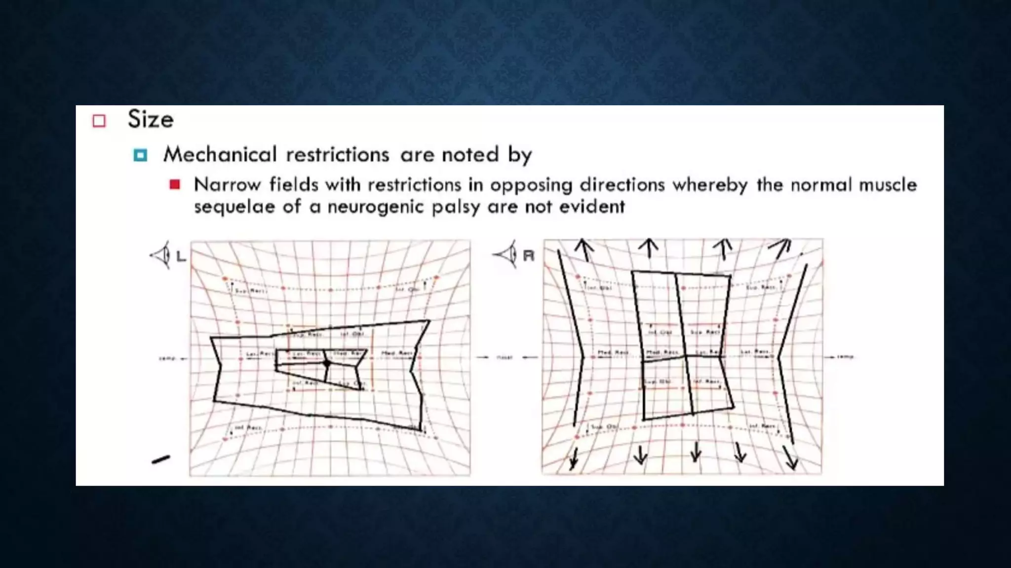
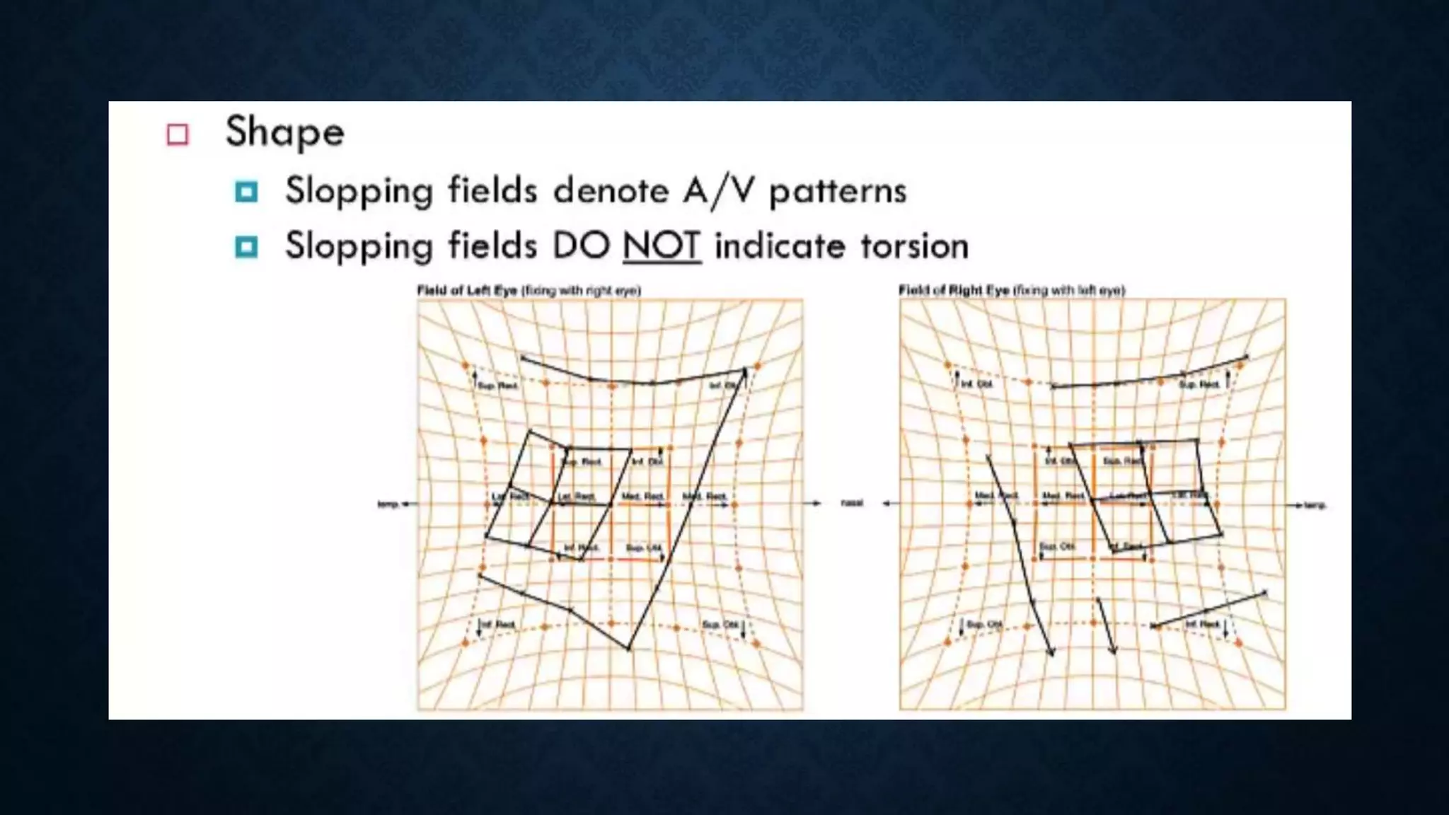
COVER TESTS
HESS CHARTING
DIPLOPIA CHARTING
Dr. Nikhil. R.P
CONTENTS
• Cover test
• Uncover test
• Alternate cover test
• Hess charting
• Diplopia charting
COVER TESTS
• Cover tests are simple and useful tests for assessing
binocular motor function
• They are entirely objective, requiring only fixation by the
patient
• Whether an ocular deviation is latent or manifest
• Direction of a deviation
PREREQUISITES
• Patient should be co- operative enough to fixate a target
• Should have sufficient vision to see the target
• Should have central fixation in both eyes
• Nystagmus shouldn’t be present
METHOD
• Test to be performed with and without glasses at distance
(6m) and at near (33cm)
• The patient is asked to fixate a 6/12 visual acuity symbol or
any object to keep his attention.
• The other eye is then covered with the help of an occlude or
palm
COVER TEST
• In this test , the movement of the uncovered eye is observed
• It confirms the presence of manifest squint
COVER - UNCOVER TEST
• It establishes the presence and type of heterophoria ( latent deviation )
• In the presence of heterophoria, the eye under the cover will deviate ( fusion
is interrupted). On uncover of the eye  movement of the eye is observed.
And speed of the movement tells whether recovery is slow or rapid
• Eg: the eyeball will move towards the nose in case of exophoria
ALTERNATE COVER TEST
• It is performed to establish phorias, alternating squints
• Also to differentiate between the comitant squint from paralytic
squint
• Occluder is placed alternatively in front of each eyes several times to
dissociate the eyes and to maximize the deviation.
• Comitant squint - Primary deviation = secondary deviation
• Paralyitc squint – secondary deviation > primary deviation
PRISM BAR COVER TEST
• Prisms of increasing strength with apex towards the deviation are
placed in front of one eye and the patient is asked to fixate a target
with other eye
• Alternate cover test is performed till there is no recovery movement of
the eye under cover  give us amount of deviation in prism dioptres.
• The test is again done for both distance and near vision and in all
positions of gaze.
LIMITATIONS
• Following deviations may be overlooked or cannot possibly be
diagnosed with cover tests-
• Small microtropia
• Small angle esotropia ( < 5deg PD )
• Monofixation syndrome
• Cyclodeviation
EXTRA OCULAR MUSCLES
EOM ACTIONS PHOTO
DIPLOPIA CHARTING
• Advantages :
• Provides a record of the
separation of diplopic images in
the 9 position of gaze
• It can be performed in any co-
operative patient experiencing
diplopia
• Assists in determining which
direction of gaze the diplopia is
worst in
• Disadvantages :
• It is only quantitative and hence
cannot comment on minor changes
• The test requires intelligent patient
to comment on separation of images
• Not possible to perform in colour
blind
• Not of use in congenital palsies and
those of long standing onset because
of suppression
DIPLOPIA CHARTING
• Diplopia charting is the record of subjective separation of double images
in the nine positions of gaze.
• Carried out in a dark room.
• A red glass is put in front of one of the eyes (red in front of right, R for R,
is a convention). It is desirable to use Armstrong goggles since these
are shaped to fit the orbital margin
• Examiner holds the torch (vertical source of light) at around ½ m or 1 m.
This source of light could be horizontal if the complain is of vertical
separation of images
• The light is held directly in front of the patient at first.
• If the patient notes a double image, the relative position of these
images is noted. The light is now carried to the right and then to the
other 8 positions of gaze.
• If there is no double vision in primary position, the position in which
double vision appears and is maximal is to be noted.
• In each gaze position, the patient must be asked whether the images
are parallel or not , distance between two images & tilt if present.
• Coloured pencils can be given to patient to show the separation.
INTERPRETATION
• If two images are joined together— no diplopia
• If images are separated—confirms diplopia.
• Position in which the separation of the two diplopic images is greatest
indicates field of gaze of paretic muscle
• The distal image ( image that is farthest away ) belongs to the paretic
eye
• The direction of the diplopic image is always opposite to the direction of the deviating
eye
• Horizontal
• Exotropia crossed
• Esotropia Uncrossed
• Vertical
• Hypertropia Down/Lower
• Hypotropia Up/ higher
• Torsion
• Excyclotropia image intorted
• Incyclotropia image extorted
• If horizontal separation with uncrossed images—esodeviation.
• If horizontal separation with crossed images—exodeviation.
• If vertical separation with uncrossed images—oblique muscles
involved.
• If vertical separation with crossed image—vertical recti muscle involved
EXAMPLES
QUANTITATIVE MEASUREMENT OF
ACTIONS OF EOM
• This is essential to comment about paretic muscles and pathological
sequelae of the paralysis – overaction, underaction, contracture
• Tests –
1. Hess screen test
2. Lees screen test
3. Lancaster red and green test
HESS CHARTING
• Walter Hess,1908.
• Principle ishaploscopic – based on Burian principle – that in presence
of normal retinal correspondence, the two test objects presented to the
two eyes will be superimposed if they stimulate foveae of the two eyes,
irrespective of the position of the two eyes.
• Chart is plotted basedon the Hering’s and Sherrington’s law of
innervation.
• Dissociation of two eyesis by the meansof colors
• Subjective assessment of deviation
– Diplopia principle: one target, dissosiation achieved
with different colour glasses
– Haploscopic principle: two target,one target pointed and
patient has to superimpose it with other target.
• Herings law of equal innervation: an equal and simultaneous
innervation flows from the brain to pair of muscles of both eyes
(yoke muscle) which contract simultaneously in different binocular
movements.
• Sherrington's law of reciprocal innervation states that: When a
muscle contracts, its direct antagonist relaxes to an equal
extent allowing smooth movement.
PREREQUISITES
• Patient should have the following:
– Full understanding about what he is supposed to do, since the
test is purely subjective.
– Good vision in both eyes.
– Central fixation.
– Normal retinal correspondence
HESS SCREEN
• Original hess screen is a single tangent screen made up of a black cloth 3 feet
height and 3 feet long, marked by horizontal and vertical lines.
• Chart includes horizontal and vertical lines that subtend a visual angle of 5 degree
• Fixation points are indicated at the centre of the screen and at the intersections
of 15 deg and 30 deg lines by red dots
• One of the modifications of the original hess screen is a wooden screen with
small red lights forming the fixation points and a green dot light projector as
the indicator. It’s the more commonly in use
HESS SCREEN
• The screen is used to map/ chart the relative positions of each eye in 9 gazes
• Inner 15 degree field – 8 dots ( testing points )
• Outer 30 degree field- 16 dots ( testing points )
• After all the points are plotted – dots are joined by lines to identify inner and
outer field
HESS SCREEN PHOTO
HESS SCREEN PHOTO
• Testis performed with eacheyefixating inturn.
• It is done at 50cm.
• Patient wears red and greenglasses.
• Eye to be tested should havegreen glassin front of it.
• Thechart haselectronically operatedboard with small redlights.
METHOD
• Patient is asked to place green light in each of points on red
light as illuminated.
• When red light controlled by the examiner
• Eye under RED goggle acts as the Fixing Eye
• Eye under GREEN goggle acts as the Indicator Eye
• Fixing right
• Plotting the deviation of the LEFT eye
• RED goggle on the right eye ( Fixed eye )
• GREEN goggle on the left eye ( Indicator eye )
• Fixing left
• Plotting the deviation of the RIGHT eye
• RED goggle on the Left eye ( Fixed eye )
• GREEN goggle on the right eye ( Indicator eye )
• Compression of spacebetween the two plotted fixation points indicates
underaction of a muscle acting in thatdirection.
• Expansion indicates overaction.
• Smaller field belongs to eyewith paretic muscle.
• Unaffected eyeshowslarger field expressing the overaction of the
contralateral synergist.
• Fieldsof similar shapeand sizeseenin comitant deviation, while dissimilar
shape and sizeindicate incomitance.
INTERPRETATION
THANK YOU

Hess chart, diplopia chart, cover tests

  • 1.
    COVER TESTS HESS CHARTING DIPLOPIACHARTING Dr. Nikhil. R.P
  • 2.
    CONTENTS • Cover test •Uncover test • Alternate cover test • Hess charting • Diplopia charting
  • 3.
    COVER TESTS • Covertests are simple and useful tests for assessing binocular motor function • They are entirely objective, requiring only fixation by the patient
  • 4.
    • Whether anocular deviation is latent or manifest • Direction of a deviation
  • 5.
    PREREQUISITES • Patient shouldbe co- operative enough to fixate a target • Should have sufficient vision to see the target • Should have central fixation in both eyes • Nystagmus shouldn’t be present
  • 6.
    METHOD • Test tobe performed with and without glasses at distance (6m) and at near (33cm) • The patient is asked to fixate a 6/12 visual acuity symbol or any object to keep his attention. • The other eye is then covered with the help of an occlude or palm
  • 7.
    COVER TEST • Inthis test , the movement of the uncovered eye is observed • It confirms the presence of manifest squint
  • 9.
    COVER - UNCOVERTEST • It establishes the presence and type of heterophoria ( latent deviation ) • In the presence of heterophoria, the eye under the cover will deviate ( fusion is interrupted). On uncover of the eye  movement of the eye is observed. And speed of the movement tells whether recovery is slow or rapid • Eg: the eyeball will move towards the nose in case of exophoria
  • 11.
    ALTERNATE COVER TEST •It is performed to establish phorias, alternating squints • Also to differentiate between the comitant squint from paralytic squint • Occluder is placed alternatively in front of each eyes several times to dissociate the eyes and to maximize the deviation. • Comitant squint - Primary deviation = secondary deviation • Paralyitc squint – secondary deviation > primary deviation
  • 12.
    PRISM BAR COVERTEST • Prisms of increasing strength with apex towards the deviation are placed in front of one eye and the patient is asked to fixate a target with other eye • Alternate cover test is performed till there is no recovery movement of the eye under cover  give us amount of deviation in prism dioptres. • The test is again done for both distance and near vision and in all positions of gaze.
  • 13.
    LIMITATIONS • Following deviationsmay be overlooked or cannot possibly be diagnosed with cover tests- • Small microtropia • Small angle esotropia ( < 5deg PD ) • Monofixation syndrome • Cyclodeviation
  • 14.
  • 15.
  • 16.
  • 17.
    • Advantages : •Provides a record of the separation of diplopic images in the 9 position of gaze • It can be performed in any co- operative patient experiencing diplopia • Assists in determining which direction of gaze the diplopia is worst in • Disadvantages : • It is only quantitative and hence cannot comment on minor changes • The test requires intelligent patient to comment on separation of images • Not possible to perform in colour blind • Not of use in congenital palsies and those of long standing onset because of suppression
  • 18.
    DIPLOPIA CHARTING • Diplopiacharting is the record of subjective separation of double images in the nine positions of gaze. • Carried out in a dark room. • A red glass is put in front of one of the eyes (red in front of right, R for R, is a convention). It is desirable to use Armstrong goggles since these are shaped to fit the orbital margin
  • 19.
    • Examiner holdsthe torch (vertical source of light) at around ½ m or 1 m. This source of light could be horizontal if the complain is of vertical separation of images • The light is held directly in front of the patient at first. • If the patient notes a double image, the relative position of these images is noted. The light is now carried to the right and then to the other 8 positions of gaze. • If there is no double vision in primary position, the position in which double vision appears and is maximal is to be noted.
  • 20.
    • In eachgaze position, the patient must be asked whether the images are parallel or not , distance between two images & tilt if present. • Coloured pencils can be given to patient to show the separation.
  • 21.
    INTERPRETATION • If twoimages are joined together— no diplopia • If images are separated—confirms diplopia. • Position in which the separation of the two diplopic images is greatest indicates field of gaze of paretic muscle • The distal image ( image that is farthest away ) belongs to the paretic eye
  • 22.
    • The directionof the diplopic image is always opposite to the direction of the deviating eye • Horizontal • Exotropia crossed • Esotropia Uncrossed • Vertical • Hypertropia Down/Lower • Hypotropia Up/ higher • Torsion • Excyclotropia image intorted • Incyclotropia image extorted
  • 23.
    • If horizontalseparation with uncrossed images—esodeviation. • If horizontal separation with crossed images—exodeviation. • If vertical separation with uncrossed images—oblique muscles involved. • If vertical separation with crossed image—vertical recti muscle involved
  • 24.
  • 30.
    QUANTITATIVE MEASUREMENT OF ACTIONSOF EOM • This is essential to comment about paretic muscles and pathological sequelae of the paralysis – overaction, underaction, contracture • Tests – 1. Hess screen test 2. Lees screen test 3. Lancaster red and green test
  • 31.
  • 32.
    • Walter Hess,1908. •Principle ishaploscopic – based on Burian principle – that in presence of normal retinal correspondence, the two test objects presented to the two eyes will be superimposed if they stimulate foveae of the two eyes, irrespective of the position of the two eyes. • Chart is plotted basedon the Hering’s and Sherrington’s law of innervation. • Dissociation of two eyesis by the meansof colors
  • 33.
    • Subjective assessmentof deviation – Diplopia principle: one target, dissosiation achieved with different colour glasses – Haploscopic principle: two target,one target pointed and patient has to superimpose it with other target.
  • 34.
    • Herings lawof equal innervation: an equal and simultaneous innervation flows from the brain to pair of muscles of both eyes (yoke muscle) which contract simultaneously in different binocular movements. • Sherrington's law of reciprocal innervation states that: When a muscle contracts, its direct antagonist relaxes to an equal extent allowing smooth movement.
  • 35.
    PREREQUISITES • Patient shouldhave the following: – Full understanding about what he is supposed to do, since the test is purely subjective. – Good vision in both eyes. – Central fixation. – Normal retinal correspondence
  • 36.
    HESS SCREEN • Originalhess screen is a single tangent screen made up of a black cloth 3 feet height and 3 feet long, marked by horizontal and vertical lines. • Chart includes horizontal and vertical lines that subtend a visual angle of 5 degree • Fixation points are indicated at the centre of the screen and at the intersections of 15 deg and 30 deg lines by red dots • One of the modifications of the original hess screen is a wooden screen with small red lights forming the fixation points and a green dot light projector as the indicator. It’s the more commonly in use
  • 37.
    HESS SCREEN • Thescreen is used to map/ chart the relative positions of each eye in 9 gazes • Inner 15 degree field – 8 dots ( testing points ) • Outer 30 degree field- 16 dots ( testing points ) • After all the points are plotted – dots are joined by lines to identify inner and outer field
  • 39.
  • 40.
  • 41.
    • Testis performedwith eacheyefixating inturn. • It is done at 50cm. • Patient wears red and greenglasses. • Eye to be tested should havegreen glassin front of it. • Thechart haselectronically operatedboard with small redlights. METHOD
  • 42.
    • Patient isasked to place green light in each of points on red light as illuminated. • When red light controlled by the examiner • Eye under RED goggle acts as the Fixing Eye • Eye under GREEN goggle acts as the Indicator Eye
  • 43.
    • Fixing right •Plotting the deviation of the LEFT eye • RED goggle on the right eye ( Fixed eye ) • GREEN goggle on the left eye ( Indicator eye ) • Fixing left • Plotting the deviation of the RIGHT eye • RED goggle on the Left eye ( Fixed eye ) • GREEN goggle on the right eye ( Indicator eye )
  • 46.
    • Compression ofspacebetween the two plotted fixation points indicates underaction of a muscle acting in thatdirection. • Expansion indicates overaction. • Smaller field belongs to eyewith paretic muscle. • Unaffected eyeshowslarger field expressing the overaction of the contralateral synergist. • Fieldsof similar shapeand sizeseenin comitant deviation, while dissimilar shape and sizeindicate incomitance. INTERPRETATION
  • 55.