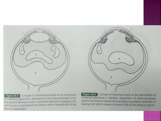
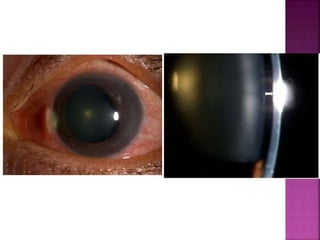
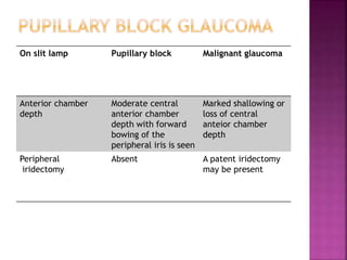
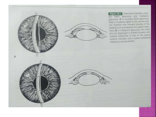
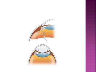
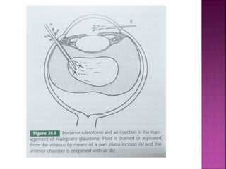
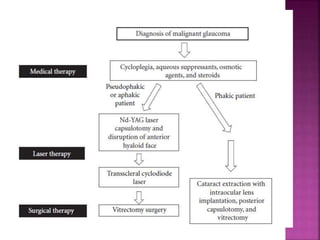
Dr Nikhil R P
Junior resident
 Introduction and definition
 Mechanism
 Clinical features
 Differential diagnosis
 Management
 Conclusion
 Malignant glaucoma is also known as –
1. Ciliary block glaucoma,
2. Aqueous misdirection syndrome,
3. Ciliovitreolenticular block,
4. Hyaloid block glauoma,
5. Posterior aqueoues entrapment
 It was first described by Von Graefe in 1869
 It is characterized by normal or increased
IOP associated with axial shallowing of
the entire anterior chamber in the
presence of a patent peripheral iridotomy
 Not responding to conventional therapy
 It is an uncommon but serious condition
occuring as a complication of intraocular
surgery
 2% to 4% - h/o of acute or chronic angle-closure glaucoma
that have undergone filtration surgery
 1.3 % - glaucoma surgery alone or combined with cataracts
 2.3%- Penetrating surgery
 Women are three times more likely than men
 Prior angle closure glaucoma
 Filtration surgeries: Trabeculectomy
 Laser treatment :
1. Peripheral laser iridotomy
2. Trabeculectomy
3. Cyclophotocoagulation
 Use of miotics
 Trabeculectomy - bleb needling
 Infection and inflammation
 Retinopathy of prematurity
 Retinal detachment
 Trauma
 Ciliolenticular block
 Anterior hyaloid obstruction
 Slackness of lens zonules
 Posterior pooling of aqueous
 The tips of the ciliary processes rotate
forward and press the lens equator or against
the anterior hyaloid (aphakic cases)
 Hence creating an obstruction to the flow of
aqueous, which pools in and behind the
vitreous with a forward shift of lens- iris
diaphragm
 Most common mechanism involved in
malignant glaucoma
 Breaks in the hyaloid near the vitreous base,
allows the posterior diversion of aqueous
 Obstruction to the aqueous flow is by the
anterior vitreous face, which is compressed
forward against the ciliary processes in
phakic and aphakic forms
 Hence anterior hyaloid may contribute to
ciliolenticular block
 Abnormal slackness or weakness of zonules of
causes forward movement of the lens – iris
diaphragm along with the pressure from the
vitreous resulting in malignant glaucoma
 Postulated by Chandler and Grant
 Shaffer hypothesized that an accumulation of
aqueous behind posterior vitreous
detachment causes the forward displacement
of iris-lens or iris – vitreous diaphragm
 This can be seen by using ultrasound bscan
demonstrating echo free zones in vitreous
from which aqueous was aspirated
 Action of miotics may produce malignant
glaucoma through contraction of ciliary
muscle or associated forward shifting of the
lens with shallowing of anterior chamber
 This mechanism have been described for
unoperated eyes recieving miotic therapy
and in eye treated with miotics after
filtering procedure
 Both inflammation and trauma are
precipitating factors of malignant glaucoma
 Endophthalmitis caused by fungal
keratomycosis and Nocardia asteroides is
associated with malignant glaucoma
 Duration – it might occur in early post op
period, delayed by some days, weeks or months
 Pain
 Congestion
 Watering
 Diminution of vision
 Myopic shift - Anterior dislocation of the
iris-lens diaphragm with secondary
improvement of near vision
 Anterior chamber is shallow or flat centrally
and peripherally even though patent
iridectomy is present
 IOP is raised (40-60mmhg)
 Medical history
1. Determination of predisposing factors
2. Symptoms
 Slit lamp examination
1. ACD - axial (central and peripheral) shallowing of the anterior
chamber
2. Patency of the iridotomy
3. Seidel test should be performed to exclude filtering bleb
leaking after filtration surgery.
4. Posterior segment : Ruling out choroidal detachment or
suprachoroidal hemorrhage
 Tonometry – usually reveals increased IOP
 The rotation of the ciliary body to the front and shallowing
of the anterior chamber
 Marked displacement of the structures of the anterior
segment
 Peripheral irido-corneal touch
 Forward shift of the lens may be noted
 Pupillary block glaucoma
 Choroidal detachment
 Suprachoroidal hemorrhage
On slit lamp Pupillary block Malignant glaucoma
Anterior chamber
depth
Moderate central
anterior chamber
depth with forward
bowing of the
peripheral iris is seen
Marked shallowing or
loss of central
anteior chamber
depth
Peripheral
iridectomy
Absent A patent iridectomy
may be present
 Choroidal separation with serous fluid is common
after glaucoma filtering procedures
 Shallow or flat anterior chamber, IOP detected is
inaccurate and cannot be relied on for diagnosis
 Anterior chamber shallowing may be secondary to
anterior uveal effusion with forward rotation of
the lens or iris diaphragm, producing secondary
angle closure glaucoma resembling malignant
glaucoma
 The presence of choroidal fluid found on
ultrasonography and ophthalmoscopic
examination of posterior segment helps in
diagnosing the condition
 Most of these resolve spontaneously as IOP
rises
 In persistent cases or massive cases, scleral
incisions are made in inferior quadrants.
Straw coloured fluid from suprachoroidal
space confirms detachment. AC is reformed
with air or saline or both
 This condition may occur hours or days after
ocular surgery
 IOP is elevated associated with pain
 Shallow anterior chamber depth
 The surgical approach is same as that of
choroidal detachment with drainage of blood
from suprachoroidal space and reformation
of anterior chamber
 Flattening or shallow AC – central and
peripheral
 IOP is greater than anticipated pressure post
filtration surgery, when malignant glaucoma
follows after filtration surgery
 Absence of pupillary block, confirmed by
patent peripheral iridectomy
 Absence of suprachoroidal effusion or
hemorrhage confirmed by B scan
 Medical
 Laser
 Surgery
 Management of the fellow eye
 Hyperosmostic agents –
20% mannitol iv reduces the pressure exerted
by vitreous or oral glycerol can also be given
 Mydriatic cycloplegic combination –
1% atropine and 10 % phenylephrine drops-
relaxation of ciliary muscle there by tightening
the zonules, break the ciliary block and push the
lens backwards helping in forming anterior
chamber
 Aqueous suppressants –
Beta blocker or alpha 2 agoinst or
carbonic anhydrase inhibitors to be used to
reduce IOP
 Maintenance therapy – the patient to be on
atropine drops to prevent recurrence
 MIOTICS – CONTRAINDICATED
 In case of no response within 5 days, laser or
surgery is employed
 Argon laser photocoagulation of the ciliary
processes
 Trans scleral diode laser
cyclophotocoagulation has been reported to
relive malignant glaucoma
 Nd: YAG laser hyaloidotomy can be
undertaken in aphakic and pseudophakic
eyes
 When medical or laser therapy fails, surgical
intervention is required
 It involves removal of aqueous pockets from
the vitreous and restoration of anterior
chamber
 Posterior sclerotomy and air injection
 Anterior pars plana vitrectomy
 Lens extraction
 Posterior sclerotomy and air injection:
Sclerotomy is done 3mm posterior to the
limbus to break the anterior hyaloid and
aspiration of liquid vitreous with reformation
of anterior chamber with an air bubble
 Post operatively – atropine eye drops is given
to prevent recurrence
 Anterior pars plana vitrectomy
Anterior vitreous including anterior
hyaloid is removed with vitrectomy
instruments
 But both posterior sclerotomy and anterior
vitrectomy have risk of serious complications
 The type of surgery depends on surgeon’s
choice
 Lens extraction(phakic cases)-
This is favoured by some surgeons when
all the three above procedure fails
 The fellow eye is at risk of developing
malignant glaucoma if it undergoes surgery
 Hence prophylactic laser iridotomy is done, if
indicated
 If angle closure glaucoma is present, every
effort should be made to break the attack
before surgery and if attack cant be broken,
mydriatic- cycloplegic therapy to be started
vigarously after iridotomy and continued
indefinitely
 Malignant glaucoma – Therapeutic challenge
 Patients with h/o MG in fellow and PACG should be closely
followed in after glaucoma filtration surgeries
 Good prognosis with current treatment modalities
Malignant glaucoma

Malignant glaucoma

  • 1.
    Dr Nikhil RP Junior resident
  • 2.
     Introduction anddefinition  Mechanism  Clinical features  Differential diagnosis  Management  Conclusion
  • 3.
     Malignant glaucomais also known as – 1. Ciliary block glaucoma, 2. Aqueous misdirection syndrome, 3. Ciliovitreolenticular block, 4. Hyaloid block glauoma, 5. Posterior aqueoues entrapment  It was first described by Von Graefe in 1869
  • 4.
     It ischaracterized by normal or increased IOP associated with axial shallowing of the entire anterior chamber in the presence of a patent peripheral iridotomy  Not responding to conventional therapy  It is an uncommon but serious condition occuring as a complication of intraocular surgery
  • 5.
     2% to4% - h/o of acute or chronic angle-closure glaucoma that have undergone filtration surgery  1.3 % - glaucoma surgery alone or combined with cataracts  2.3%- Penetrating surgery  Women are three times more likely than men
  • 6.
     Prior angleclosure glaucoma  Filtration surgeries: Trabeculectomy  Laser treatment : 1. Peripheral laser iridotomy 2. Trabeculectomy 3. Cyclophotocoagulation  Use of miotics  Trabeculectomy - bleb needling  Infection and inflammation  Retinopathy of prematurity  Retinal detachment  Trauma
  • 7.
     Ciliolenticular block Anterior hyaloid obstruction  Slackness of lens zonules  Posterior pooling of aqueous
  • 8.
     The tipsof the ciliary processes rotate forward and press the lens equator or against the anterior hyaloid (aphakic cases)  Hence creating an obstruction to the flow of aqueous, which pools in and behind the vitreous with a forward shift of lens- iris diaphragm
  • 9.
     Most commonmechanism involved in malignant glaucoma
  • 11.
     Breaks inthe hyaloid near the vitreous base, allows the posterior diversion of aqueous  Obstruction to the aqueous flow is by the anterior vitreous face, which is compressed forward against the ciliary processes in phakic and aphakic forms  Hence anterior hyaloid may contribute to ciliolenticular block
  • 13.
     Abnormal slacknessor weakness of zonules of causes forward movement of the lens – iris diaphragm along with the pressure from the vitreous resulting in malignant glaucoma  Postulated by Chandler and Grant
  • 14.
     Shaffer hypothesizedthat an accumulation of aqueous behind posterior vitreous detachment causes the forward displacement of iris-lens or iris – vitreous diaphragm  This can be seen by using ultrasound bscan demonstrating echo free zones in vitreous from which aqueous was aspirated
  • 15.
     Action ofmiotics may produce malignant glaucoma through contraction of ciliary muscle or associated forward shifting of the lens with shallowing of anterior chamber  This mechanism have been described for unoperated eyes recieving miotic therapy and in eye treated with miotics after filtering procedure
  • 16.
     Both inflammationand trauma are precipitating factors of malignant glaucoma  Endophthalmitis caused by fungal keratomycosis and Nocardia asteroides is associated with malignant glaucoma
  • 17.
     Duration –it might occur in early post op period, delayed by some days, weeks or months  Pain  Congestion  Watering  Diminution of vision
  • 18.
     Myopic shift- Anterior dislocation of the iris-lens diaphragm with secondary improvement of near vision  Anterior chamber is shallow or flat centrally and peripherally even though patent iridectomy is present  IOP is raised (40-60mmhg)
  • 20.
     Medical history 1.Determination of predisposing factors 2. Symptoms  Slit lamp examination 1. ACD - axial (central and peripheral) shallowing of the anterior chamber 2. Patency of the iridotomy 3. Seidel test should be performed to exclude filtering bleb leaking after filtration surgery. 4. Posterior segment : Ruling out choroidal detachment or suprachoroidal hemorrhage  Tonometry – usually reveals increased IOP
  • 21.
     The rotationof the ciliary body to the front and shallowing of the anterior chamber  Marked displacement of the structures of the anterior segment  Peripheral irido-corneal touch  Forward shift of the lens may be noted
  • 22.
     Pupillary blockglaucoma  Choroidal detachment  Suprachoroidal hemorrhage
  • 23.
    On slit lampPupillary block Malignant glaucoma Anterior chamber depth Moderate central anterior chamber depth with forward bowing of the peripheral iris is seen Marked shallowing or loss of central anteior chamber depth Peripheral iridectomy Absent A patent iridectomy may be present
  • 26.
     Choroidal separationwith serous fluid is common after glaucoma filtering procedures  Shallow or flat anterior chamber, IOP detected is inaccurate and cannot be relied on for diagnosis  Anterior chamber shallowing may be secondary to anterior uveal effusion with forward rotation of the lens or iris diaphragm, producing secondary angle closure glaucoma resembling malignant glaucoma
  • 27.
     The presenceof choroidal fluid found on ultrasonography and ophthalmoscopic examination of posterior segment helps in diagnosing the condition  Most of these resolve spontaneously as IOP rises  In persistent cases or massive cases, scleral incisions are made in inferior quadrants. Straw coloured fluid from suprachoroidal space confirms detachment. AC is reformed with air or saline or both
  • 28.
     This conditionmay occur hours or days after ocular surgery  IOP is elevated associated with pain  Shallow anterior chamber depth  The surgical approach is same as that of choroidal detachment with drainage of blood from suprachoroidal space and reformation of anterior chamber
  • 29.
     Flattening orshallow AC – central and peripheral  IOP is greater than anticipated pressure post filtration surgery, when malignant glaucoma follows after filtration surgery  Absence of pupillary block, confirmed by patent peripheral iridectomy  Absence of suprachoroidal effusion or hemorrhage confirmed by B scan
  • 30.
     Medical  Laser Surgery  Management of the fellow eye
  • 31.
     Hyperosmostic agents– 20% mannitol iv reduces the pressure exerted by vitreous or oral glycerol can also be given  Mydriatic cycloplegic combination – 1% atropine and 10 % phenylephrine drops- relaxation of ciliary muscle there by tightening the zonules, break the ciliary block and push the lens backwards helping in forming anterior chamber
  • 32.
     Aqueous suppressants– Beta blocker or alpha 2 agoinst or carbonic anhydrase inhibitors to be used to reduce IOP  Maintenance therapy – the patient to be on atropine drops to prevent recurrence  MIOTICS – CONTRAINDICATED  In case of no response within 5 days, laser or surgery is employed
  • 33.
     Argon laserphotocoagulation of the ciliary processes  Trans scleral diode laser cyclophotocoagulation has been reported to relive malignant glaucoma  Nd: YAG laser hyaloidotomy can be undertaken in aphakic and pseudophakic eyes
  • 35.
     When medicalor laser therapy fails, surgical intervention is required  It involves removal of aqueous pockets from the vitreous and restoration of anterior chamber  Posterior sclerotomy and air injection  Anterior pars plana vitrectomy  Lens extraction
  • 36.
     Posterior sclerotomyand air injection: Sclerotomy is done 3mm posterior to the limbus to break the anterior hyaloid and aspiration of liquid vitreous with reformation of anterior chamber with an air bubble  Post operatively – atropine eye drops is given to prevent recurrence
  • 38.
     Anterior parsplana vitrectomy Anterior vitreous including anterior hyaloid is removed with vitrectomy instruments  But both posterior sclerotomy and anterior vitrectomy have risk of serious complications  The type of surgery depends on surgeon’s choice  Lens extraction(phakic cases)- This is favoured by some surgeons when all the three above procedure fails
  • 40.
     The felloweye is at risk of developing malignant glaucoma if it undergoes surgery  Hence prophylactic laser iridotomy is done, if indicated  If angle closure glaucoma is present, every effort should be made to break the attack before surgery and if attack cant be broken, mydriatic- cycloplegic therapy to be started vigarously after iridotomy and continued indefinitely
  • 41.
     Malignant glaucoma– Therapeutic challenge  Patients with h/o MG in fellow and PACG should be closely followed in after glaucoma filtration surgeries  Good prognosis with current treatment modalities

Editor's Notes

  • #42 Whatever the true mechanism, the fact that it is relieved when a direct communication is made between the anterior chamber and vitreous cavity supports the theory that the lens, anterior vitreous, and ciliary processes are intimately involved in the pathogenesis