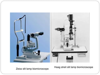
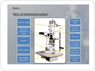
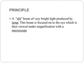
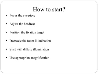
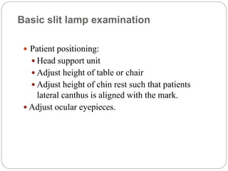
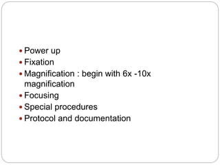
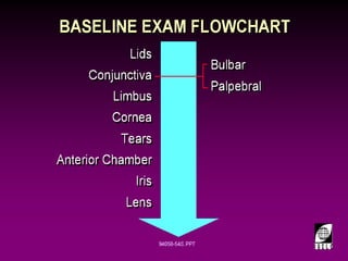
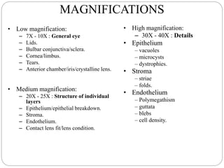
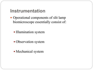
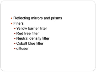
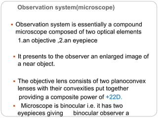
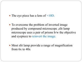
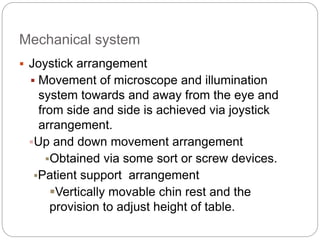
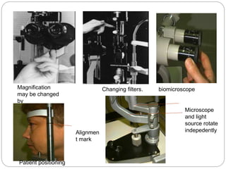
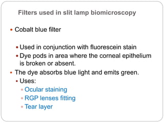
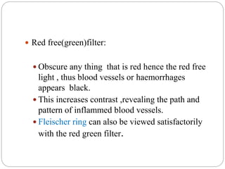
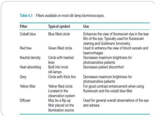
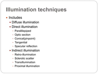
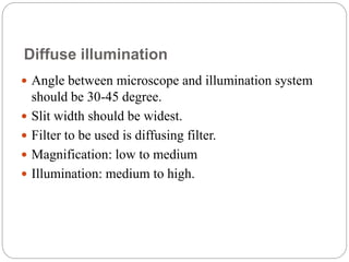
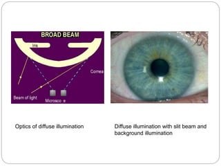
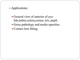
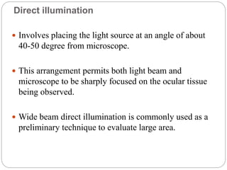
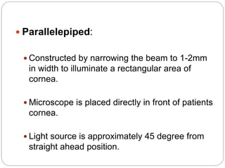
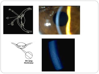
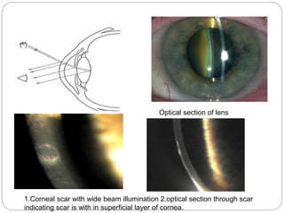
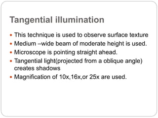
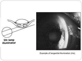
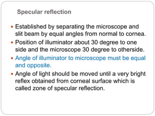
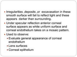
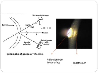
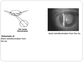
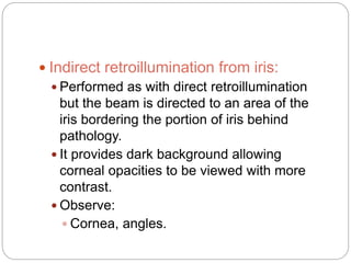
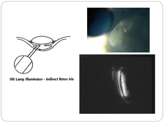
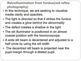
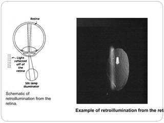
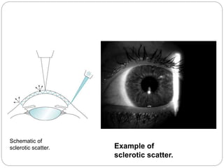
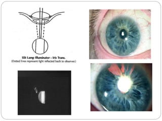
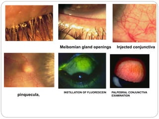
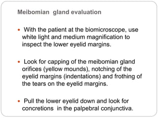
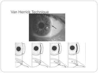
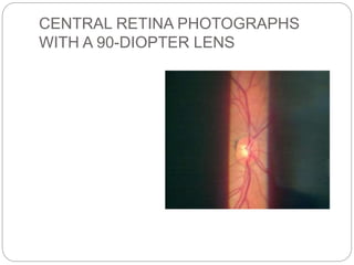
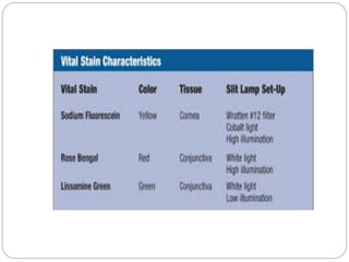
The document discusses slit lamp examinations, which use a high-intensity light source focused as a slit and viewed through a microscope to examine the anterior segment of the eye. It describes the basic components and principles of the slit lamp biomicroscope, various illumination techniques used to examine different ocular structures, and historical developments of the slit lamp.
To know Humphrey visual field analyser
To know about various types of perimetry
To identify field defect
To recognize that field defect is due to glaucoma or neurological lesion
To know that field defect is progressive or not
Interpretation of HVFA
To know Humphrey visual field analyser
To know about various types of perimetry
To identify field defect
To recognize that field defect is due to glaucoma or neurological lesion
To know that field defect is progressive or not
Interpretation of HVFA
a comprehensive presentation on Slit-lamp in which i have described about the history, optics and also the uses of different illuminations by Dr. Paresh Nichlani
A Classroom presentation, showing the various types of slit-lamps, their parts, and other accessory instruments that can be used with it for enhanced optometric clinical examination.
Utilizzo di una Scheimpflug Camera in Banca Cornee per Pachimetria pre e post...Luca Avoni
Questa Presentazione ha ricevuto l'attestazione di Best Paper in occasione del 12° Congresso Internazionale SOI - Società Oftalmologica Italiana, tenutosi a Milano dal 21 al 24 maggio 2014
La pachimetria corneale (spessore della cornea) nel trapianto endoteliale di cornea (DSAEK) è un esame molto importante sia per la Banca delle Cornee, sia per il chirurgo oculista.
In Banca Cornee è utile per poter scegliere la testina di spessore più idonea per il taglio della cornea con microcheratomo al fine di ottenere lembi endoteliali molto sottili senza perforare la cornea durante il taglio. Al chirurgo è utile per sapere le caratteristiche del lembo endoteliale pretagliato dalla Banca delle Cornee per una ottimizzazione dell'intervento e del post operatorio.
La strumentazione per effettuare la pachimetria in Banca Cornee può essere diversa: scheimpflug camera, OCT della cornea, pachimetri ad ultrasuoni. Ogni strumentazione va testata, verificata e il processo va validato.
Presso la Banca delle Cornee dell'Emilia Romagna si è effettuata una ricerca sull'utilizzo di una scheimpflug camera per ottenere le informazioni sopracitate ottenendo risultati riproducibili e validando il processo. Altre strumentazioni che si valuteranno per la pachimetria corneale saranno i pachimetri ad ultrasuoni.
Contenuto a cura del Dottor Luca Avoni
. Introduction Biomicroscope derives its name from the fact that it enables the practitioner to observe the living tissue of eye under magnification. It not only provides magnified view of every part of eye but also allows quantitative measurements and photography of every part for documentation.
3. • The lamp facilitates an examination which looks at anterior segment, or frontal structures, of the human eye, which includes the –Eyelid –Cornea –Sclera –Conjunctiva –Iris –Aqueous –Natural crystalline lens and –Anterior vitreous.
4. Important historical landmarks De Wecker 1863 devised a portable ophthalmomicroscope . Albert and Greenough 1891,developed a binocular microscope which provided stereoscopic view. Gullstrand ,1911 introduced the illumination system which had for the first time a slit diapharm in it Therefore Gullstrand is credited with the invention of slit lamp.
SLIT LAMP AND ITS DIFFERENT ILLUMINATION TECHNIQUES.pptxAbhishek Kashyap
This presentation explains in detail about different illumination techniques and filters used in slit lamp examination and the procedure to perform slit lamp examination.
Slit lamp biomicroscopy and illumination techniquesLoknath Goswami
It is a presentation on slitlamp for beginner, shown the parts and different illumination techniques both for eye and contact lens and it have short history
Explore natural remedies for syphilis treatment in Singapore. Discover alternative therapies, herbal remedies, and lifestyle changes that may complement conventional treatments. Learn about holistic approaches to managing syphilis symptoms and supporting overall health.
Ozempic: Preoperative Management of Patients on GLP-1 Receptor Agonists Saeid Safari
Preoperative Management of Patients on GLP-1 Receptor Agonists like Ozempic and Semiglutide
ASA GUIDELINE
NYSORA Guideline
2 Case Reports of Gastric Ultrasound
Tom Selleck Health: A Comprehensive Look at the Iconic Actor’s Wellness Journeygreendigital
Tom Selleck, an enduring figure in Hollywood. has captivated audiences for decades with his rugged charm, iconic moustache. and memorable roles in television and film. From his breakout role as Thomas Magnum in Magnum P.I. to his current portrayal of Frank Reagan in Blue Bloods. Selleck's career has spanned over 50 years. But beyond his professional achievements. fans have often been curious about Tom Selleck Health. especially as he has aged in the public eye.
Follow us on: Pinterest
Introduction
Many have been interested in Tom Selleck health. not only because of his enduring presence on screen but also because of the challenges. and lifestyle choices he has faced and made over the years. This article delves into the various aspects of Tom Selleck health. exploring his fitness regimen, diet, mental health. and the challenges he has encountered as he ages. We'll look at how he maintains his well-being. the health issues he has faced, and his approach to ageing .
Early Life and Career
Childhood and Athletic Beginnings
Tom Selleck was born on January 29, 1945, in Detroit, Michigan, and grew up in Sherman Oaks, California. From an early age, he was involved in sports, particularly basketball. which played a significant role in his physical development. His athletic pursuits continued into college. where he attended the University of Southern California (USC) on a basketball scholarship. This early involvement in sports laid a strong foundation for his physical health and disciplined lifestyle.
Transition to Acting
Selleck's transition from an athlete to an actor came with its physical demands. His first significant role in "Magnum P.I." required him to perform various stunts and maintain a fit appearance. This role, which he played from 1980 to 1988. necessitated a rigorous fitness routine to meet the show's demands. setting the stage for his long-term commitment to health and wellness.
Fitness Regimen
Workout Routine
Tom Selleck health and fitness regimen has evolved. adapting to his changing roles and age. During his "Magnum, P.I." days. Selleck's workouts were intense and focused on building and maintaining muscle mass. His routine included weightlifting, cardiovascular exercises. and specific training for the stunts he performed on the show.
Selleck adjusted his fitness routine as he aged to suit his body's needs. Today, his workouts focus on maintaining flexibility, strength, and cardiovascular health. He incorporates low-impact exercises such as swimming, walking, and light weightlifting. This balanced approach helps him stay fit without putting undue strain on his joints and muscles.
Importance of Flexibility and Mobility
In recent years, Selleck has emphasized the importance of flexibility and mobility in his fitness regimen. Understanding the natural decline in muscle mass and joint flexibility with age. he includes stretching and yoga in his routine. These practices help prevent injuries, improve posture, and maintain mobilit
MANAGEMENT OF ATRIOVENTRICULAR CONDUCTION BLOCK.pdfJim Jacob Roy
Cardiac conduction defects can occur due to various causes.
Atrioventricular conduction blocks ( AV blocks ) are classified into 3 types.
This document describes the acute management of AV block.
micro teaching on communication m.sc nursing.pdfAnurag Sharma
Microteaching is a unique model of practice teaching. It is a viable instrument for the. desired change in the teaching behavior or the behavior potential which, in specified types of real. classroom situations, tends to facilitate the achievement of specified types of objectives.
New Directions in Targeted Therapeutic Approaches for Older Adults With Mantl...i3 Health
i3 Health is pleased to make the speaker slides from this activity available for use as a non-accredited self-study or teaching resource.
This slide deck presented by Dr. Kami Maddocks, Professor-Clinical in the Division of Hematology and
Associate Division Director for Ambulatory Operations
The Ohio State University Comprehensive Cancer Center, will provide insight into new directions in targeted therapeutic approaches for older adults with mantle cell lymphoma.
STATEMENT OF NEED
Mantle cell lymphoma (MCL) is a rare, aggressive B-cell non-Hodgkin lymphoma (NHL) accounting for 5% to 7% of all lymphomas. Its prognosis ranges from indolent disease that does not require treatment for years to very aggressive disease, which is associated with poor survival (Silkenstedt et al, 2021). Typically, MCL is diagnosed at advanced stage and in older patients who cannot tolerate intensive therapy (NCCN, 2022). Although recent advances have slightly increased remission rates, recurrence and relapse remain very common, leading to a median overall survival between 3 and 6 years (LLS, 2021). Though there are several effective options, progress is still needed towards establishing an accepted frontline approach for MCL (Castellino et al, 2022). Treatment selection and management of MCL are complicated by the heterogeneity of prognosis, advanced age and comorbidities of patients, and lack of an established standard approach for treatment, making it vital that clinicians be familiar with the latest research and advances in this area. In this activity chaired by Michael Wang, MD, Professor in the Department of Lymphoma & Myeloma at MD Anderson Cancer Center, expert faculty will discuss prognostic factors informing treatment, the promising results of recent trials in new therapeutic approaches, and the implications of treatment resistance in therapeutic selection for MCL.
Target Audience
Hematology/oncology fellows, attending faculty, and other health care professionals involved in the treatment of patients with mantle cell lymphoma (MCL).
Learning Objectives
1.) Identify clinical and biological prognostic factors that can guide treatment decision making for older adults with MCL
2.) Evaluate emerging data on targeted therapeutic approaches for treatment-naive and relapsed/refractory MCL and their applicability to older adults
3.) Assess mechanisms of resistance to targeted therapies for MCL and their implications for treatment selection
Ethanol (CH3CH2OH), or beverage alcohol, is a two-carbon alcohol
that is rapidly distributed in the body and brain. Ethanol alters many
neurochemical systems and has rewarding and addictive properties. It
is the oldest recreational drug and likely contributes to more morbidity,
mortality, and public health costs than all illicit drugs combined. The
5th edition of the Diagnostic and Statistical Manual of Mental Disorders
(DSM-5) integrates alcohol abuse and alcohol dependence into a single
disorder called alcohol use disorder (AUD), with mild, moderate,
and severe subclassifications (American Psychiatric Association, 2013).
In the DSM-5, all types of substance abuse and dependence have been
combined into a single substance use disorder (SUD) on a continuum
from mild to severe. A diagnosis of AUD requires that at least two of
the 11 DSM-5 behaviors be present within a 12-month period (mild
AUD: 2–3 criteria; moderate AUD: 4–5 criteria; severe AUD: 6–11 criteria).
The four main behavioral effects of AUD are impaired control over
drinking, negative social consequences, risky use, and altered physiological
effects (tolerance, withdrawal). This chapter presents an overview
of the prevalence and harmful consequences of AUD in the U.S.,
the systemic nature of the disease, neurocircuitry and stages of AUD,
comorbidities, fetal alcohol spectrum disorders, genetic risk factors, and
pharmacotherapies for AUD.
Couples presenting to the infertility clinic- Do they really have infertility...Sujoy Dasgupta
Dr Sujoy Dasgupta presented the study on "Couples presenting to the infertility clinic- Do they really have infertility? – The unexplored stories of non-consummation" in the 13th Congress of the Asia Pacific Initiative on Reproduction (ASPIRE 2024) at Manila on 24 May, 2024.
Lung Cancer: Artificial Intelligence, Synergetics, Complex System Analysis, S...Oleg Kshivets
RESULTS: Overall life span (LS) was 2252.1±1742.5 days and cumulative 5-year survival (5YS) reached 73.2%, 10 years – 64.8%, 20 years – 42.5%. 513 LCP lived more than 5 years (LS=3124.6±1525.6 days), 148 LCP – more than 10 years (LS=5054.4±1504.1 days).199 LCP died because of LC (LS=562.7±374.5 days). 5YS of LCP after bi/lobectomies was significantly superior in comparison with LCP after pneumonectomies (78.1% vs.63.7%, P=0.00001 by log-rank test). AT significantly improved 5YS (66.3% vs. 34.8%) (P=0.00000 by log-rank test) only for LCP with N1-2. Cox modeling displayed that 5YS of LCP significantly depended on: phase transition (PT) early-invasive LC in terms of synergetics, PT N0—N12, cell ratio factors (ratio between cancer cells- CC and blood cells subpopulations), G1-3, histology, glucose, AT, blood cell circuit, prothrombin index, heparin tolerance, recalcification time (P=0.000-0.038). Neural networks, genetic algorithm selection and bootstrap simulation revealed relationships between 5YS and PT early-invasive LC (rank=1), PT N0—N12 (rank=2), thrombocytes/CC (3), erythrocytes/CC (4), eosinophils/CC (5), healthy cells/CC (6), lymphocytes/CC (7), segmented neutrophils/CC (8), stick neutrophils/CC (9), monocytes/CC (10); leucocytes/CC (11). Correct prediction of 5YS was 100% by neural networks computing (area under ROC curve=1.0; error=0.0).
CONCLUSIONS: 5YS of LCP after radical procedures significantly depended on: 1) PT early-invasive cancer; 2) PT N0--N12; 3) cell ratio factors; 4) blood cell circuit; 5) biochemical factors; 6) hemostasis system; 7) AT; 8) LC characteristics; 9) LC cell dynamics; 10) surgery type: lobectomy/pneumonectomy; 11) anthropometric data. Optimal diagnosis and treatment strategies for LC are: 1) screening and early detection of LC; 2) availability of experienced thoracic surgeons because of complexity of radical procedures; 3) aggressive en block surgery and adequate lymph node dissection for completeness; 4) precise prediction; 5) adjuvant chemoimmunoradiotherapy for LCP with unfavorable prognosis.
2. Introduction
• An instrument consisting of a high-intensity light source that
can be focused to shine as a slit.
• Used in conjunction with a microscope.
• The lamp facilitates an examination which looks at anterior
segment, or frontal structures, of the human eye, which
includes the
– Eyelid
– Cornea
– Sclera
– Conjunctiva
– Iris
– Anterior chamber
– Natural crystalline lens and
– Anterior vitreous.
3. Important historical landmarks
De Wecker 1863 devised a portable ophthalmomicroscope .
Albert and Greenough 1891,developed a binocular
microscope which provided stereoscopic view.
Gullstrand ,1911 introduced the illumination system which
had for the first time a slit diapharm in it
Therefore Gullstrand is credited with the invention of slit
lamp.
4. TYPES
There are 2 types of slit lamp biomicroscope
1)Zeiss slit lamp biomicroscope
2)Haag streit slit lamp biomicroscope
In Zeiss type light source is at the base of the instrument while
in Haag streit type it is at the top of the instrument.
7. PRINCIPLE
A "slit" beam of very bright light produced by
lamp. This beam is focused on to the eye which is
then viewed under magnification with a
microscope
8. How to start?
• Focus the eye piece
• Adjust the headrest
• Position the fixation target
• Decrease the room illumination
• Start with diffuse illumination
• Use appropriate magnification
9. Basic slit lamp examination
Patient positioning:
Head support unit
Adjust height of table or chair
Adjust height of chin rest such that patients
lateral canthus is aligned with the mark.
Adjust ocular eyepieces.
10. Power up
Fixation
Magnification : begin with 6x -10x
magnification
Focusing
Special procedures
Protocol and documentation
14. Illumination system
It consist of:
A bright ,focal source of light with a slit
mechanism
Provides an illumination of 2*10^5 to 4*10^5
lux.
The beam of light can be changed in
intensity,height,width,direction or angle and
color during the examination with the flick of
lever.
15. Condensing lens system:
Consist of a couple of planoconvex lenses with their
convex surface in apposition.
Slit and other diapharm:
Height and width of slit can be varied by using
knobs.
16. Projection lens:
Form an image of slit at eye.
Advantages,
1.keeps the aberration of lens down.
2.increase the depth of focus of slit.
17. Reflecting mirrors and prisms
Filters
Yellow barrier filter
Red free filter
Neutral density filter
Cobalt blue filter
diffuser
18. Observation system(microscope)
Observation system is essentially a compound
microscope composed of two optical elements
1.an objective ,2.an eyepiece
It presents to the observer an enlarged image of
a near object.
The objective lens consists of two planoconvex
lenses with their convexities put together
providing a composite power of +22D.
Microscope is binocular i.e. it has two
eyepieces giving binocular observer a
19. The eye piece has a lens of +10D.
To overcome the problem of inverted image
produced by compound microscope ,slit lamp
microscope uses a pair of prisms b/w the objective
and eyepiece to reinvert the image.
Most slit lamp provide a range of magnification
from 6x to 40x
20. Mechanical system
Joystick arrangement
Movement of microscope and illumination
system towards and away from the eye and
from side and side is achieved via joystick
arrangement.
Up and down movement arrangement
Obtained via some sort or screw devices.
Patient support arrangement
Vertically movable chin rest and the
provision to adjust height of table.
21. Fixation target:
A movable fixation target greatly faciliates the
examination under some conditions.
Mechanical coupling :
Provides a coupling of microscope and the
illumination system along a common axis of
rotation that coincides their focal planes.
This ensures that light falls on the point where
the microscope is focused
Has advantages when using the slit lamp for
routine examination of anterior segment of eye.
22. Magnification control :
Including two or pair of readily changeable
objective lenses and two sets of eyepieces.
An on and off switch and illumination control .
23. Magnification
may be changed
by
flipping a lever...
Changing filters. biomicroscope
Patient positioning
Alignmen
t mark
Microscope
and light
source rotate
indepedently
24. Filters used in slit lamp biomicroscopy
Cobalt blue filter
Used in conjunction with fluorescein stain
Dye pods in area where the corneal epithelium
is broken or absent.
The dye absorbs blue light and emits green.
Uses:
Ocular staining
RGP lenses fitting
Tear layer
25. Red free(green)filter:
Obscure any thing that is red hence the red free
light , thus blood vessels or haemorrhages
appears black.
This increases contrast ,revealing the path and
pattern of inflammed blood vessels.
Fleischer ring can also be viewed satisfactorily
with the red green filter.
28. Diffuse illumination
Angle between microscope and illumination system
should be 30-45 degree.
Slit width should be widest.
Filter to be used is diffusing filter.
Magnification: low to medium
Illumination: medium to high.
29. Optics of diffuse illumination Diffuse illumination with slit beam and
background illumination
30. Applications:
General view of anterior of eye:
lids,lashes,sclera,cornea ,iris, pupil,
Gross pathology and media opacities
Contact lens fitting.
31. Direct illumination
Involves placing the light source at an angle of about
40-50 degree from microscope.
This arrangement permits both light beam and
microscope to be sharply focused on the ocular tissue
being observed.
Wide beam direct illumination is commonly used as a
preliminary technique to evaluate large area.
32. Parallelepiped:
Constructed by narrowing the beam to 1-2mm
in width to illuminate a rectangular area of
cornea.
Microscope is placed directly in front of patients
cornea.
Light source is approximately 45 degree from
straight ahead position.
33. Applications:
Used to detect and examine corneal
structures and defects.
Used to detect corneal striae that develop
when corneal edema occurs with hydrogel
lens wear and in keratoconus.
Higher magnification than that used with wide
beam illumination is preferred to evaluate
both depth and extent of corneal ,scarring or
foreign bodies.
it is particularly suitable for assessment of
cataracts,scars,nerves,vessels etc.
34.
35. Conical beam(pinpoint)
Produced by narrowing the vertical height of a
parallelepiped to produce a small circular or square
spot of light.
Light source is 45-60 degree temporally and
directed into pupil.
Biomicroscope: directly in front of eye.
Magnification: high(16-25x)
Intensity of light source to heighest setting.
36. Focusing:
Beam is focused between cornea and anterior
lens surface and dark zone between cornea
and anterior lens observed.
Principle is same as that of beam of sun light
streaming through a room ,illuminating airborne
dust particles.This occurance is called tyndall
phenomenon.
Most useful when examining the transparency of
anterior chamber for evidence of floating cells and
flare seen in anterior uveitis.
37. Tyndall phenomenon
Cells, pigment or proteins in the
aqueous humour reflect the light like a
faint fog.
To visualise this the slit illuminator is
adjusted to the smallest circular beam
and is projected through the anterior
chamber from a 42° to 90° angle.
The strongest reflection is possible at
90°.
38.
39. Optic section
Optic section is a very thin parallelepiped and
optically cuts a very thin slice of the cornea.
Axes of illuminating and viewing path intersect in
the area of anterior eye media to be examined e.g.
the individual corneal layers.
Angle between illuminating and viewing path is 45
degree.
Slit length should be kept small to minimize
dazzling the patient.
40. With narrow slit the depth and portion of different
objects(penetration depth of foreign bodies,
shape of lens etc) can be resolved more easily.
With wider slit their extension and shape are
visible more clearly.
Magnification: maximum.
Examination of AC depth is performed by wider
slit width .1-.3mm .
41. Used to localize:
Nerve fibers
Blood vessels
Infiltrates
Cataracts
AC depth.
42. Optical section of lens
1.Corneal scar with wide beam illumination 2.optical section through scar
indicating scar is with in superficial layer of cornea.
43. Tangential illumination
This technique is used to observe surface texture
Medium –wide beam of moderate height is used.
Microscope is pointing straight ahead.
Tangential light(projected from a oblique angle)
creates shadows
Magnification of 10x,16x,or 25x are used.
44. Observe:
Anterior and posterior cornea
Iris is best viewed without dilation by this
method.
Anterior lens (especially useful for viewing
pseudoexfolation).
46. Specular reflection
Established by separating the microscope and
slit beam by equal angles from normal to cornea.
Position of illuminator about 30 degree to one
side and the microscope 30 degree to otherside.
Angle of illuminator to microscope must be equal
and opposite.
Angle of light should be moved until a very bright
reflex obtained from corneal surface which is
called zone of specular reflection.
47. Irregularities ,deposits ,or excavasation in these
smooth surface will fail to reflect light and these
appears darker than surrounding.
Under specular reflection anterior corneal
surface appears as white uniform surface and
corneal endothelium takes on a mosaic pattern.
Used to observe:
Evaluate general appearance of corneal
endothelium
Lens surfaces
Corneal epithelium
49. Indirect illumination
The beam is focused in an area adjacent to
ocular tissue to be observed.
Main application:
Examination of objects in direct vicinity of
corneal areas of reduced transparency e,g,
infiltrates,corneal scars,deposits,epithelial and
stromal defects
Illumination:
Narrow to medium slit beam
Decentred beam
Magnification: approx. m=12x (depending upon
object size)
50. Retroillumination
Retroillumination is used to evaluate the optical
qualities of a structure
The light strikes the object of interest from a
point behind the object and is then reflected back
to the observer
A vertical slit beam 1-4mm wide can be used.
Purpose:
Place object of regard against a bright
background allowing object to appear dark or
black.
51. Used most often in searching for keratic
precipitates and other debris on corneal
endothelium.
The crystalline lens can also be
retroilluminated for viewing of water clefts and
vacuoles of anterior lens and posterior
subcapsular cataract
52. Direct retroillumination from iris:
Used to view corneal pathology.
A moderately wide slit beam is aimed towards
the iris directly behind the corneal anomaly.
Use magnification of 16x to 25x and direct the
light from 45 degree.
Microscope is directed straight ahead .
54. Indirect retroillumination from iris:
Performed as with direct retroillumination
but the beam is directed to an area of the
iris bordering the portion of iris behind
pathology.
It provides dark background allowing
corneal opacities to be viewed with more
contrast.
Observe:
Cornea, angles.
55.
56. Retroillumination from fundus(red reflex
photography)
In this technique, we are seeking to visualize
media clarity and opacities.
The light is directed so that it strikes the fundus
and creates a glow behind the abnormality
The defect creates a shadow in the light
The slit illuminator is positioned in an almost
coaxial position with the biomicroscope.
A wide slit beam is decentered and adjusted to a
half circle by using the slit width
The decentred slit beam is projected near the
pupil margin through a dilated pupil.
57. Focus the microscope directly on the
pathology using 10X to 16X
magnification
Observe:
cornea, lens, vitreous
59. Sclerotic scatter
A tall, wide beam is directed onto the limbal
area.
Such an illumination technique causes cornea
to take on total internal reflection.
The slit beam should be placed approximately
40-60 degree from the microscope.
When properly positioned this technique will
produce halo glow of light around the limbus
as the light is transmitted around the cornea.
Corneal changes or abnormalities can be
visualized by reflecting the scattered light.
60. Used to observe:
Central corneal epithelial edema
Corneal abrasions
Corneal opacities
62. Proximal illumination
This illumination technique is used to observe
internal detail, depth, and density.
Use a short,fairly narrow slit beam.
Place the beam at the border of the structure
or pathology.
The light will be scattered into the
surrounding tissue, creating a light
background that highlights the edges of the
abnormality.
63. Depending on the density of the abnormality,
the light from behind may reflect through,
allowing detailed examination of the internal
structure of the pathology.
Observe: corneal opacities (edema,
infiltrates, vessels, foreign bodies), lens,
iris
64. Transillumination
In transillumination, a structure (in the eye, the
iris) is evaluated by how light passes through
it.
Iris transillumination:
This technique also takes advantage of the
red reflex.
The pupil must be at mid mydriasis (3to 4 mm
when light stimulated).
Place the light source coaxial (directly in line)
with the microscope.
.
65. Use a full circle beam of light equal to the size of
the pupil.
Project the light through the pupil and into the eye
.
Focus the microscope on the iris.
Magnification of 10X to 16X is adequate
Normally the iris pigment absorbs the light, but
pigmentation defects let the red fundus light pass
through..
Observe: iris defects (they will glow with
the orange light reflected from the
fundus)
66.
67. Uses of slit lamp biomicroscopy
Diagnostic:
OCT
FFA
Anterior segment and posterior segment
diseases
Dry eye
70. Meibomian gland evaluation
With the patient at the biomicroscope, use
white light and medium magnification to
inspect the lower eyelid margins.
Look for capping of the meibomian gland
orifices (yellow mounds), notching of the
eyelid margins (indentations) and frothing of
the tears on the eyelid margins.
Pull the lower eyelid down and look for
concretions in the palpebral conjunctiva.
71. With mild pressure, press on the eyelid
margins near the eyelashes and watch the
meibomian gland orifices.
Clear fluid should be expressed.
Capping of the orifices, a cheesy secretion on
expression and frothing of the eyelid margins
indicates meibomian gland dysfunction.
72. Van Herrick Technique
Use to evaluate anterior chamber angle without gonioscopy
Medium magnification
Angle 60 degrees
Narrow beam close to limbus
Depth of anterior chamber is evaluated it to the thickness of
cornea:
4. grade – open anterior chamber angle 1:1 ratio
3. grade – open anterior chamber angle 1:2 ratio
2. grade – narrow anterior chamber angle1:4 ratio
1. grade – risky narrow anterior chamber angle less
than 1:4 ratio
0. grade – closed anterior chamber , cornea “sits” on iris