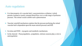
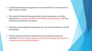
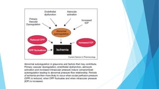
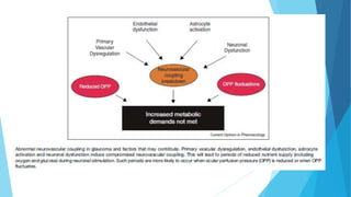
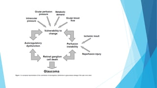
This document discusses ocular blood flow in glaucoma. It provides details on the arterial blood supply and venous drainage of the eye. It describes how glaucoma may be related to low ocular perfusion pressure and abnormalities in ocular blood flow autoregulation. Several studies linking low ocular perfusion pressure to glaucoma risk are summarized. Possible mechanisms like atherosclerosis, vasospasm and endothelial dysfunction that could contribute to impaired ocular blood flow in glaucoma are also outlined.
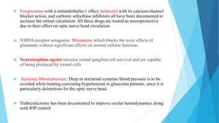
This document provides guidance on how to draw fundus diagrams. It lists the requisites needed which include an examination table, indirect ophthalmoscope, 20D lens, scleral depressor, and colored pencils. It then provides detailed instructions on how to represent various retinal structures, abnormalities, tumors, detachments and other findings using different colors and markings. Remember that choroidal tumors are drawn in brown, retinoblastomas are outlined in blue and colored yellow, and retinoschisis involves outlining the inner layer in blue and cross hatching or coloring the open portions.
USG B scan is a noninvasive imaging technique used to assess ocular structures. It works by emitting high frequency sound waves into the eye, which are reflected back to a probe and converted into an image. Key principles include sound traveling faster in solids than liquids, stronger reflections occurring at interfaces of different densities, and perpendicular angle of incidence providing best images. Clinical applications include evaluating conditions that prevent normal examination like corneal scarring or dense cataracts. It can differentiate pathologies like vitreous hemorrhage from asteroid hyalosis.
Looking deep into retina : indirect ophthalmoscopy and fundus drawingPrachir Agarwal
Indirect ophthalmoscopy provides a panoramic view of the fundus and is essential for a complete examination. It works by forming an aerial image of the retina between the condensing lens and observer. The power of the condensing lens determines the working distance, magnification, and field of view. Indirect ophthalmoscopy has advantages over direct including an unaffected view by refractive error and better illumination, resolution, and peripheral visualization. Proper technique involves adjusting the device, positioning the patient, and using scleral depression for stereoscopic viewing. Fundus drawings document examination findings and utilize various colors and line styles to depict different retinal structures and pathologies.
This document discusses keratorefractive surgeries. It begins by describing the anatomy of the cornea, including the epithelium, Bowman's layer, stroma, Descemet's membrane, and endothelium. It then provides a brief history of keratorefractive surgeries and classifications including location, addition, subtraction, relaxation, and compression techniques. Common keratorefractive surgeries discussed include PRK, LASEK, LASIK, epikeratophakia, keratophakia, and excimer laser procedures. Complications, indications, and the role of techniques like corneal topography and wavefront analysis are also summarized.
This document discusses various methods for assessing the anterior chamber angle, including subjective tests like the oblique flashlight test and Van Herrick's technique, as well as objective tests like gonioscopy, ultrasound biomicroscopy (UBM), and anterior segment optical coherence tomography (AS-OCT). Gonioscopy is considered the reference standard but can be subjective, while UBM and AS-OCT provide high resolution cross-sectional images of the angle but have limitations like requiring specialized equipment. No single test is perfect, and gonioscopy remains essential for glaucoma evaluation and management despite advances in imaging technology.
This document discusses using optical coherence tomography (OCT) to analyze the macula, retinal nerve fiber layer (RNFL), and optic nerve head in patients with glaucoma or suspected glaucoma. It describes how OCT can measure macular thickness, RNFL thickness, and optic disc parameters. Five case studies are presented showing how structural changes seen on OCT correlate with functional defects on visual field tests or clinical findings. The document concludes by mentioning Doppler OCT may help understand the role of blood flow in glaucoma and other optic neuropathies.
Gonioscopy allows visualization of the anterior chamber angle to evaluate for angle closure and diagnose glaucoma. It was pioneered in the early 20th century with the introduction of contact lenses to eliminate total internal reflection at the cornea. Direct gonioscopy uses contact lenses for a straight view, while indirect gonioscopy uses prisms for an inverted image at the slit lamp. Examination of angle structures like the trabecular meshwork and classification systems help diagnose angle closure and glaucoma. Gonioscopy is used for diagnostic and therapeutic purposes like laser and surgery.
The document discusses paralytic strabismus, including:
1) Hering's law of equal innervation and Sherington's law of reciprocal innervation which are important in diagnosing paralytic strabismus.
2) The sequelae of ocular muscle palsy including overactions and underactions of muscles.
3) Methods for investigating incomitant strabismus including cover tests, motility examination, and Hess screen plots to identify the affected muscle.
Most retinal surgeons are trained to create formal retinal drawings of the fundus.
Retinal drawings are useful to document pathology, although more and more people now prefer fundus photographs.
Can be used for serial follow up of patients to document changes in the pathology.
This document discusses intermittent exotropia, including its theories, presentation, examination, classification, treatment, and surgical management. The key points are:
1. Intermittent exotropia is thought to be caused by an imbalance between convergence and divergence muscles. It typically begins as exophoria in infancy and progresses to intermittent exotropia.
2. Examination includes measuring the deviation at distance and near with and without lenses to classify the type. Non-surgical treatment aims to improve vergence control through patching, lenses, and orthoptics.
3. Surgical treatment is indicated for deviations over 20 prism diopters, worsening control, or failure of conservative therapy.
Pigment dispersion syndrome is characterized by the dispersion of pigment granules from the iris pigment epithelium throughout the anterior segment of the eye. This can lead to elevated intraocular pressure and pigmentary glaucoma. It typically affects young, myopic white males and is caused by rubbing of the iris pigment epithelium against the lens zonules due to posterior bowing of the peripheral iris. Diagnosis is based on finding the classic triad of Krukenberg spindle on the cornea, midperipheral iris transillumination defects, and dense trabecular pigmentation on gonioscopy.
1. Corneal wound healing involves an epithelial phase, stromal phase, and endothelial phase. The epithelial phase begins within 12-48 hours as the surface epithelium slides and replicates to form a plug. The stromal phase lasts several weeks as keratocytes transform and synthesize new collagen to bridge the wound. The endothelial phase can take up to 30 days as the monolayer remodels to form a functional barrier.
Optical coherence tomography in glaucoma - Dr Shylesh DabkeShylesh Dabke
This document discusses optical coherence tomography (OCT) in evaluating glaucoma. It begins by outlining the importance of early glaucoma detection to prevent vision loss. OCT is described as the most appropriate technology for detecting glaucoma as it can assess retinal nerve fiber layer (RNFL) thickness before visual field or optic disc changes occur. RNFL thinning is an early sign of glaucoma. The document then provides details on OCT technology and analysis of RNFL thickness, optic nerve head, and macula to diagnose and monitor glaucoma. RNFL analysis, especially of the inferior quadrant, is highlighted as the most useful OCT assessment for detecting early glaucoma.
The document discusses patterns of strabismus, specifically the A pattern and V pattern. The A pattern involves relative convergence on upgaze and divergence on downgaze, while the V pattern is the opposite with relative divergence on upgaze and convergence on downgaze. Variants include the X, Y, lambda, and diamond patterns. The etiology of these patterns involves dysfunction of the horizontal, vertical, or oblique eye muscles. Clinical features may include anomalous head posture, amblyopia, and abnormal retinal correspondence. Diagnosis involves measuring alignment in upgaze and downgaze while preventing accommodation.
LIMBUS… • The limbus forms the border between the transparent cornea and opaque sclera, contains the pathways of aqueous humour outflow, and is the site of surgical incisions for cataract and glaucoma
2. Anatomical Limbus: Circumcorneal transitional zone of the conjunctivocorneal & corneoscleral junction Conjunctivo-corneal junction: • Bulbar conjunctiva is firmly adherent to underlying structures • Substantia propria of the conjunctiva stops here but its epithelium continues with that of the cornea. Sclero-corneal junction: • Transparent corneal lamellae become continuous • With the oblique, circular and opaque fibres of sclera
3. CONTINUE…. • In the area near limbus, the conjunctiva, tenon’s capsule & the episcleral tissue are fused into a dense tissue which is strongly adherent to corneo scleral junction.It is preferred site for obtaining a firm hold of the eyeball during ocular surgery. • The limbus is a common site for the occurrence of corneal epithelial neoplasm. • The Limbus contains radially oriented fibrovascular ridge known as the palisades of Vogt that may harbour a stem cell population. The palisades of Vogt are more common in the superior and inferior quadrants around the eye
Pseudoexfoliation syndrome is a systemic condition characterized by grey-white fibrillar deposits that can lead to open-angle glaucoma. It involves the trabecular meshwork, lens, ciliary body and other ocular tissues, and is a major risk factor for glaucoma. Treatment involves managing elevated intraocular pressure through medications, laser trabeculoplasty, trabeculectomy or cataract surgery due to the increased risk of complications from zonular weakness.
Indirect ophthalmoscopy has evolved since its introduction in the 1850s to become an indispensable tool for examining the retina. It allows examination of the peripheral retina through the use of a condensing lens held close to the eye. The observer views an enlarged, inverted image of the retina. Several advantages include the ability to compensate for a patient's refractive error, good illumination, and use with scleral indentation to examine the far periphery. Adjustments of the lens diopter and observation distance allow viewing different areas of the retina with varying magnification and field of view. Proper technique involves adjusting the headband-mounted binocular scope and positioning the condensing lens.
1) Biometry is the process of measuring the eye to determine the ideal intraocular lens power for cataract surgery. It involves measuring the corneal power and axial length of the eye.
2) Traditional A-scan ultrasound biometry measures axial length using sound waves, but has limitations like variable corneal compression. Newer devices like the IOL Master use optical interferometry and are non-contact.
3) Proper technique and accounting for factors like intraocular lens material are important for accurate biometry and intraocular lens power calculation. Inaccuracies can result in postoperative refractive surprises.
It describes about the procedure of Hess charting. it serves as a great tool to understand the concepts involved. Suitable for optometry course. This is not a routine procedure but an important procedure which is used in diagnosis.
UBM and ASOCT provide high-resolution cross-sectional images of the anterior segment including the cornea, anterior chamber, angle, and iris. ASOCT uses optical coherence tomography with a wavelength of 1310nm for improved penetration and reduced retinal damage compared to posterior segment OCT. It allows high-speed imaging of dynamic structures. ASOCT has applications in assessing corneal diseases and procedures, glaucoma (including angle anatomy and iridotomy evaluation), and intraocular lens implantation. Measurements of angle width parameters help evaluate angle closure risk. While valuable for objective angle assessment, ASOCT cannot image all anatomical structures involved in glaucoma.
Ultrasound biomicroscopy (UBM) provides high-resolution imaging of ocular structures in the anterior segment of the eye using 50 MHz ultrasound. UBM allows visualization of tissues like the ciliary body and zonules that are not visible by slit lamp examination. UBM can be used to qualitatively and quantitatively evaluate the anterior segment structures and has applications in diagnosing and monitoring conditions like glaucoma, corneal diseases, tumors, and intraocular lenses. While UBM provides excellent detail, it has limitations including only being able to image about 5mm into the eye and requiring contact with the eye, unlike anterior segment OCT which is non-contact.
Diplopia, or double vision, occurs when more than one image of an object is seen simultaneously. It can be caused by abnormalities in the eyes themselves or issues with eye movement coordination. A diplopia chart is used to evaluate the type and location of double vision by having the patient report the appearance of light sources in different gaze positions. Interpretation of the chart provides clues to which eye muscles may be affected and whether the cause is neurogenic, restrictive, or myogenic in nature. Treatment options include glasses, prisms, eye patching, or strabismus surgery depending on the deviation and goal of eliminating diplopia.
Iol power calculation in pediatric patientsAnisha Rathod
- Many factors affect intraocular lens (IOL) power calculation in pediatric patients including age at surgery, laterality, amblyopia, axial length, keratometry, and expected myopic shift due to ongoing eye growth.
- Normal eye development involves rapid growth of the axial length and changes in lens power in the first years of life.
- Target postoperative refraction must account for this myopic shift and generally involves undercorrecting more in younger patients.
- Accurate biometry using immersion ultrasound or optical techniques is important to minimize errors from corneal compression.
- Formulas, IOL type and position can further influence outcomes.
1. Monocular elevation deficiency (MED), also known as double elevator palsy, is characterized by an inability to elevate one eye in all fields of gaze, resulting in hypotropia of the affected eye.
2. The condition can be congenital or acquired, with causes including superior rectus palsy, inferior rectus restriction, and supranuclear lesions.
3. Surgical management of MED depends on forced duction test results and may include inferior rectus recession, superior rectus resection, or Knapp's procedure to transpose the horizontal rectus muscles. The goal is to improve eye position and increase binocular vision.
Gonioscopy refers to techniques for examining the anterior chamber angle of the eye to evaluate and classify normal and abnormal angle structures. It involves using specialized lenses and lights to view the iridocorneal angle. There are two main methods: direct gonioscopy uses large contact lenses requiring saline, while indirect uses smaller lenses with mirrors or prisms that utilize tear film. Gonioscopy is used to diagnose angle-closure glaucoma and other conditions by allowing visualization of the trabecular meshwork, scleral spur, and other angle structures. Indentation gonioscopy, where the lens is pressed against the cornea, can differentiate appositional from synechial angle closure. Proper technique and
Optical coherence tomography (OCT) is useful for imaging both the anterior and posterior segments in glaucoma. Posterior segment OCT allows quantification of retinal nerve fiber layer thickness, optic nerve head parameters, and ganglion cell layer thickness. Changes in these measurements over time can help detect glaucomatous progression. Anterior segment OCT visualizes angle anatomy and structures after glaucoma surgery. OCT provides objective data but results must be interpreted carefully while considering limitations such as variability between devices and lack of representation in normative databases.
This document discusses glaucomatous optic atrophy (GOA), an irreversible end-stage condition caused by glaucoma that results in severe vision loss. GOA can be caused by uncontrolled primary or secondary glaucoma over many years. The case report describes a patient who developed GOA in one eye due to non-compliance with treatment for chronic open-angle glaucoma, resulting in advanced cupping and pallor of the optic nerve. Aggressive treatment including multiple medications and laser surgery was able to slow progression in the other eye but vision could not be restored in the affected eye. Genetic factors may also contribute to glaucoma types like normal tension glaucoma that can lead to GOA.
Binocular single vision refers to simultaneous vision with two eyes that occurs when an individual fixates on an object. There are three grades of binocular vision: simultaneous perception, fusion, and stereopsis. Fusion is the ability to see a composite picture from two similar images, while stereopsis provides the impression of depth by superimposing images from slightly different angles. Tests for binocular vision include those for simultaneous perception, fusion, and stereopsis using instruments like the synaptophore. Binocular vision develops through infancy and childhood as the visual axes become coordinated to direct each fovea at the object of regard.
Correlation Between Intraocular pressure(IOP)and Intracranial pressure(ICP) i...Alireza Sheikhi
This document discusses several studies that have investigated the relationship between intraocular pressure (IOP) and intracranial pressure (ICP). The studies have shown mixed results, with some finding a correlation between IOP and ICP, and others finding no correlation. The current study aims to further explore this relationship by experimentally reducing IOP in dogs through medication or surgery and measuring the impact on ICP over time. Fifteen healthy dogs were divided into groups that received eye drops, eye surgery, or no treatment to the right eye. IOP and ICP were measured at baseline and 2 and 4 weeks later to analyze any changes and potential correlation between the pressures.
TeezCorrelation Between Intraocular pressure(IOP)and Intracranial pressure(IC...Alireza Sheikhi
This document discusses several studies that have investigated the relationship between intraocular pressure (IOP) and intracranial pressure (ICP). Some studies found a significant correlation between IOP and ICP, while others did not find a clear relationship. The current study aims to help address limitations of prior studies by experimentally reducing IOP in animal models to observe the effects on ICP.
Ocular/ orbital auscultation-Technique | Ocular Bruits| Conditions Useful | ...martinshaji
Ocular auscultation is a commonly neglected step of routine physical examination. An adequate ocular auscultation can be helpful in discovering an ocular bruit, which is an important diagnostic finding for a broad spectrum of pathologic conditions, some of which are potentially fatal.
this is a brief study on orbital auscultation
please comment
thank you
Postoperative vision loss (POVL) can occur after certain surgical procedures and is associated with patient positioning and other factors that compromise blood flow to the optic nerve. The document discusses POVL, including reported incidence rates after different surgeries. It reviews anatomy of the eye, normal blood flow, and how compromised flow can lead to vision loss through ischemic optic neuropathy or retinal ischemia. Risk factors discussed include prone positioning, steep Trendelenberg positioning, surgery duration over 6 hours, blood loss over 1000mL, obesity, and male sex. Interventions to reduce risk include careful patient positioning, limiting crystalloid fluids, using colloids, taking breaks in steep Trendelenberg every 4 hours, and applying eye drops
Glaucoma is defined as a group of diseases characterized by optic nerve damage and visual field loss. It is associated with elevated intraocular pressure but this is not required for diagnosis. The aqueous humor is produced by the ciliary body and drains through two pathways - the trabecular meshwork and Schlemm's canal or through the uveoscleral pathway. Investigations for glaucoma include tonometry, gonioscopy, optic nerve examination, and visual field testing. Glaucomas are classified as open-angle, angle-closure, childhood, and secondary to other conditions.
Association between Blood Pressure and Intraocular Pressure in Relation to Gl...AI Publications
High blood pressure is not only associated with systemic cardiovascular complications, but also, ocular complications such as glaucoma. This study sought to determine the association between blood pressure, intraocular pressure and glaucoma. In this cross-sectional study, 162 hypertensive patients (with mean age of 57 (±14.26) years) visiting the St. Dominic’s hospital, in Ghana were purposively sampled to participate in the study. Data on patient’s history, blood pressure, intraocular pressure and cup-to-disc ratio were collected and analysed using Statistical Package for Social Sciences (SPSS) version 20. Descriptive statistics was computed and thestatistical significance of associations was assessed at a significance level of 0.01. The outcome of the study revealed a positive correlation of 0.364 and 0.309 between systolic blood pressure and the intraocular pressure of the right and left eye respectively.The correlation between diastolic blood pressure and intraocular pressure of the right and left eyes were 0.334 and 0.239 respectively. Out of the 104 patients with systolic blood pressure exceeding 139mmHg, 54 of them (51.9%) had intraocular pressures greater than 21mmHg and 58 of them (55.8%) had cup-to-disc ratio in the range of 0.7–1.0. A weak but positive correlation between systolic blood pressure and cup-to-disc was also established.Last but not least,a correlation of 0.441 between intraocular pressure and cup-to-disc ratio of the right eye of participants as against a correlation 0.256 for the left eye was reported.The susceptibility to glaucomawas thus, relatively high in patients withuncontrolled hypertension and higher intraocular pressure.Hence, pragmatic measures should be takenby all stakeholders towards managing hypertension and intraocular pressurein order to safeguard the health and sight of all.
Ophthalmodynamometry is a clinical procedure that measures the pressure in the ophthalmic artery to assess patency of the internal carotid artery. It involves applying pressure to the eye until the central retinal artery collapses, noting the diastolic and systolic pressures. This provides information about blood flow through the ophthalmic artery and can detect carotid artery occlusive diseases, helping prevent strokes. The procedure has been used since the early 1900s and modern methods include compression or suction ophthalmodynamometry. Precautions must be taken and it can detect conditions affecting the carotid or cerebral vasculature.
Acute Promyelocytic Leukemia with Intracerebral Bleed and ARDSMedicalintensivist
This document discusses the history, anatomy, physiology and indications for intracranial pressure (ICP) monitoring. It describes various methods for directly measuring ICP including ventricular catheters and parenchymal monitors. It also discusses indirect monitoring techniques like transcranial Doppler ultrasound, jugular venous oximetry and brain tissue oxygen monitoring. The document outlines the components of normal and abnormal ICP waveforms and how analysis can provide information on intracranial compliance and pressure.
A patent ductus arteriosus (PDA) is common in preterm infants, occurring in 15-65% of very low birth weight infants. The ductus arteriosus normally closes after birth to direct blood flow to the lungs, but in preterm infants closure may be delayed. A PDA can cause left-to-right shunting, increasing cardiac output but reducing blood flow and oxygenation to vital organs. Echocardiography is the gold standard for diagnosing and assessing the severity of a PDA, but treatment should not be based on echocardiographic criteria alone and requires consideration of clinical symptoms. While PDA closure interventions are effective, evidence for long term benefits is inconclusive, so
This document provides an overview of glaucoma, including its definition, types, symptoms, risk factors, diagnostic techniques, and treatment approaches. Primary open-angle glaucoma is the most common type of glaucoma. It is characterized by optic nerve damage and visual field loss associated with elevated intraocular pressure. While increased IOP is a major risk factor, some patients can have glaucoma despite normal IOP levels. Diagnosis involves measuring IOP, examining the optic nerve head and retinal nerve fiber layer, and testing visual fields. Treatment may involve eye drops or surgery to reduce IOP and prevent further optic nerve damage.
Linking HFpEF and Chronic kidney disease magdy elmasry
Cardio-renal interactions
Introducing nephro-cardiology
{ or cardio-nephrology }
Where are we in 2022 with HFpEF ?CKD in HFpEF { or HFpEF in CKD } Cardiorenal
Syndrome .Four-step
HFA-PEFF diagnostic algorithm
heterogeneity in patients with HFpEF.Phenotyping HFpEF :
Beyond EF.Management of HFpEF .patients with HF on dialysis
This document discusses ocular hypertension. It begins by defining ocular hypertension and noting that distinguishing it from early glaucoma can be difficult. It then discusses the epidemiology, risk factors, diagnosis, investigations, treatment, and the Ocular Hypertension Treatment Study (OHTS). The OHTS was a landmark study that showed topical ocular hypotensive medication can delay or prevent the onset of glaucoma in patients with ocular hypertension. It identified several risk factors for progression to glaucoma, including older age, higher eye pressure, larger optic discs, thinner corneas, and abnormal visual fields.
Partial Exchange Transfussion In Polycythemia Secondary To Complex Cyanotic H...AR Muhamad Na'im
This patient is a 12-year-old girl with pulmonary atresia and ventricular septal defect who presents with symptoms of hyperviscosity due to polycythemia. She has undergone partial exchange transfusions every 4 months to manage her polycythemia. Examination revealed central cyanosis, clubbing, and a heart murmur. Laboratory tests showed hemoglobin of 21.9 g/dL and hematocrit of 68.7%. She underwent a successful partial exchange transfusion of 400 mL to reduce her hematocrit and symptoms. Partial exchange transfusion is an established treatment for polycythemia in cyanotic heart disease patients to reduce viscosity and improve oxygen delivery.
This document summarizes a presentation on normal tension glaucoma (NTG). It begins with an introduction defining NTG as open-angle glaucoma with characteristic optic nerve damage and visual field defects in patients with consistently low intraocular pressure (IOP) below 21 mmHg. It then describes a case presentation of a 47-year-old female patient. The remainder of the document discusses the history, examination, investigations, differential diagnosis, management, pathogenesis involving both IOP-dependent and independent factors, and epidemiology of NTG. Key points are that lowering IOP through medication or surgery can help prevent progression even in NTG, and that NTG may have an underlying vascular component involving low ocular perfusion pressure
Vasopressor and inotropic_support_in_septic_shock_an_evidence_based_review_cr...Gaston Droguett
This document discusses vasopressor and inotropic support in septic shock. It begins by describing the pathophysiology of septic shock and how it differs from other forms of shock. It then reviews the available vasopressor agents used in clinical practice for septic shock, including norepinephrine, dopamine, epinephrine, and phenylephrine. The document discusses the challenges in making recommendations due to the lack of controlled trials. It then outlines the end points used to guide resuscitation in septic shock, including arterial blood pressure, cardiac output, mixed venous oxygen saturation, blood lactate levels, and gut tonometry.
This document provides an overview of cerebrospinal fluid (CSF) anatomy and physiology, as well as disorders of CSF circulation. Key points include: CSF is formed in the choroid plexus and circulates through the ventricles before being absorbed into venous sinuses; intracranial pressure is regulated by CSF production, circulation and absorption; hydrocephalus is an excess accumulation of CSF within the brain that can be communicating or non-communicating; management involves medical treatment, temporary procedures like external ventricular drainage, and surgical options like shunts or endoscopic third ventriculostomy.
This document provides information on anterior ischemic optic neuropathy (AION), which is the most common cause of acute optic neuropathy in older age groups. It can be divided into two types: arteritic AION, which is due to giant cell arteritis; and non-arteritic AION, which makes up most cases. Both types present with sudden painless vision loss and optic disc swelling. Arteritic AION carries a worse prognosis and requires high-dose steroid treatment to prevent loss of vision in the fellow eye. Non-arteritic AION has a variable course but generally a poor rate of recovery without any proven effective treatments.
This document provides an overview of branched retinal vein occlusion (BRVO). It discusses the classification, epidemiology, risk factors, pathogenesis, signs and symptoms, diagnostic evaluation, and management of BRVO. Key points include that BRVO is the most common type of retinal vein occlusion and risk factors include hypertension, glaucoma, hyperlipidemia, and advancing age. Diagnostic testing includes fluorescein angiography and OCT to evaluate for macular edema, capillary nonperfusion, and neovascularization. Laser photocoagulation and anti-VEGF injections are common treatment approaches for complications such as macular edema.
1) Obstructive sleep apnea (OSA) is a common risk factor for cardiovascular disease (CVD) that is often underdiagnosed. OSA causes intermittent hypoxia during sleep that can activate the sympathetic nervous system and increase inflammation, potentially contributing to the development of CVD over time.
2) Studies in India have found high rates of OSA, including moderate to severe OSA, in patients who have experienced acute coronary syndrome. However, the relationship between OSA and CVD is complex and not fully understood.
3) While OSA is generally seen as harmful for CVD, a few studies have found that people with OSA may experience less severe cardiac injury during a heart attack, possibly due to ischemic
The document discusses parameters for analyzing corneal topography scans from the Pentacam. It describes various indices and maps generated by the Pentacam that are used to evaluate corneal shape, thickness, and biomechanical properties. Key parameters include the ISV, IVA, IHA, and IHD indices, BAD-D values, PPI and ART values, and the 13-point algorithm classification. Together these provide a comprehensive overview of corneal shape and help distinguish normal corneas from those with conditions like keratoconus.
This document discusses malignant glaucoma, beginning with definitions and history. It describes the mechanisms behind malignant glaucoma as abnormal forward shifting of the iris-lens diaphragm. Clinical features include increased eye pressure despite patent iridotomy and shallowing of the anterior chamber. Differential diagnosis and management options are provided, including medical therapy, laser treatment, and surgeries like posterior sclerotomy with air injection. The fellow eye is also at risk and may require prophylactic treatment.
This document discusses choroidal neovascularization (CNV), which is the abnormal growth of blood vessels from the choroid into the retina or subretinal space. It is a cause of vision loss and the main feature of exudative age-related macular degeneration. The document defines CNV and lists various conditions that can cause it. It then focuses on CNV caused by age-related macular degeneration, covering risk factors, pathogenesis, symptoms, diagnostic findings on fluorescein angiography and OCT, and various treatment options including anti-VEGF drugs, photodynamic therapy, and laser photocoagulation.
This document discusses various tests used to evaluate binocular vision, including cover tests, Hess charting, and diplopia charting. Cover tests are used to detect manifest or latent strabismus and determine deviation direction. Hess charting maps eye positions in 9 gazes using colored lenses to dissociate vision between eyes. It identifies muscle under or overaction. Diplopia charting records double vision separation in 9 gazes to localize affected muscles. These objective tests evaluate binocular function and strabismus type and localization.
This document provides information about artificial cornea or keratoprosthesis surgery. It discusses the history and indications for the procedure, describes common designs for artificial corneas including biointegrated and non-biointegrated options, and outlines some of the major keratoprosthesis designs including the Boston KPro, AlphaCor KPro, and modified osteo-odonto keratoprosthesis. It also covers the preoperative evaluation, surgical procedure, postoperative management, prognosis, and complications for keratoprosthesis surgery.
Tractional retinal detachment occurs when fibrovascular membranes exert traction on the retina, most commonly due to proliferative diabetic retinopathy or posterior segment trauma. Static traction from these membranes causes the retina to detach in a shallow, concave configuration without visible retinal breaks. Diagnosis is based on finding reduced retinal mobility and the absence of breaks, with traction visible from fibrovascular tissue. Ultrasound can help diagnose tractional detachments when the media is opaque.
The document discusses the anatomy and examination of the peripheral retina. It provides definitions for key terms related to retinal detachment such as the vitreous base, vitreoretinal traction, posterior vitreous detachment, and retinal breaks. It also describes techniques for indirect ophthalmoscopy and scleral indentation which are used to clinically examine the peripheral retina.
Pneumatic retinopexy is an office-based procedure for repairing retinal detachments without surgery. It involves applying cryotherapy or laser around retinal breaks, then injecting an intraocular gas bubble. The gas bubble seals the breaks and pushes subretinal fluid back into the vitreous cavity. Pneumatic retinopexy is less expensive than other surgical options. Sulfur hexafluoride is commonly used as the intraocular gas due to its longevity. The procedure aims to position the gas bubble over breaks for 5 days to seal them before the gas is absorbed. Precise technique is required to ensure the gas bubble seals breaks without detaching the macula.
OCT provides high-resolution, cross-sectional images of the retina and anterior eye using low-coherence interferometry. It allows detection of morphological changes and measurement of retinal thickness, volume, and nerve fiber layer thickness. Newer variants such as ultra-high resolution OCT, Doppler OCT, and anterior segment OCT provide additional structural and functional information. OCT is a non-invasive imaging technique that has become an essential tool for diagnosing and managing retinal diseases.
The white dot syndromes are a group of diseases characterized by inflammation of the outer retina, retinal pigment epithelium, and choroid. They include birdshot retinochoroidopathy, multifocal choroiditis and panuveitis, punctate inner choroidopathy, subretinal fibrosis and uveitis syndrome, multiple evanescent white dot syndrome, acute posterior multifocal placoid pigment epitheliopathy, and serpiginous choroidopathy. The etiology is unknown but may be autoimmune or infectious. Patients typically present with blurred vision, photopsias, and floaters. Examinations reveal multiple cream-colored lesions throughout the fundus. Treatment involves corticosteroids and immunosuppress
This document contains slides for a university practical exam covering various tissues and organs of the human body, including loose connective tissue, adipose tissue, three types of cartilage tissue, bone, skeletal muscle, cardiac muscle, peripheral and optic nerves, arteries and veins of different sizes, serous and mixed salivary glands, lymph node, thymus, tonsil, spleen, sympathetic and spinal ganglia, hairy and non-hairy skin, placenta, and umbilical cord. It wishes students the best of luck on their practical exams.
The document summarizes ocular viscosurgical devices used in ophthalmic procedures. It discusses the history and ideal properties of viscoelastic substances. Commonly used families include sodium hyaluronate, chondroitin sulfate, and hydroxypropyl methylcellulose. Physical properties like viscosity, pseudoplasticity, and coatability are described. Clinical applications involve cataract surgery, glaucoma surgery, and keratoplasty. Complications from use include increased intraocular pressure and capsular block syndrome.
Muktapishti is a traditional Ayurvedic preparation made from Shoditha Mukta (Purified Pearl), is believed to help regulate thyroid function and reduce symptoms of hyperthyroidism due to its cooling and balancing properties. Clinical evidence on its efficacy remains limited, necessitating further research to validate its therapeutic benefits.
ABDOMINAL TRAUMA in pediatrics part one.drhasanrajab
Abdominal trauma in pediatrics refers to injuries or damage to the abdominal organs in children. It can occur due to various causes such as falls, motor vehicle accidents, sports-related injuries, and physical abuse. Children are more vulnerable to abdominal trauma due to their unique anatomical and physiological characteristics. Signs and symptoms include abdominal pain, tenderness, distension, vomiting, and signs of shock. Diagnosis involves physical examination, imaging studies, and laboratory tests. Management depends on the severity and may involve conservative treatment or surgical intervention. Prevention is crucial in reducing the incidence of abdominal trauma in children.
Local Advanced Lung Cancer: Artificial Intelligence, Synergetics, Complex Sys...Oleg Kshivets
Overall life span (LS) was 1671.7±1721.6 days and cumulative 5YS reached 62.4%, 10 years – 50.4%, 20 years – 44.6%. 94 LCP lived more than 5 years without cancer (LS=2958.6±1723.6 days), 22 – more than 10 years (LS=5571±1841.8 days). 67 LCP died because of LC (LS=471.9±344 days). AT significantly improved 5YS (68% vs. 53.7%) (P=0.028 by log-rank test). Cox modeling displayed that 5YS of LCP significantly depended on: N0-N12, T3-4, blood cell circuit, cell ratio factors (ratio between cancer cells-CC and blood cells subpopulations), LC cell dynamics, recalcification time, heparin tolerance, prothrombin index, protein, AT, procedure type (P=0.000-0.031). Neural networks, genetic algorithm selection and bootstrap simulation revealed relationships between 5YS and N0-12 (rank=1), thrombocytes/CC (rank=2), segmented neutrophils/CC (3), eosinophils/CC (4), erythrocytes/CC (5), healthy cells/CC (6), lymphocytes/CC (7), stick neutrophils/CC (8), leucocytes/CC (9), monocytes/CC (10). Correct prediction of 5YS was 100% by neural networks computing (error=0.000; area under ROC curve=1.0).
Promoting Wellbeing - Applied Social Psychology - Psychology SuperNotesPsychoTech Services
A proprietary approach developed by bringing together the best of learning theories from Psychology, design principles from the world of visualization, and pedagogical methods from over a decade of training experience, that enables you to: Learn better, faster!
Osteoporosis - Definition , Evaluation and Management .pdfJim Jacob Roy
Osteoporosis is an increasing cause of morbidity among the elderly.
In this document , a brief outline of osteoporosis is given , including the risk factors of osteoporosis fractures , the indications for testing bone mineral density and the management of osteoporosis
8 Surprising Reasons To Meditate 40 Minutes A Day That Can Change Your Life.pptxHolistified Wellness
We’re talking about Vedic Meditation, a form of meditation that has been around for at least 5,000 years. Back then, the people who lived in the Indus Valley, now known as India and Pakistan, practised meditation as a fundamental part of daily life. This knowledge that has given us yoga and Ayurveda, was known as Veda, hence the name Vedic. And though there are some written records, the practice has been passed down verbally from generation to generation.
Adhd Medication Shortage Uk - trinexpharmacy.comreignlana06
The UK is currently facing a Adhd Medication Shortage Uk, which has left many patients and their families grappling with uncertainty and frustration. ADHD, or Attention Deficit Hyperactivity Disorder, is a chronic condition that requires consistent medication to manage effectively. This shortage has highlighted the critical role these medications play in the daily lives of those affected by ADHD. Contact : +1 (747) 209 – 3649 E-mail : sales@trinexpharmacy.com
2. Glaucoma is a family of multifactorial optic neuropathies characterized by loss
of RGCs leading to ONH damage and distinctive visual field defects.
Pathogeneis of glaucoma is unknown, it is well established that the main risk
factor for glaucoma is elevated IOP
Several studies implicated vascular risk factors in the pathogenesis of
glaucoma of which blood pressure (BP) and Low Ocular perfusion
pressure(OPP) being most studied (1)
Both systemic hypertension and hypotension are risk factors for development
and progression of glaucoma (1)
1) A Popa C, et al., Ocular perfusion pressure and ocular blood flow in glaucoma. Current Opinion in Pharmacology 2013, 13:36-42
4. Arterial blood supply:
Internal carotid artery:
It supplies the structures within the cranium
, including the eye and related structures.
External carotid artery:
It supplies the superficial areas of the head
and neck, and also small portion of ocular
adnexa.
Eyelids supplied by both internal and external
artery.
5. Branches of ophthalmic artery:
• Central retinal artery.
• Lacrimal artery (terminates into zygomatic branches)
• Supra – orbital artery.
• Posteriorciliary artery
-long posterior ciliary arteries ( 2 arteries)
- short posterior ciliary arteries (10 to 20 arteries )
• Muscular arteries
• Ethmoidal arteries
• Anterior ciliary arteries
• Palpabral arteries
• Dorsonasal artery
• Supratrochlear artery
6. Circle of Zinn
Circular anastomosis between short
posterior ciliary arteries when they are
piercing the sclera
Gives branches to choriod, optic nerve
head and pial network
Cilio-retinal artery – helps to maintain
vision in case of CRA occlusion
7. Blood supply of Optic Nerve:
Intraocular part/ optic nerve head
Surface nerve
fiber layer and
Prelaminar part
•
•
•
Cilioretinal artery
Peripapillary choroidal vessels
Vessels from zinn and heller
Lamina cribrosa
region
• Short Posterior Ciliary Arteries
•
•
Central retinal artery
Pial vesselsRetrolaminar
8. Venous drainage of Eye
No valves
Tortuous & freely anastomose with one another
Veins
Superior ophthalmic vein
Inferior ophthalmic vein
Middle ophthalmic vein
Medial ophthalmic vein
Vortex veins
Angular Vein
Cavernous Sinus
9. Venous drainage :
part
cerebral &
Intracranial
• Pial plexus
which ends
in anterior
basal vein
Orbital part
• Peripheral
pial plexus
• Central
retinal vein
Optic nerve
head
• Central
retinal vein
10. Ocular blood Flow
Blood flow is laminar. The pressure and shearing stresses in the fluid vary smoothly from point
to point.
The mean retinal circulation time has been determined to be 4.7+/-1.1 s (2)
The retinal blood flow and blood volume in healthy adult have been estimated to be about
170ml/100g/min and 14ml/100g (2)
Choroid receives about 65- 85 %
Iris and ciliary body receives about 10-35%
Retina receives about 5 or less than 5 % of total ocular blood flow (3)
The oxygen utilisation from choroid is less (about 3 %) as compared to retina (38%) (2)
As the IOP is raised , the blood flow ceases in the following order – anterior uvea , choroid and
retina (4)
2) Hickam J et . Al., A study of retinal venous blood oxygen saturation in human subjects by photographic means . Circulation
27: 375
3) Henkind P et al., Ocular circulation in records RE, ed: Physiology of the human eye and visual system, Harper Row
1979,p98-155
4) Masket et al., Vascular perfusion pressure gradients in the eye . Invest Ophthal 12: 198
11.
12. Colour Doppler imaging
It is a method of detecting changes in the frequency of sound reflected from
flowing blood, allowing estimation of flow velocity.
The technology of duplex scanning allows simultaneous B-mode imaging and
pulse wave Doppler facility
To relate the systolic and diastolic velocities to each other, a ratio, the resistive
index (Pourcelot’s index), is used. This ratio is angle independent and is
regarded as a good method to quantify the vascular resistance of the
circulation.
Ocular perfusion pressure (OPP) in the sitting position is defined by the
equation: OPP=2/3 (MAP-IOP), where MAP= mean arterial pressure
This equation shows that in normotensive patients increased IOP leads to decreased
ocular perfusion.
13. Ophthalmic artery imaged as over the optic nerve.
Note the tall wave forms at the bottom, indicating high flow
14. Central retinal artery imaged in the substance of optic nerve.
Note the short wave forms at the bottom, indicating low flow
15. Posterior ciliary arteries (PCA) imaged as they pierce the ocular wall around the
optic nerve head. CRA is in the substance of Optic nerve.
16. Perfusion pressure
Perfusion pressure is defined as the difference in the pressure between the
arteries entering the tissue and veins leaving it.
In the eye, PP refers to the mean arterial pressure(MAP) minus the intraocular
pressure (IOP)
Therefore, a fall in the MAP or a rise in IOP should lower the Perfusion
pressure and thus lowering ocular blood flow
However with the progressive increase in IOP, there in no decrease in retinal
blood flow upto a certain level because of autoregulation
Bill A et al., and their collegues concluded that for a given BP, the OPP in the
supine position is actually higher than in erect position because the height
difference between the heart and eye is eliminated
Bill A Et al., Circulation in the eye. In: Renkin EM- handbook of physiology pp1001-1034
17. Mean arterial BP (MAP) was calculated as DBP + ⅓(SBP − DBP).
Mean ocular perfusion pressure (MOPP) was defined as ⅔(MAP −
IOP),
Systolic perfusion pressure (SPP) as SBP − IOP,
Diastolic perfusion pressure (DPP) as DBP − IOP.
In a study done by Y Zheng et al., in the year 2010, they showed that
MOPP and DPP were significantly associated with OAG risk in Asian
Malay persons, consistent with the findings from population-based
studies in white, black, and Hispanic populations. They concluded that
low DBP, low MOPP, and low DPP are statistically significant and
independent risk factors for OAG
18. Example - How the BP will affect OPP
1. When BP 140/90 IOP – 14 mmhg
MAP = 90+ 1/3( 140-90)= 90+20 = 110
OPP = 110-14 = 96 Normal Autoregulation
2. When BP 80/ 60 , IOP- 20
MAP= 60 + 1/3( 80-60)= 60+ 7= 67
OPP = 67- 20 = 47 Altered Autoregulation
20. Prevalence, incidence and progression studies
investigating Ocular perfusion pressure and
glaucoma
1) Baltimore eye survey Tielsch et al. (1995)
DOPP < 30 mmHg had a 69 increased risk of developing POAG compared to
individuals with DOPP > 56 mmHg
2) Blue mountains eye study (Mitchell et al. 2004)
Higher SOPP (for each 10 mmHg) had a 10% increase in OAG prevalence (OR =
1.09; p = 0.05)
3)The Beijing eye study(Xu et al. 2009)
No association between OPP and OAG risk
21. 4) The Singapore Malay eye study (Zheng et al. 2010)
Lower MOPP and DOPP were associated with increased prevalence of POAG For
each ↓ 10 mmHg in MOPP and DOPP – OR = 1.22
5) Barbados eye study (Leske et al. 2008)
Lower OPP was associated with increased risk of OAG
6) Los angeles latino eye study (Memarzadeh et al. 2010)
Lower MOPP; DOPP and SOPP and higher SOPP were associated with increased
prevalence of OAG
7) The rotterdam study (Ramdas et al. 2011)
No independent significant effect of MOPP on OAG
22. Auto regulation
It is that property of a vascular bed ( vasoconstriction or dilation ) which
permits constant or nearly constant blood flow over a wide range of perfusion
pressure. The retinal vessels exhibit such a phenomenon
Vascular waterfall mechanism explains that the pressure perfusing the retinal
vascular bed is dependent upon the mean arterial pressure and IOP.
In retina and ONH – myogenic and metabolic mechanisms
In the choroid – Parasympathetic, sympathetic, intrinsic neurons play a role in
autoregulation
23. In glaucoma abnormal autoregulation of ocular blood flow was observed in a
large variety of studies.
The reason for abnormal autoregularities in glaucoma patients is not fully
understood but increased variability of OPP and nocturnal dipping of BP have
indeed been identified as risk factors.
Alteration in autoregulation in glaucoma may also arise from primary vascular
dysregulation
Various underlying medical conditions have been proposed which may
contribute to ocular vascular regulatory dysfunction including atherosclerosis,
vasospasm and endothelial dysfunction
24.
25.
26.
27. Atherosclerosis
Atherosclerosis is a chronic, progressive disease affecting many vascular beds
including the ocular circulation
Rotterdam eye study found neither atherosclerosis nor serum C-reactive
protein (a marker of inflammation associated with atherosclerosis) to be
important risk factors for the development of OAG (de Voogd et al 2006).
Although a relationship between certain atherosclerotic risk factors and OAG
has been reported, the direct influence of atherosclerosis on the progression of
glaucoma is undetermined.
28. Vasospasm
Vasospasm is characterized as a sharp, exaggerated, and often persistent
contraction of a blood vessel, resulting in reduction in luminal diameter and
respective blood flow. This vasoconstriction or insufficient dilatation of the
microcirculation may lead to inadequate blood flow to the surrounding tissue
and consequent ischemia (Mchedlishvili 1981).
Vasospasm can create an environment of blood flow dysregulation which
increases the vulnerability of the ONH to vascular challenges, leading to
perfusion instability, ischemic changes, axonal loss, and changes of the ONH.
It has been shown that blood pressure variations away from normal physiologic
nocturnal dips are correlated with vasospasm, OAG, and NTG (Werne et al
2008).
In summary, vasospasm is associated with multiple disease states including
OAG with NTG patients and women appearing to be the most at risk for
vasospastic contributions to disease processes
29. Endothelial dysfunction
It is known that NO activity contributes to ocular autoregulation and can
protect the endothelium and nerve fiber layer against pathologic stressors
implicated in glaucoma, ischemia, and diabetes
A number of studies have investigated the role of ET-1 in glaucoma. ET-1 has
been shown to decrease blood flow to the anterior optic nerve when directly
applied to this region (Orgul et al 1996)
In summary endothelial dysfunction is likely related to glaucoma, but the exact
role of mediators such as NO, NOS, ET-1, and ETA/ETB in OAG
pathophysiology has yet to be fully appreciated. Endothelial dysfunction may
therefore be primary or secondary to vascular diseases including
atherosclerosis and vasospasm in its contribution to OAG pathology.
30. Blood pressure changes and blood flow
Fluctuations in IOP, blood pressure, ocular perfusion pressure, and thus blood flow
are implicated in OAG. A patient with vasospastic syndrome may not be able to
compensate for elevations of IOP and/or blood pressure dips, while a healthy patient
can autoregulate to maintain consistent ocular perfusion.
Choi and colleagues (2007) investigated 24-hour IOP, blood pressure, and perfusion
pressure changes associated with clinically relevant visual field outcomes. The study
determined that both anatomic (retinal nerve fiber layer thickness) and functional
(visual field) outcome variables were significantly worse in glaucoma patients with
wider circadian perfusion pressure fluctuations
In the Thessaloniki Eye Study, patients without glaucoma having a diastolic blood
pressure of 90 mm Hg with antihypertensive treatment was associated with increased
cupping and decreased rim area of the optic disk (Topouzis et al 2006).
31. Nocturnal Hypertension
Systemic blood pressure lowers or dips physiologically at night
Graham et al., and Hayreh SS et al., concluded that in some glaucomatous patients , the
level of dipping is exaggerated compared to non- glaucoma subjects , with the potential
of hypoperfusion of the optic nerve head contributing to glaucomatous optic
neuropathy.
Study done by Choi et al., and collegues showed that fluctuations in mean OPP were
associated with nocturnal dipping and that the level of fluctuation was related to the
level of visual field damage at diagnosis
Choii J et al., Effect of nocturnal blood pressure reduction on circardian fluctuation of mean OPP Visc Sci 47: 831-836
32. Therapeutic implications
Modalities or substances which improve ocular blood flow would have a
definitive role in halting glaucoma progression independent of their effect on
IOP
• Aspirin – by stabilizing microcirculatory flow aspirin improves optic nerve
head perfusion.
• Ginkgo biloba – by increasing ocular blood flow and platelet activating factor
inhibitory activity it has been demonstrated to improve ocular blood flow. This
drug is now being extensively studied for improving vascular perfusion in
glaucomatous eyes.
• Calcium channel blockers also act by improving ocular perfusion.
33. Unoprostone with a antiendothelin-1 effect, betaxolol with its calcium-channel
blocker action, and carbonic anhydrase inhibitors all have been documented to
increase the retinal circulation. All these drugs are touted as neuroprotective
due to their effect on optic nerve head circulation.
NMDA receptor antagonist Memantine which blocks the toxic effects of
glutamate without significant effects on normal cellular function.
Neurotrophins agents increase retinal ganglion cell survival and are capable
of being produced by retinal cells
Systemic Blood pressure: Drop in nocturnal systemic blood pressure is to be
avoided while treating coexisting hypertension in glaucoma patients, since it is
particularly deleterious for the optic nerve head.
Trabeculectomy has been documented to improve ocular hemodynamics along
with IOP control
34. Conclusion
Glaucoma is a heterogenous disease comprising multiple etiologies. Alterations in
ocular blood flow have become interestingly implicated in open angle glaucoma
disease pathology.
One of the reasons why our understanding of the relation between OPP and glaucoma
is still limited, lies in the difficulties to measure retinal and ONH BF [5, 6].
Doppler optical coherence tomography may become a technique capable of measuring
BF in a valid and reproducible way.
Continuing research is needed to discern the significance of the vascular factors
responsible for glaucoma.
5) Sugiyama T, Araie M, Riva CE, Schmetterer L, Orgul S: Use of laser speckle flowgraphy in ocular blood flow research. Acta
Ophthalmol 2010, 88:723-729.
6)Riva CE, Geiser M, Petrig BL: Ocular blood flow assessment using continuous laser Doppler flowmetry. Acta Ophthalmol
2010, 88:622-629.