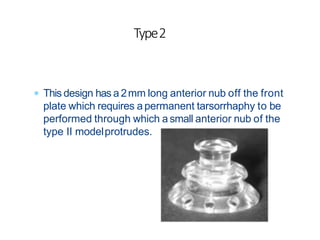
SlideShare a Scribd company logo
Artificial cornea
or
Keratoprosthesis
Dr Nikhil R P
 Keratoprosthesis is asurgical procedure where a
severely damaged ordiseased cornea is replaced with
an artificial cornea to restore useful vision
 It is usually the last option for the surgeon and the
patient who has visual potential in an eye with
severely compromised cornea
History
 Bilateral Blindness with Visual acuity of hand movements
or less with normal Bscan
 Severe Debilitating but inactive anterior segment disease
such asSteven Johnson Syndrome, Chemical Burns or
Trachoma
 Multipleprevious failed corneal grafts
 Ocular cicatricial phemphigoid
 Vascularized corneas with complete stem cell loss and
dryness
Indications
Designs
• Consists of Optical cylinder and Supporting flange
Non Biointegrated(Optical cylinder materials)
• Polymethyl methacrylate (PMMA) – most commonly used material
• Glass, Ceramic, Quartz , Silicon
Non Biointegrated(Supporting flange)
• Methacrylate
• Teflon
• Dacron mesh
• Polycarbon
Biointegrated Supporting Flange (Autologous tissue)
• Tooth and bone (osteo-odonto-keratoprosthesis)
• Cartilage (chondro-keratoprosthesis)
• Nail (onycho-keratoprosthesis)
Keratoprosthesis designs have primarily been variations of 3 main
types.
• First Type PMMA stem with skirt embedded within the cornea
• Second Type Transparent membrane with porous edges inserted
• into the cornea
• Third Type PMMA ‘collar button’ with cornea between the plates
First type: It is most commonly used. The optical core is stabilized
by a permanently attached supporting plate or skirt implanted in
a pocket created in the collagen lamellae of the corneal stroma.
Consists of
• Central Optical Cylinder
• Supporting Flange
• PMMA stem
• Skirt placed intralamellarly in the stroma(made of
perforated grids of PMMA, nylon, Dacron, proplast/
covered by transplanted autologous tissue/ lid skin
Second Type: Consists of transparent plate with a porous periphery,
allowing tissue ingrowth into the pores. Such designs have been
made of polytetraflouroethylene, polyurethane, or modified gels.
By using suitable pore size, these devices have been well colonized
with tissue elements which might help to anchor the device and
prevent future extrusion.
Third Type: has a collar button shaped device consisting of two plates
joined by a stem, which constitutes the optical portion. This is inserted
into the patient’s cornea or a donor cornea so that the plates sandwich
the corneal tissue between them. The optical stem is short, allowing a
generous field and wide plates stabilize the stem so that it cannot
easily deviate from the axis to the macula.
 BOSTON KPRO(TYPE 1 AND 2) :-
 The Boston Type I Kpro is the most widely used device.
 The Boston Type II Kpro
 AlphaCor Kpro
 Modified OSTEO-ODONTO KPRO(MOOKP)
CommonlyUsedProcedures
 In development since1960
 Made of PMMA
 Also called Dohlman-DoaneProcedure
 2Types – Type 1 and Type2
BostonKeratoprosthesis
 2Types
 Type 1:-More commonly
used. Used in eyes that have
sufficient wetting function
to maintain the corneal
tissue in which the Kpro is
placed.
 Type 2:-Used in very dryeye
with minimal or no tear
production.
Boston KeratoprosthesisTypes
TYPE 1
• Front plate -6-7 mm dia
• Stem- 3.35mm
• Back plate – 7-8.5mm-16 holes
• Locked with titanium plate
• Length – AP -3.7mm – visual
field of 60 deg
TYPE 2
• 2mm long anterior nub off the
front plate
• Front plate -6 mm dia
• Back plate of 8.5mm dia -8 holes
• Length – AP-4.7mm
• 40 deg field of vision
 Indication
 Two failed grafts, with poor prognosis for further grafting
 Vision less than 20/400 in the affected eye
 Minimum vision of Light Perception
 Lower than optimal vision in the opposite eye
 Advantages
 Long-term (many years) stability and safety
 Excellent optics
 Canprovide excellent vision if the rest of the eye is undamaged
 Contraindications
 Unilateral visionloss
 End-stage glaucoma or uncontrolledglaucoma
 Posterior segmentpathology
 Presence of afunctioning KPro in the fellow eye
Partsof aBostonProsthesis
 Collar Button Design
 Front Plate and Back Plate
Sandwiching a fresh donor
graft
 Titanium locking ring is
used tosecure front and
back plates.
 Back plate holes are
important to improve
corneal nutrition.
 Electroretinography (ERG) and Visually Evoked
Response (VER) helpful in predicting the visual
potential.
 Pre operative Ultrasound examination to rule out
presence of Retinal Detachment and other posterior
segment abnormalities.
 IOPshould be maximally controlled before surgery
PreoperativeEvaluation
Procedure
 7.5-8.5mm donor corneal buttonis prepared
 3mm dermal punch used to punch central
donor corneal button
• Front plate placed face down on
adhesive to stabilize during
assembly
• Corneal donor button placedover
KPro stem
• Back plate carefully pressed into
position with
• Titanium locking ring is
snapped into position
• A snap sound signifies , it is in
proper position
• Graft and Type I BostonKPro
fully assembled
 The recipient cornea is then trephined (9mm)
similar to conventional PKP
 Natural lens or pseudophakia( anterior or iris supported
lens) are removed and total iridectomy and anterior
vitrectomy are done to reduce retro prosthetic membranes
andglaucoma.
 The donor graft with the KPro is then sutured in place with
interrupted 10–0nylon, using the same technique asa
standard PKP.
 Intravitreal injection of 0.4 mg dexamethasone.
 Bandage contact lens is applied
 Topical Antibiotics daily for3-4 weeks.
 Oral antibiotics for 1 week
 Steroid eye drops – 4-6 weeks and then tapered.
 Sub-Tenons injection of 20-40mg Triamcinalone if
eye shows an inflammatory reaction.
 Initially weekly followup, after 6 months once every 2
months.
PostoperativeManagement
Best:
•Multiple Graft failure in a relatively non-inflamed eye
with intact tear and blink mechanisms (following
dystrophies, infections, etc)
• Aniridia and other limbal stem cell failure cases
Intermediate:
•Chemical burns, HSV
Worst:
•Autoimmune diseases
•Mucous membrane pemphigoid
•Stevens-Johnson syndrome
• Chronic uveitis
Prognosis
 This design has a 2mm long anterior nub off the front
plate which requires apermanent tarsorrhaphy to be
performed through which a small anterior nub of the
type II modelprotrudes.
Type2
 Developed by Traian Chirila research group from Australia.
 Biocompatible, flexible, one-piece 7mm artificial cornea
designed to replace a scarred or diseased native cornea.
 Refractive Power close to that of human cornea .
AlphaCor
 Consists of
 the outer opaque porous skirt made from high water content
poly 2-hydroxyethyl methacrylatePHEMA.
 Atransparent central optic made from low-water content
PHEMA.
 Interpenetrating polymer network (IPN) – junction between
the skirt and central optic and is apermanentbond
 Principle :- The ability of the outer skirt to be colonized by
invading keratocytes resulting in integration of the device
with surroundingtissues
 VAfrom <6/60 to light perception
 Previous failed grafts with a poor chance with further PKP
 Functioning retina
 Absence of evidence of advanced glaucomatous optic
neuropathy or well-controlled glaucoma on medication
INDICATIONS
 Stage 1:-
 360 deg conjunctival peritomy is done
 Superficial keratectmoy
 Acorneal limbal incision is made for 5 clock hours and a
corneal stromal dissection throughout the areaan intra
lamellarpocket.
 AnAlphaCor is inserted into the interlamellar pocket followed
by removal of the posterior disc using an intra stromal
trephine.
 The surfacce is then covered with an conjunctival flap.
 Stage 2:-
 2months after Stage 1,tissues superficial tothe AlphaCor
optic are removed (trephination of central 4mm)
Procedure
 Trephination of Cornea
 AlphaCor KPro after stage 1
 Insertion ofAlphaCor
 AlphaCor KPro after stage
2
 The OOKPwas first described by Strampelli in 1963.
 Later modifiedby Falcinelli and Coll.
 It uses the patient’s own tooth root and surrounding
alveolar bone to support acentrally cemented optical
cylinder.
 Multi staged procedure, surgery in mouth and eye.
 Use of a wide single rooted tooth with surrounding alveolar
bone acts ascarrier for aPMMA optical cylinder, which is
covered by buccal mucousmembrane,
Modified Osteo-Odento
Keratoprosthesis
Indications
 Full thickness Mucous MembraneGraft
harvested from thebuccal mucosa.
 Graft is sutured over damaged cornea at
insertion of 4 recti muscles and sclera In
four quadrnts with 6-0 vicryl. The extent of
Graft should be extend from upper to
lower fornix and measures around 3-4 cm
in diameter.
 Ithas stem cells,high proliferating Capacity
and adapted tohigh Bacterial load
Stage1
to promote the growth
 Followed by Preparation of the Osteodentalacrylic Lamina
(ODAL)
 Asingle rooted tooth, preferably the upper canine is chosen
for preparation of the lamina.
 The tooth with the surrounding alveolar bone is extracted.
 Then sliced sagitally and Central hole is drilled
 customized PMMA optical cylinderis cemented
 ODALis then placed deep to orbicularis oculi of lower lid
in the fellow eye for next 3months to develop
vascularization and of connective
tissue.
 This is performed 3months after stage 1
 The Graft is dissected off from the
muscle area and examined for its
integrity.
 The central cornea is trephinedaccording
to the posterior diameter of the cylinder.
 The Graft is placed with the cylinder
centered over the corneal trephination and
sutured.
 Iris is removed with forceps and lens
extraction by ICCE or ECCE. Anterior
vitrectomy is then done
 The Mucous Membrane Graft is finally
reflected back on the lamina with acentral
trephination through which the anterior
cylinder protrudes out.
STAGE 2
Corneal Diseases
causing
Blindness
Low Risk for
Penetrating
Keratoplasty
One or more
Multiple Failures
Type 1Boston
Keratoprosthesis
HighPenetrating
Keratoplasty
Dry Surface
GoodLid
MOOKPor Type
2Boston
Keratoprosthesis
Insufficient lid
for ccomplete
tarsorrhapy
Type 2Boston
Keratoprosthesis
WetSurface
Type 1Boston
Keratoprosthesis
 MELTSAND EXTRUSION
 INFECTIOUSENDOPHTHALMITIS
 GLAUCOMA
 RETROPROSTHETIC MEMBRANES
 RETINALDETACHMENT
 OTHERS
Complications
 Most commonlyreported
 Occurs in 25-64%of pts in 1yr follow up
 These fibrous membranes originate from activated host stromal
cornea cells that migrate through gaps in the posterior graft–host
junction
 More prevalent in individual with chronic inflammation such as
autoimmune diseases anduveitis
 Treatment:-
 Majority may notrequire treatment
 Nd yag capsultomy following by steroids in 90%cases
 Ifmembrane thick, leathery and vascularised -Sxmanagement
 For Boston kpro membranectomy can be performed
 Removal of prosthesis and replacement with new one is preferred
RETRO PROSTHETIC MEMBRANES
 Occur at the base of Boston Kpro
 Slit lamp examination and anterior segment OCTare
helpful in detection of corneal thinning aroundKPro
 If melts are seen then replace the whole thing with fresh
graft and put new KPro
 In MOOKP,resorption of buccal mucosa can occur,new
graft can beplaced
 Resoprtion of osteo odonto lamina can occur
 If resorption of dentine has occurred it should be replaced
MELTSANDEXTRUSION
 Dreadful complication following kpro surgery
 Treatment:- includes leak repair,injectionof
antibiotics and topicalantibiotics
 Fungal infection suspected -change contactlens and
give topical amphotericin and systemic anti fungals
required
INFECTIOUS ENDOPHTHALMITIS
 Single most serious complication following surgery leading
to irreversible loss of vision due to chronic low grade
inflammation, progressive angle closure, anterior
displacement ofiris have been implicated.
 Topical Treatment is effective in Boston type 1Kpro.
 Systemic Treatments can be used with Boston type 2and
MOOKP.
 Tube shunts and endoscopic cyclophotocoagulation have
been successfully used.
GLAUCOMA
 Most common posterior segment complication, an
incidence of 16.9%
 Surgical Rx with buckle orvitrectomy
 Choroidal detachments can also develop in eyes with
KPro, in asmany as17%ofpatients
RETINALDETACHMENT
 Stanford Keratoprosthesis :-
 Kpro is based onamechanically enhanced Hydrogel
called Duoptix
 It supports the growth of epithelial cells.
 Surrounding the optic is a microperforated rim designed
to promote peripheral tissue integration
 Collagen BasedKeratoprosthesis
 Designed to mimic the extra cellular Matrix of corneal
stroma
RecentAdvances
ThankYou

More Related Content

What's hot

Interventions to Reduce Myopia Progression in Children (Journal Club) (health...
Interventions to Reduce Myopia Progression in Children (Journal Club) (health...Interventions to Reduce Myopia Progression in Children (Journal Club) (health...
Interventions to Reduce Myopia Progression in Children (Journal Club) (health...
Bikash Sapkota
 
collagen crosslinking.pptx
collagen crosslinking.pptxcollagen crosslinking.pptx
collagen crosslinking.pptx
piyush tewari
 
Transpupillary Thermotherapy (TTT)
Transpupillary Thermotherapy (TTT)Transpupillary Thermotherapy (TTT)
Transpupillary Thermotherapy (TTT)
Pushkar Dhir
 
Intra Oular Lenses
Intra Oular LensesIntra Oular Lenses
Intra Oular Lenses
slidenka
 
Anophthalmic socket.pptx
Anophthalmic socket.pptxAnophthalmic socket.pptx
Anophthalmic socket.pptx
SHAYRI PILLAI
 
Assessment of corneal endothelium
Assessment of corneal endotheliumAssessment of corneal endothelium
Assessment of corneal endothelium
drvasant162
 
Lamellar keratoplasty
Lamellar keratoplastyLamellar keratoplasty
Lamellar keratoplasty
Akshay Nayak
 
keratoconus
keratoconuskeratoconus
keratoconus
أنس القاضي
 
Retinal breaks
Retinal breaksRetinal breaks
Retinal breaks
Mae William- Ganaca
 
Epiretinal membrane
Epiretinal membraneEpiretinal membrane
Epiretinal membrane
Pushkal Katara
 
RECENT ADVANCES IN INTRAOCULAR LENS
RECENT ADVANCES IN INTRAOCULAR LENSRECENT ADVANCES IN INTRAOCULAR LENS
RECENT ADVANCES IN INTRAOCULAR LENS
Dr Laltanpuia Chhangte
 
Minimally invasive Glaucoma surgery MIGS
Minimally invasive Glaucoma surgery MIGSMinimally invasive Glaucoma surgery MIGS
Minimally invasive Glaucoma surgery MIGS
ankita mahapatra
 
IOL power calculation formulae
IOL power calculation formulaeIOL power calculation formulae
IOL power calculation formulae
pujarai
 
Femtosecond laser assistedcataractsurgery
Femtosecond laser assistedcataractsurgeryFemtosecond laser assistedcataractsurgery
Femtosecond laser assistedcataractsurgery
solinskyeyecare
 
Keratoconus
Keratoconus Keratoconus
Keratoconus
Tushya Parkash
 
Limbal Stem Cell Deficiency & its management
Limbal Stem Cell Deficiency & its  managementLimbal Stem Cell Deficiency & its  management
Limbal Stem Cell Deficiency & its management
Karan Bhatia
 
Microspherophakia
MicrospherophakiaMicrospherophakia
Microspherophakia
ikramdr01
 
Contracted eye socket reconstruction
Contracted eye socket reconstructionContracted eye socket reconstruction
Contracted eye socket reconstruction
Mohammed Aljodah
 
LAMELLAR KERATOPLASTY
LAMELLAR KERATOPLASTYLAMELLAR KERATOPLASTY
LAMELLAR KERATOPLASTY
Smith Snehal Sute
 
Current Trend in Management of Amblyopia (Amblyopia Therapy)/ Amblyopia Treat...
Current Trend in Management of Amblyopia (Amblyopia Therapy)/ Amblyopia Treat...Current Trend in Management of Amblyopia (Amblyopia Therapy)/ Amblyopia Treat...
Current Trend in Management of Amblyopia (Amblyopia Therapy)/ Amblyopia Treat...
Bikash Sapkota
 

What's hot (20)

Interventions to Reduce Myopia Progression in Children (Journal Club) (health...
Interventions to Reduce Myopia Progression in Children (Journal Club) (health...Interventions to Reduce Myopia Progression in Children (Journal Club) (health...
Interventions to Reduce Myopia Progression in Children (Journal Club) (health...
 
collagen crosslinking.pptx
collagen crosslinking.pptxcollagen crosslinking.pptx
collagen crosslinking.pptx
 
Transpupillary Thermotherapy (TTT)
Transpupillary Thermotherapy (TTT)Transpupillary Thermotherapy (TTT)
Transpupillary Thermotherapy (TTT)
 
Intra Oular Lenses
Intra Oular LensesIntra Oular Lenses
Intra Oular Lenses
 
Anophthalmic socket.pptx
Anophthalmic socket.pptxAnophthalmic socket.pptx
Anophthalmic socket.pptx
 
Assessment of corneal endothelium
Assessment of corneal endotheliumAssessment of corneal endothelium
Assessment of corneal endothelium
 
Lamellar keratoplasty
Lamellar keratoplastyLamellar keratoplasty
Lamellar keratoplasty
 
keratoconus
keratoconuskeratoconus
keratoconus
 
Retinal breaks
Retinal breaksRetinal breaks
Retinal breaks
 
Epiretinal membrane
Epiretinal membraneEpiretinal membrane
Epiretinal membrane
 
RECENT ADVANCES IN INTRAOCULAR LENS
RECENT ADVANCES IN INTRAOCULAR LENSRECENT ADVANCES IN INTRAOCULAR LENS
RECENT ADVANCES IN INTRAOCULAR LENS
 
Minimally invasive Glaucoma surgery MIGS
Minimally invasive Glaucoma surgery MIGSMinimally invasive Glaucoma surgery MIGS
Minimally invasive Glaucoma surgery MIGS
 
IOL power calculation formulae
IOL power calculation formulaeIOL power calculation formulae
IOL power calculation formulae
 
Femtosecond laser assistedcataractsurgery
Femtosecond laser assistedcataractsurgeryFemtosecond laser assistedcataractsurgery
Femtosecond laser assistedcataractsurgery
 
Keratoconus
Keratoconus Keratoconus
Keratoconus
 
Limbal Stem Cell Deficiency & its management
Limbal Stem Cell Deficiency & its  managementLimbal Stem Cell Deficiency & its  management
Limbal Stem Cell Deficiency & its management
 
Microspherophakia
MicrospherophakiaMicrospherophakia
Microspherophakia
 
Contracted eye socket reconstruction
Contracted eye socket reconstructionContracted eye socket reconstruction
Contracted eye socket reconstruction
 
LAMELLAR KERATOPLASTY
LAMELLAR KERATOPLASTYLAMELLAR KERATOPLASTY
LAMELLAR KERATOPLASTY
 
Current Trend in Management of Amblyopia (Amblyopia Therapy)/ Amblyopia Treat...
Current Trend in Management of Amblyopia (Amblyopia Therapy)/ Amblyopia Treat...Current Trend in Management of Amblyopia (Amblyopia Therapy)/ Amblyopia Treat...
Current Trend in Management of Amblyopia (Amblyopia Therapy)/ Amblyopia Treat...
 

Similar to Keratoprsthesis

Keratprosthesis
KeratprosthesisKeratprosthesis
Keratprosthesis
Dr Samarth Mishra
 
KERATOPROSTHESIS
KERATOPROSTHESISKERATOPROSTHESIS
KERATOPROSTHESIS
SSSIHMS-PG
 
Keratoprothesis &amp; osteoodonto keraoprosthesis
Keratoprothesis &amp; osteoodonto keraoprosthesisKeratoprothesis &amp; osteoodonto keraoprosthesis
Keratoprothesis &amp; osteoodonto keraoprosthesis
Kishor Badhe
 
Osteo odonto kerato prosthesis
Osteo odonto kerato prosthesisOsteo odonto kerato prosthesis
Osteo odonto kerato prosthesis
Asok Kumar
 
Apeceoctomy traditional and new concepts
Apeceoctomy traditional and new conceptsApeceoctomy traditional and new concepts
Apeceoctomy traditional and new concepts
Ahmed Alrashedi
 
KERATOPLASTY by arthur mohan and niko.pptx
KERATOPLASTY by arthur mohan and niko.pptxKERATOPLASTY by arthur mohan and niko.pptx
KERATOPLASTY by arthur mohan and niko.pptx
TarakeeshCH
 
Perioscope
PerioscopePerioscope
Perioscope
DrDeepali Jain
 
Lamellar keratoplasty
Lamellar keratoplastyLamellar keratoplasty
Lamellar keratoplasty
SubhraSarita
 
Keratoplasty
KeratoplastyKeratoplasty
Keratoplasty
Pravda Chaturvedi
 
Keratoplasty , Dr M SAQUIB
Keratoplasty , Dr M SAQUIBKeratoplasty , Dr M SAQUIB
Keratoplasty , Dr M SAQUIB
MEDICS india
 
penetrating keratoplasty seminar ...pptx
penetrating keratoplasty seminar ...pptxpenetrating keratoplasty seminar ...pptx
penetrating keratoplasty seminar ...pptx
SrishtiAhuja7
 
Non incisional, non laser refractive surgery
Non incisional, non laser refractive surgeryNon incisional, non laser refractive surgery
Non incisional, non laser refractive surgery
Ankit Gupta
 
Management of cataract
Management of cataractManagement of cataract
Management of cataract
Shuhadah Ros
 
Keratoplasty
KeratoplastyKeratoplasty
Keratoplasty
Karan Bhatia
 
Ocular Prosthesis
Ocular ProsthesisOcular Prosthesis
Ocular Prosthesis
Fahmida Hoque
 
Pediatric Keratoplasty 10 minutes.pptx
Pediatric Keratoplasty 10 minutes.pptxPediatric Keratoplasty 10 minutes.pptx
Pediatric Keratoplasty 10 minutes.pptx
Mohammad Bawtag
 
The anophthalmic socket
The anophthalmic socketThe anophthalmic socket
The anophthalmic socket
Niwar Ameen
 
Ferrara ring review (2017)
Ferrara ring review (2017)Ferrara ring review (2017)
Ferrara ring review (2017)
Ferrara Ophthalmics
 
Reconstructive periodontal therapy
Reconstructive periodontal therapyReconstructive periodontal therapy
Reconstructive periodontal therapy
Dr. Abhishek Ashok Sharma
 
Art of iris repair ppt
Art of iris repair pptArt of iris repair ppt
Art of iris repair ppt
DiyarAlzubaidy
 

Similar to Keratoprsthesis (20)

Keratprosthesis
KeratprosthesisKeratprosthesis
Keratprosthesis
 
KERATOPROSTHESIS
KERATOPROSTHESISKERATOPROSTHESIS
KERATOPROSTHESIS
 
Keratoprothesis &amp; osteoodonto keraoprosthesis
Keratoprothesis &amp; osteoodonto keraoprosthesisKeratoprothesis &amp; osteoodonto keraoprosthesis
Keratoprothesis &amp; osteoodonto keraoprosthesis
 
Osteo odonto kerato prosthesis
Osteo odonto kerato prosthesisOsteo odonto kerato prosthesis
Osteo odonto kerato prosthesis
 
Apeceoctomy traditional and new concepts
Apeceoctomy traditional and new conceptsApeceoctomy traditional and new concepts
Apeceoctomy traditional and new concepts
 
KERATOPLASTY by arthur mohan and niko.pptx
KERATOPLASTY by arthur mohan and niko.pptxKERATOPLASTY by arthur mohan and niko.pptx
KERATOPLASTY by arthur mohan and niko.pptx
 
Perioscope
PerioscopePerioscope
Perioscope
 
Lamellar keratoplasty
Lamellar keratoplastyLamellar keratoplasty
Lamellar keratoplasty
 
Keratoplasty
KeratoplastyKeratoplasty
Keratoplasty
 
Keratoplasty , Dr M SAQUIB
Keratoplasty , Dr M SAQUIBKeratoplasty , Dr M SAQUIB
Keratoplasty , Dr M SAQUIB
 
penetrating keratoplasty seminar ...pptx
penetrating keratoplasty seminar ...pptxpenetrating keratoplasty seminar ...pptx
penetrating keratoplasty seminar ...pptx
 
Non incisional, non laser refractive surgery
Non incisional, non laser refractive surgeryNon incisional, non laser refractive surgery
Non incisional, non laser refractive surgery
 
Management of cataract
Management of cataractManagement of cataract
Management of cataract
 
Keratoplasty
KeratoplastyKeratoplasty
Keratoplasty
 
Ocular Prosthesis
Ocular ProsthesisOcular Prosthesis
Ocular Prosthesis
 
Pediatric Keratoplasty 10 minutes.pptx
Pediatric Keratoplasty 10 minutes.pptxPediatric Keratoplasty 10 minutes.pptx
Pediatric Keratoplasty 10 minutes.pptx
 
The anophthalmic socket
The anophthalmic socketThe anophthalmic socket
The anophthalmic socket
 
Ferrara ring review (2017)
Ferrara ring review (2017)Ferrara ring review (2017)
Ferrara ring review (2017)
 
Reconstructive periodontal therapy
Reconstructive periodontal therapyReconstructive periodontal therapy
Reconstructive periodontal therapy
 
Art of iris repair ppt
Art of iris repair pptArt of iris repair ppt
Art of iris repair ppt
 

More from Nikhil Rp

Pentacam analysis
Pentacam analysisPentacam analysis
Pentacam analysis
Nikhil Rp
 
Ocular blood flow in glaucoma
Ocular  blood flow in glaucomaOcular  blood flow in glaucoma
Ocular blood flow in glaucoma
Nikhil Rp
 
Malignant glaucoma
Malignant glaucomaMalignant glaucoma
Malignant glaucoma
Nikhil Rp
 
Choroidal neovascularisation(cnv)
Choroidal neovascularisation(cnv)Choroidal neovascularisation(cnv)
Choroidal neovascularisation(cnv)
Nikhil Rp
 
Hess chart, diplopia chart, cover tests
Hess chart, diplopia chart, cover testsHess chart, diplopia chart, cover tests
Hess chart, diplopia chart, cover tests
Nikhil Rp
 
Tractional RD
Tractional RD Tractional RD
Tractional RD
Nikhil Rp
 
Retinal detachment
Retinal detachment Retinal detachment
Retinal detachment
Nikhil Rp
 
Pneumatic retinopexy
Pneumatic retinopexyPneumatic retinopexy
Pneumatic retinopexy
Nikhil Rp
 
Oct
OctOct
White dot syndromes
White dot syndromesWhite dot syndromes
White dot syndromes
Nikhil Rp
 
General slides
General slidesGeneral slides
General slides
Nikhil Rp
 
Ocular viscosurgical devices
Ocular viscosurgical devicesOcular viscosurgical devices
Ocular viscosurgical devices
Nikhil Rp
 

More from Nikhil Rp (12)

Pentacam analysis
Pentacam analysisPentacam analysis
Pentacam analysis
 
Ocular blood flow in glaucoma
Ocular  blood flow in glaucomaOcular  blood flow in glaucoma
Ocular blood flow in glaucoma
 
Malignant glaucoma
Malignant glaucomaMalignant glaucoma
Malignant glaucoma
 
Choroidal neovascularisation(cnv)
Choroidal neovascularisation(cnv)Choroidal neovascularisation(cnv)
Choroidal neovascularisation(cnv)
 
Hess chart, diplopia chart, cover tests
Hess chart, diplopia chart, cover testsHess chart, diplopia chart, cover tests
Hess chart, diplopia chart, cover tests
 
Tractional RD
Tractional RD Tractional RD
Tractional RD
 
Retinal detachment
Retinal detachment Retinal detachment
Retinal detachment
 
Pneumatic retinopexy
Pneumatic retinopexyPneumatic retinopexy
Pneumatic retinopexy
 
Oct
OctOct
Oct
 
White dot syndromes
White dot syndromesWhite dot syndromes
White dot syndromes
 
General slides
General slidesGeneral slides
General slides
 
Ocular viscosurgical devices
Ocular viscosurgical devicesOcular viscosurgical devices
Ocular viscosurgical devices
 

Recently uploaded

CHEMOTHERAPY_RDP_CHAPTER 3_ANTIFUNGAL AGENT.pdf
CHEMOTHERAPY_RDP_CHAPTER 3_ANTIFUNGAL AGENT.pdfCHEMOTHERAPY_RDP_CHAPTER 3_ANTIFUNGAL AGENT.pdf
CHEMOTHERAPY_RDP_CHAPTER 3_ANTIFUNGAL AGENT.pdf
rishi2789
 
Aortic Association CBL Pilot April 19 – 20 Bern
Aortic Association CBL Pilot April 19 – 20 BernAortic Association CBL Pilot April 19 – 20 Bern
Aortic Association CBL Pilot April 19 – 20 Bern
suvadeepdas911
 
Adhd Medication Shortage Uk - trinexpharmacy.com
Adhd Medication Shortage Uk - trinexpharmacy.comAdhd Medication Shortage Uk - trinexpharmacy.com
Adhd Medication Shortage Uk - trinexpharmacy.com
reignlana06
 
Cell Therapy Expansion and Challenges in Autoimmune Disease
Cell Therapy Expansion and Challenges in Autoimmune DiseaseCell Therapy Expansion and Challenges in Autoimmune Disease
Cell Therapy Expansion and Challenges in Autoimmune Disease
Health Advances
 
OCT Training Course for clinical practice Part 1
OCT Training Course for clinical practice Part 1OCT Training Course for clinical practice Part 1
OCT Training Course for clinical practice Part 1
KafrELShiekh University
 
Promoting Wellbeing - Applied Social Psychology - Psychology SuperNotes
Promoting Wellbeing - Applied Social Psychology - Psychology SuperNotesPromoting Wellbeing - Applied Social Psychology - Psychology SuperNotes
Promoting Wellbeing - Applied Social Psychology - Psychology SuperNotes
PsychoTech Services
 
TEST BANK For Community and Public Health Nursing: Evidence for Practice, 3rd...
TEST BANK For Community and Public Health Nursing: Evidence for Practice, 3rd...TEST BANK For Community and Public Health Nursing: Evidence for Practice, 3rd...
TEST BANK For Community and Public Health Nursing: Evidence for Practice, 3rd...
Donc Test
 
Abortion PG Seminar Power point presentation
Abortion PG Seminar Power point presentationAbortion PG Seminar Power point presentation
Abortion PG Seminar Power point presentation
AksshayaRajanbabu
 
8 Surprising Reasons To Meditate 40 Minutes A Day That Can Change Your Life.pptx
8 Surprising Reasons To Meditate 40 Minutes A Day That Can Change Your Life.pptx8 Surprising Reasons To Meditate 40 Minutes A Day That Can Change Your Life.pptx
8 Surprising Reasons To Meditate 40 Minutes A Day That Can Change Your Life.pptx
Holistified Wellness
 
Histopathology of Rheumatoid Arthritis: Visual treat
Histopathology of Rheumatoid Arthritis: Visual treatHistopathology of Rheumatoid Arthritis: Visual treat
Histopathology of Rheumatoid Arthritis: Visual treat
DIVYANSHU740006
 
Cardiac Assessment for B.sc Nursing Student.pdf
Cardiac Assessment for B.sc Nursing Student.pdfCardiac Assessment for B.sc Nursing Student.pdf
Cardiac Assessment for B.sc Nursing Student.pdf
shivalingatalekar1
 
K CỔ TỬ CUNG.pdf tự ghi chép, chữ hơi xấu
K CỔ TỬ CUNG.pdf tự ghi chép, chữ hơi xấuK CỔ TỬ CUNG.pdf tự ghi chép, chữ hơi xấu
K CỔ TỬ CUNG.pdf tự ghi chép, chữ hơi xấu
HongBiThi1
 
Hemodialysis: Chapter 4, Dialysate Circuit - Dr.Gawad
Hemodialysis: Chapter 4, Dialysate Circuit - Dr.GawadHemodialysis: Chapter 4, Dialysate Circuit - Dr.Gawad
Hemodialysis: Chapter 4, Dialysate Circuit - Dr.Gawad
NephroTube - Dr.Gawad
 
Histololgy of Female Reproductive System.pptx
Histololgy of Female Reproductive System.pptxHistololgy of Female Reproductive System.pptx
Histololgy of Female Reproductive System.pptx
AyeshaZaid1
 
TEST BANK For Community Health Nursing A Canadian Perspective, 5th Edition by...
TEST BANK For Community Health Nursing A Canadian Perspective, 5th Edition by...TEST BANK For Community Health Nursing A Canadian Perspective, 5th Edition by...
TEST BANK For Community Health Nursing A Canadian Perspective, 5th Edition by...
Donc Test
 
Ketone bodies and metabolism-biochemistry
Ketone bodies and metabolism-biochemistryKetone bodies and metabolism-biochemistry
Ketone bodies and metabolism-biochemistry
Dhayanithi C
 
Journal Article Review on Rasamanikya
Journal Article Review on RasamanikyaJournal Article Review on Rasamanikya
Journal Article Review on Rasamanikya
Dr. Jyothirmai Paindla
 
CHEMOTHERAPY_RDP_CHAPTER 2 _LEPROSY.pdf1
CHEMOTHERAPY_RDP_CHAPTER 2 _LEPROSY.pdf1CHEMOTHERAPY_RDP_CHAPTER 2 _LEPROSY.pdf1
CHEMOTHERAPY_RDP_CHAPTER 2 _LEPROSY.pdf1
rishi2789
 
Ear and its clinical correlations By Dr. Rabia Inam Gandapore.pptx
Ear and its clinical correlations By Dr. Rabia Inam Gandapore.pptxEar and its clinical correlations By Dr. Rabia Inam Gandapore.pptx
Ear and its clinical correlations By Dr. Rabia Inam Gandapore.pptx
Dr. Rabia Inam Gandapore
 
Vestibulocochlear Nerve by Dr. Rabia Inam Gandapore.pptx
Vestibulocochlear Nerve by Dr. Rabia Inam Gandapore.pptxVestibulocochlear Nerve by Dr. Rabia Inam Gandapore.pptx
Vestibulocochlear Nerve by Dr. Rabia Inam Gandapore.pptx
Dr. Rabia Inam Gandapore
 

Recently uploaded (20)

CHEMOTHERAPY_RDP_CHAPTER 3_ANTIFUNGAL AGENT.pdf
CHEMOTHERAPY_RDP_CHAPTER 3_ANTIFUNGAL AGENT.pdfCHEMOTHERAPY_RDP_CHAPTER 3_ANTIFUNGAL AGENT.pdf
CHEMOTHERAPY_RDP_CHAPTER 3_ANTIFUNGAL AGENT.pdf
 
Aortic Association CBL Pilot April 19 – 20 Bern
Aortic Association CBL Pilot April 19 – 20 BernAortic Association CBL Pilot April 19 – 20 Bern
Aortic Association CBL Pilot April 19 – 20 Bern
 
Adhd Medication Shortage Uk - trinexpharmacy.com
Adhd Medication Shortage Uk - trinexpharmacy.comAdhd Medication Shortage Uk - trinexpharmacy.com
Adhd Medication Shortage Uk - trinexpharmacy.com
 
Cell Therapy Expansion and Challenges in Autoimmune Disease
Cell Therapy Expansion and Challenges in Autoimmune DiseaseCell Therapy Expansion and Challenges in Autoimmune Disease
Cell Therapy Expansion and Challenges in Autoimmune Disease
 
OCT Training Course for clinical practice Part 1
OCT Training Course for clinical practice Part 1OCT Training Course for clinical practice Part 1
OCT Training Course for clinical practice Part 1
 
Promoting Wellbeing - Applied Social Psychology - Psychology SuperNotes
Promoting Wellbeing - Applied Social Psychology - Psychology SuperNotesPromoting Wellbeing - Applied Social Psychology - Psychology SuperNotes
Promoting Wellbeing - Applied Social Psychology - Psychology SuperNotes
 
TEST BANK For Community and Public Health Nursing: Evidence for Practice, 3rd...
TEST BANK For Community and Public Health Nursing: Evidence for Practice, 3rd...TEST BANK For Community and Public Health Nursing: Evidence for Practice, 3rd...
TEST BANK For Community and Public Health Nursing: Evidence for Practice, 3rd...
 
Abortion PG Seminar Power point presentation
Abortion PG Seminar Power point presentationAbortion PG Seminar Power point presentation
Abortion PG Seminar Power point presentation
 
8 Surprising Reasons To Meditate 40 Minutes A Day That Can Change Your Life.pptx
8 Surprising Reasons To Meditate 40 Minutes A Day That Can Change Your Life.pptx8 Surprising Reasons To Meditate 40 Minutes A Day That Can Change Your Life.pptx
8 Surprising Reasons To Meditate 40 Minutes A Day That Can Change Your Life.pptx
 
Histopathology of Rheumatoid Arthritis: Visual treat
Histopathology of Rheumatoid Arthritis: Visual treatHistopathology of Rheumatoid Arthritis: Visual treat
Histopathology of Rheumatoid Arthritis: Visual treat
 
Cardiac Assessment for B.sc Nursing Student.pdf
Cardiac Assessment for B.sc Nursing Student.pdfCardiac Assessment for B.sc Nursing Student.pdf
Cardiac Assessment for B.sc Nursing Student.pdf
 
K CỔ TỬ CUNG.pdf tự ghi chép, chữ hơi xấu
K CỔ TỬ CUNG.pdf tự ghi chép, chữ hơi xấuK CỔ TỬ CUNG.pdf tự ghi chép, chữ hơi xấu
K CỔ TỬ CUNG.pdf tự ghi chép, chữ hơi xấu
 
Hemodialysis: Chapter 4, Dialysate Circuit - Dr.Gawad
Hemodialysis: Chapter 4, Dialysate Circuit - Dr.GawadHemodialysis: Chapter 4, Dialysate Circuit - Dr.Gawad
Hemodialysis: Chapter 4, Dialysate Circuit - Dr.Gawad
 
Histololgy of Female Reproductive System.pptx
Histololgy of Female Reproductive System.pptxHistololgy of Female Reproductive System.pptx
Histololgy of Female Reproductive System.pptx
 
TEST BANK For Community Health Nursing A Canadian Perspective, 5th Edition by...
TEST BANK For Community Health Nursing A Canadian Perspective, 5th Edition by...TEST BANK For Community Health Nursing A Canadian Perspective, 5th Edition by...
TEST BANK For Community Health Nursing A Canadian Perspective, 5th Edition by...
 
Ketone bodies and metabolism-biochemistry
Ketone bodies and metabolism-biochemistryKetone bodies and metabolism-biochemistry
Ketone bodies and metabolism-biochemistry
 
Journal Article Review on Rasamanikya
Journal Article Review on RasamanikyaJournal Article Review on Rasamanikya
Journal Article Review on Rasamanikya
 
CHEMOTHERAPY_RDP_CHAPTER 2 _LEPROSY.pdf1
CHEMOTHERAPY_RDP_CHAPTER 2 _LEPROSY.pdf1CHEMOTHERAPY_RDP_CHAPTER 2 _LEPROSY.pdf1
CHEMOTHERAPY_RDP_CHAPTER 2 _LEPROSY.pdf1
 
Ear and its clinical correlations By Dr. Rabia Inam Gandapore.pptx
Ear and its clinical correlations By Dr. Rabia Inam Gandapore.pptxEar and its clinical correlations By Dr. Rabia Inam Gandapore.pptx
Ear and its clinical correlations By Dr. Rabia Inam Gandapore.pptx
 
Vestibulocochlear Nerve by Dr. Rabia Inam Gandapore.pptx
Vestibulocochlear Nerve by Dr. Rabia Inam Gandapore.pptxVestibulocochlear Nerve by Dr. Rabia Inam Gandapore.pptx
Vestibulocochlear Nerve by Dr. Rabia Inam Gandapore.pptx
 

Keratoprsthesis

  • 2.  Keratoprosthesis is asurgical procedure where a severely damaged ordiseased cornea is replaced with an artificial cornea to restore useful vision  It is usually the last option for the surgeon and the patient who has visual potential in an eye with severely compromised cornea
  • 4.  Bilateral Blindness with Visual acuity of hand movements or less with normal Bscan  Severe Debilitating but inactive anterior segment disease such asSteven Johnson Syndrome, Chemical Burns or Trachoma  Multipleprevious failed corneal grafts  Ocular cicatricial phemphigoid  Vascularized corneas with complete stem cell loss and dryness Indications
  • 5. Designs • Consists of Optical cylinder and Supporting flange Non Biointegrated(Optical cylinder materials) • Polymethyl methacrylate (PMMA) – most commonly used material • Glass, Ceramic, Quartz , Silicon Non Biointegrated(Supporting flange) • Methacrylate • Teflon • Dacron mesh • Polycarbon
  • 6. Biointegrated Supporting Flange (Autologous tissue) • Tooth and bone (osteo-odonto-keratoprosthesis) • Cartilage (chondro-keratoprosthesis) • Nail (onycho-keratoprosthesis)
  • 7. Keratoprosthesis designs have primarily been variations of 3 main types. • First Type PMMA stem with skirt embedded within the cornea • Second Type Transparent membrane with porous edges inserted • into the cornea • Third Type PMMA ‘collar button’ with cornea between the plates
  • 8. First type: It is most commonly used. The optical core is stabilized by a permanently attached supporting plate or skirt implanted in a pocket created in the collagen lamellae of the corneal stroma. Consists of • Central Optical Cylinder • Supporting Flange • PMMA stem • Skirt placed intralamellarly in the stroma(made of perforated grids of PMMA, nylon, Dacron, proplast/ covered by transplanted autologous tissue/ lid skin
  • 9. Second Type: Consists of transparent plate with a porous periphery, allowing tissue ingrowth into the pores. Such designs have been made of polytetraflouroethylene, polyurethane, or modified gels. By using suitable pore size, these devices have been well colonized with tissue elements which might help to anchor the device and prevent future extrusion.
  • 10. Third Type: has a collar button shaped device consisting of two plates joined by a stem, which constitutes the optical portion. This is inserted into the patient’s cornea or a donor cornea so that the plates sandwich the corneal tissue between them. The optical stem is short, allowing a generous field and wide plates stabilize the stem so that it cannot easily deviate from the axis to the macula.
  • 11.  BOSTON KPRO(TYPE 1 AND 2) :-  The Boston Type I Kpro is the most widely used device.  The Boston Type II Kpro  AlphaCor Kpro  Modified OSTEO-ODONTO KPRO(MOOKP) CommonlyUsedProcedures
  • 12.  In development since1960  Made of PMMA  Also called Dohlman-DoaneProcedure  2Types – Type 1 and Type2 BostonKeratoprosthesis
  • 13.  2Types  Type 1:-More commonly used. Used in eyes that have sufficient wetting function to maintain the corneal tissue in which the Kpro is placed.  Type 2:-Used in very dryeye with minimal or no tear production. Boston KeratoprosthesisTypes
  • 14. TYPE 1 • Front plate -6-7 mm dia • Stem- 3.35mm • Back plate – 7-8.5mm-16 holes • Locked with titanium plate • Length – AP -3.7mm – visual field of 60 deg TYPE 2 • 2mm long anterior nub off the front plate • Front plate -6 mm dia • Back plate of 8.5mm dia -8 holes • Length – AP-4.7mm • 40 deg field of vision
  • 15.  Indication  Two failed grafts, with poor prognosis for further grafting  Vision less than 20/400 in the affected eye  Minimum vision of Light Perception  Lower than optimal vision in the opposite eye  Advantages  Long-term (many years) stability and safety  Excellent optics  Canprovide excellent vision if the rest of the eye is undamaged  Contraindications  Unilateral visionloss  End-stage glaucoma or uncontrolledglaucoma  Posterior segmentpathology  Presence of afunctioning KPro in the fellow eye
  • 16. Partsof aBostonProsthesis  Collar Button Design  Front Plate and Back Plate Sandwiching a fresh donor graft  Titanium locking ring is used tosecure front and back plates.  Back plate holes are important to improve corneal nutrition.
  • 17.  Electroretinography (ERG) and Visually Evoked Response (VER) helpful in predicting the visual potential.  Pre operative Ultrasound examination to rule out presence of Retinal Detachment and other posterior segment abnormalities.  IOPshould be maximally controlled before surgery PreoperativeEvaluation
  • 18. Procedure  7.5-8.5mm donor corneal buttonis prepared  3mm dermal punch used to punch central donor corneal button • Front plate placed face down on adhesive to stabilize during assembly • Corneal donor button placedover KPro stem
  • 19. • Back plate carefully pressed into position with • Titanium locking ring is snapped into position • A snap sound signifies , it is in proper position • Graft and Type I BostonKPro fully assembled
  • 20.  The recipient cornea is then trephined (9mm) similar to conventional PKP  Natural lens or pseudophakia( anterior or iris supported lens) are removed and total iridectomy and anterior vitrectomy are done to reduce retro prosthetic membranes andglaucoma.  The donor graft with the KPro is then sutured in place with interrupted 10–0nylon, using the same technique asa standard PKP.  Intravitreal injection of 0.4 mg dexamethasone.  Bandage contact lens is applied
  • 21.  Topical Antibiotics daily for3-4 weeks.  Oral antibiotics for 1 week  Steroid eye drops – 4-6 weeks and then tapered.  Sub-Tenons injection of 20-40mg Triamcinalone if eye shows an inflammatory reaction.  Initially weekly followup, after 6 months once every 2 months. PostoperativeManagement
  • 22. Best: •Multiple Graft failure in a relatively non-inflamed eye with intact tear and blink mechanisms (following dystrophies, infections, etc) • Aniridia and other limbal stem cell failure cases Intermediate: •Chemical burns, HSV Worst: •Autoimmune diseases •Mucous membrane pemphigoid •Stevens-Johnson syndrome • Chronic uveitis Prognosis
  • 23.  This design has a 2mm long anterior nub off the front plate which requires apermanent tarsorrhaphy to be performed through which a small anterior nub of the type II modelprotrudes. Type2
  • 24.  Developed by Traian Chirila research group from Australia.  Biocompatible, flexible, one-piece 7mm artificial cornea designed to replace a scarred or diseased native cornea.  Refractive Power close to that of human cornea . AlphaCor
  • 25.  Consists of  the outer opaque porous skirt made from high water content poly 2-hydroxyethyl methacrylatePHEMA.  Atransparent central optic made from low-water content PHEMA.  Interpenetrating polymer network (IPN) – junction between the skirt and central optic and is apermanentbond  Principle :- The ability of the outer skirt to be colonized by invading keratocytes resulting in integration of the device with surroundingtissues
  • 26.  VAfrom <6/60 to light perception  Previous failed grafts with a poor chance with further PKP  Functioning retina  Absence of evidence of advanced glaucomatous optic neuropathy or well-controlled glaucoma on medication INDICATIONS
  • 27.  Stage 1:-  360 deg conjunctival peritomy is done  Superficial keratectmoy  Acorneal limbal incision is made for 5 clock hours and a corneal stromal dissection throughout the areaan intra lamellarpocket.  AnAlphaCor is inserted into the interlamellar pocket followed by removal of the posterior disc using an intra stromal trephine.  The surfacce is then covered with an conjunctival flap.  Stage 2:-  2months after Stage 1,tissues superficial tothe AlphaCor optic are removed (trephination of central 4mm) Procedure
  • 28.  Trephination of Cornea  AlphaCor KPro after stage 1  Insertion ofAlphaCor  AlphaCor KPro after stage 2
  • 29.  The OOKPwas first described by Strampelli in 1963.  Later modifiedby Falcinelli and Coll.  It uses the patient’s own tooth root and surrounding alveolar bone to support acentrally cemented optical cylinder.  Multi staged procedure, surgery in mouth and eye.  Use of a wide single rooted tooth with surrounding alveolar bone acts ascarrier for aPMMA optical cylinder, which is covered by buccal mucousmembrane, Modified Osteo-Odento Keratoprosthesis
  • 30.
  • 32.  Full thickness Mucous MembraneGraft harvested from thebuccal mucosa.  Graft is sutured over damaged cornea at insertion of 4 recti muscles and sclera In four quadrnts with 6-0 vicryl. The extent of Graft should be extend from upper to lower fornix and measures around 3-4 cm in diameter.  Ithas stem cells,high proliferating Capacity and adapted tohigh Bacterial load Stage1
  • 33. to promote the growth  Followed by Preparation of the Osteodentalacrylic Lamina (ODAL)  Asingle rooted tooth, preferably the upper canine is chosen for preparation of the lamina.  The tooth with the surrounding alveolar bone is extracted.  Then sliced sagitally and Central hole is drilled  customized PMMA optical cylinderis cemented  ODALis then placed deep to orbicularis oculi of lower lid in the fellow eye for next 3months to develop vascularization and of connective tissue.
  • 34.  This is performed 3months after stage 1  The Graft is dissected off from the muscle area and examined for its integrity.  The central cornea is trephinedaccording to the posterior diameter of the cylinder.  The Graft is placed with the cylinder centered over the corneal trephination and sutured.  Iris is removed with forceps and lens extraction by ICCE or ECCE. Anterior vitrectomy is then done  The Mucous Membrane Graft is finally reflected back on the lamina with acentral trephination through which the anterior cylinder protrudes out. STAGE 2
  • 35. Corneal Diseases causing Blindness Low Risk for Penetrating Keratoplasty One or more Multiple Failures Type 1Boston Keratoprosthesis HighPenetrating Keratoplasty Dry Surface GoodLid MOOKPor Type 2Boston Keratoprosthesis Insufficient lid for ccomplete tarsorrhapy Type 2Boston Keratoprosthesis WetSurface Type 1Boston Keratoprosthesis
  • 36.  MELTSAND EXTRUSION  INFECTIOUSENDOPHTHALMITIS  GLAUCOMA  RETROPROSTHETIC MEMBRANES  RETINALDETACHMENT  OTHERS Complications
  • 37.  Most commonlyreported  Occurs in 25-64%of pts in 1yr follow up  These fibrous membranes originate from activated host stromal cornea cells that migrate through gaps in the posterior graft–host junction  More prevalent in individual with chronic inflammation such as autoimmune diseases anduveitis  Treatment:-  Majority may notrequire treatment  Nd yag capsultomy following by steroids in 90%cases  Ifmembrane thick, leathery and vascularised -Sxmanagement  For Boston kpro membranectomy can be performed  Removal of prosthesis and replacement with new one is preferred RETRO PROSTHETIC MEMBRANES
  • 38.  Occur at the base of Boston Kpro  Slit lamp examination and anterior segment OCTare helpful in detection of corneal thinning aroundKPro  If melts are seen then replace the whole thing with fresh graft and put new KPro  In MOOKP,resorption of buccal mucosa can occur,new graft can beplaced  Resoprtion of osteo odonto lamina can occur  If resorption of dentine has occurred it should be replaced MELTSANDEXTRUSION
  • 39.  Dreadful complication following kpro surgery  Treatment:- includes leak repair,injectionof antibiotics and topicalantibiotics  Fungal infection suspected -change contactlens and give topical amphotericin and systemic anti fungals required INFECTIOUS ENDOPHTHALMITIS
  • 40.  Single most serious complication following surgery leading to irreversible loss of vision due to chronic low grade inflammation, progressive angle closure, anterior displacement ofiris have been implicated.  Topical Treatment is effective in Boston type 1Kpro.  Systemic Treatments can be used with Boston type 2and MOOKP.  Tube shunts and endoscopic cyclophotocoagulation have been successfully used. GLAUCOMA
  • 41.  Most common posterior segment complication, an incidence of 16.9%  Surgical Rx with buckle orvitrectomy  Choroidal detachments can also develop in eyes with KPro, in asmany as17%ofpatients RETINALDETACHMENT
  • 42.  Stanford Keratoprosthesis :-  Kpro is based onamechanically enhanced Hydrogel called Duoptix  It supports the growth of epithelial cells.  Surrounding the optic is a microperforated rim designed to promote peripheral tissue integration  Collagen BasedKeratoprosthesis  Designed to mimic the extra cellular Matrix of corneal stroma RecentAdvances