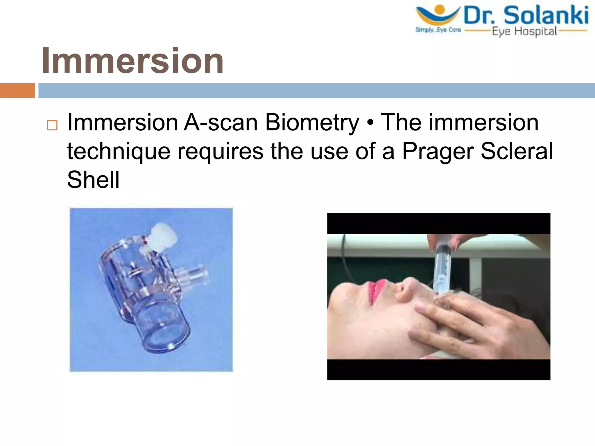
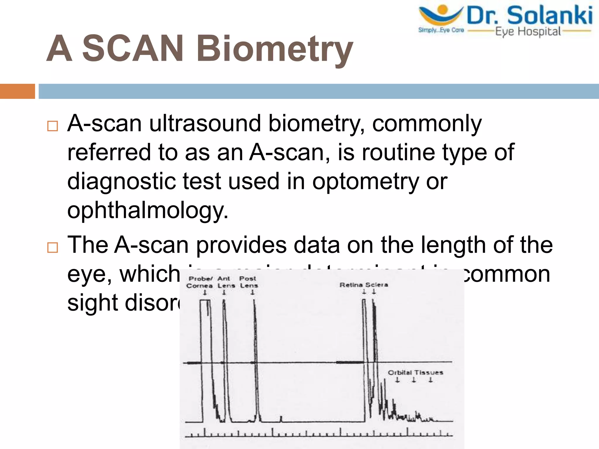
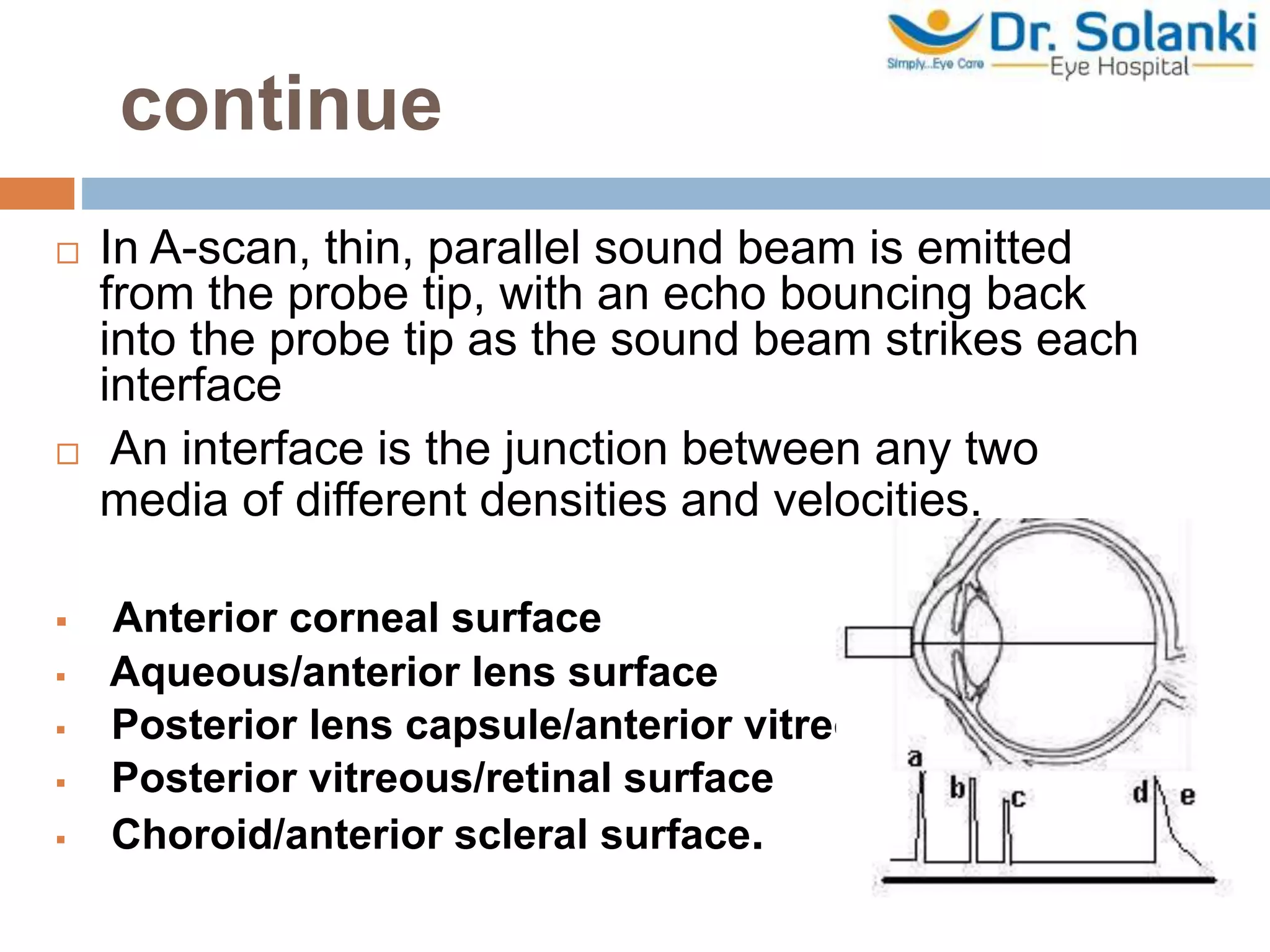
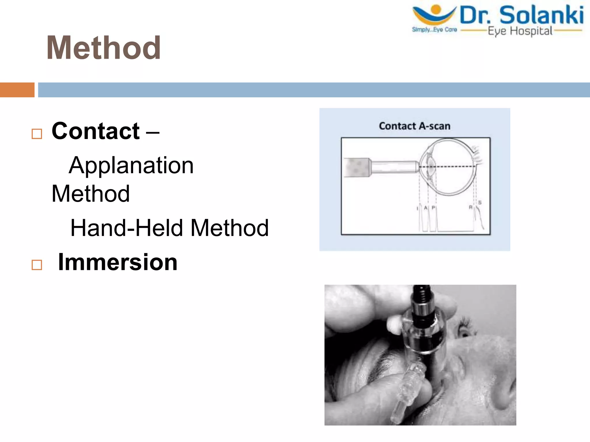
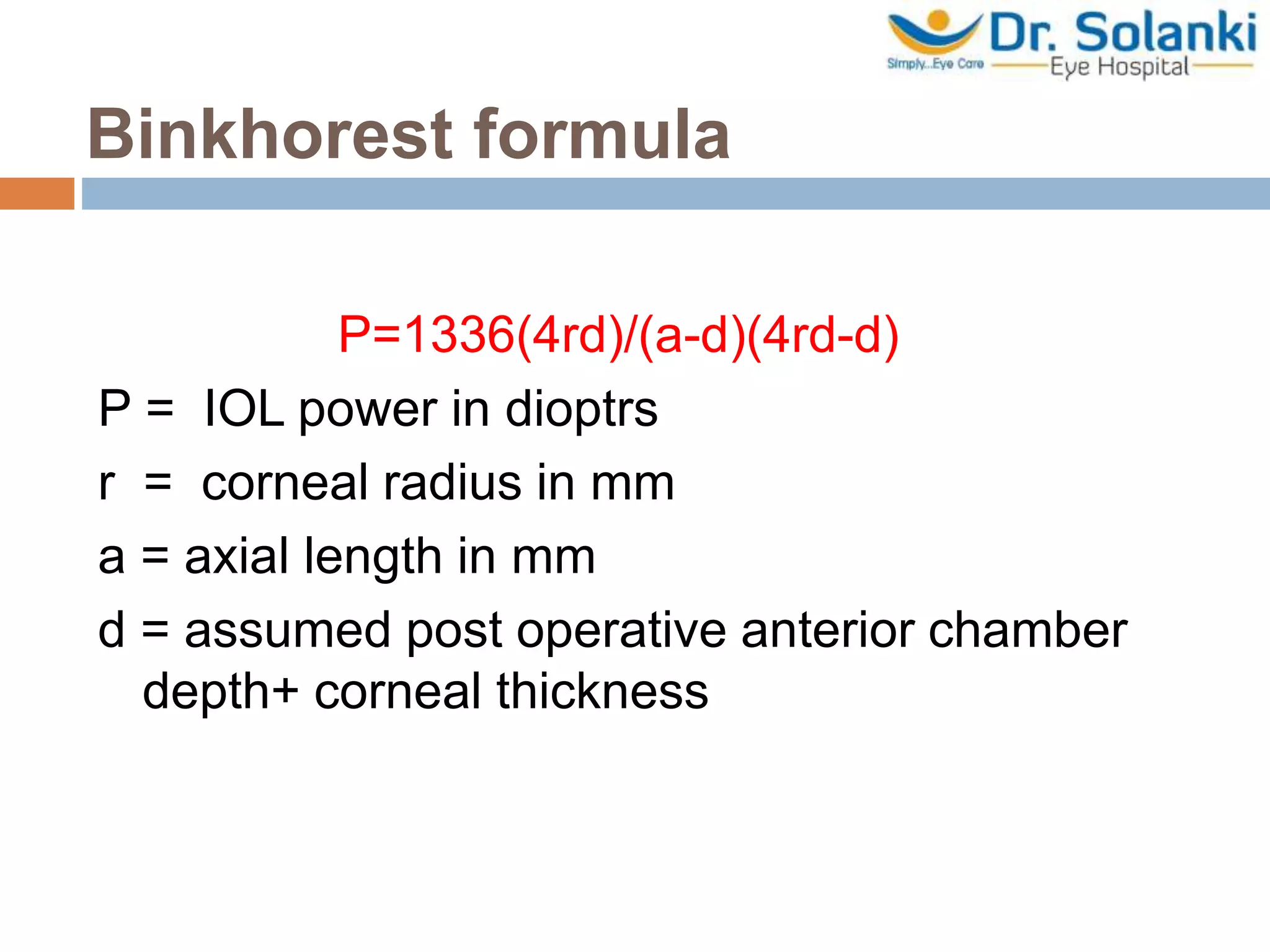
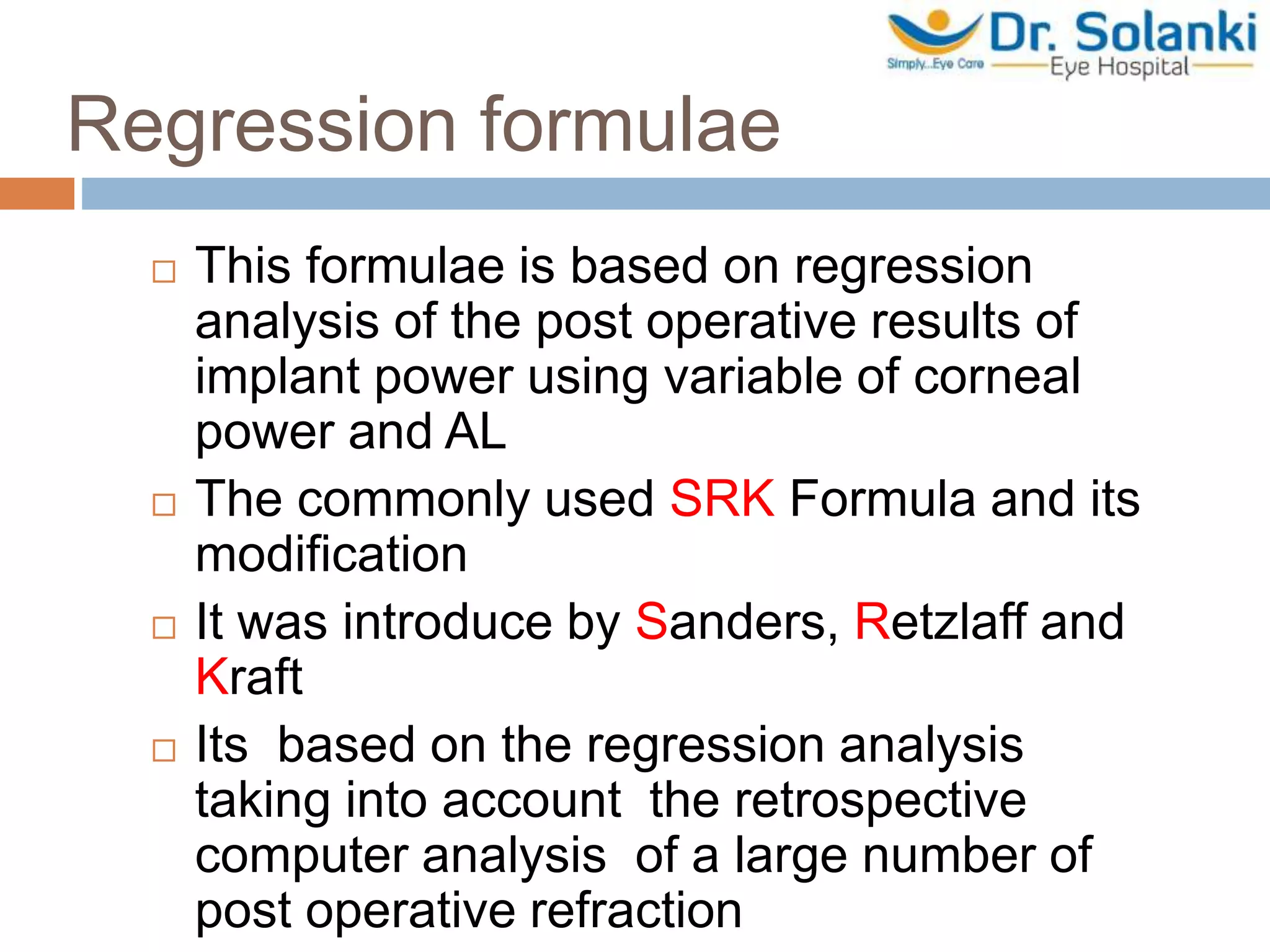
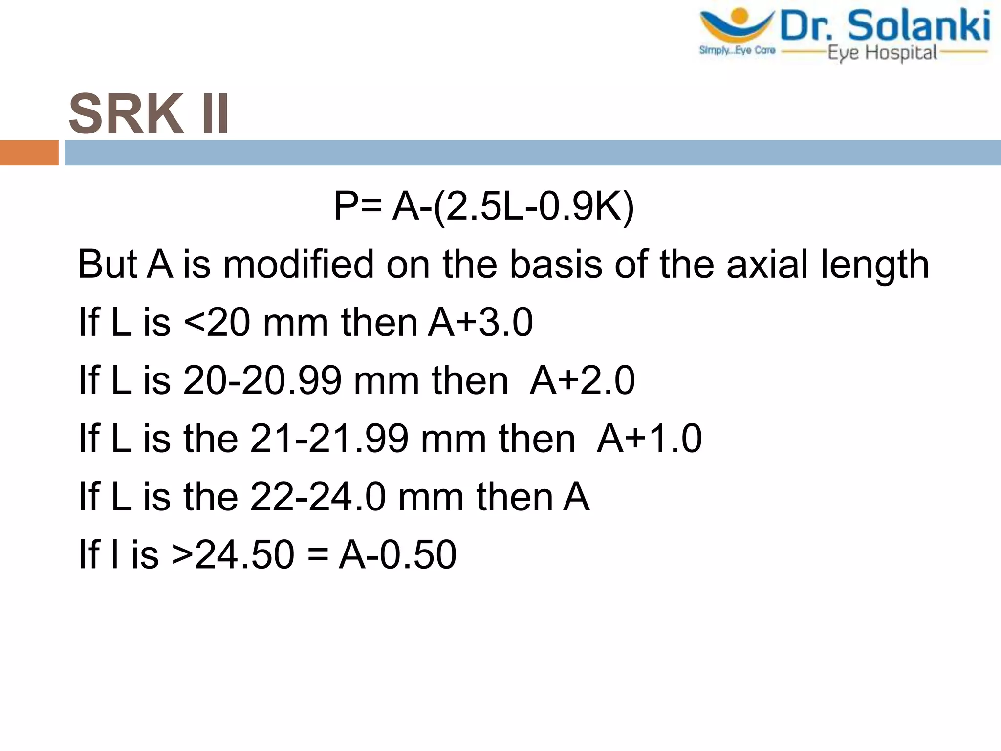
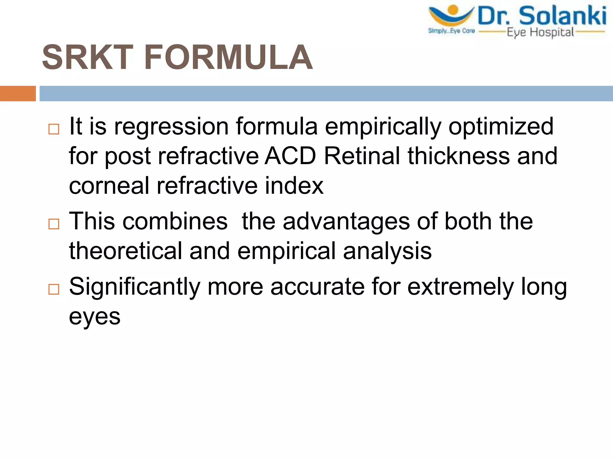
A-scan biometry is an ultrasound test used to measure the length of the eye, which is important for determining treatments for sight disorders. It works by emitting a sound beam into the eye and measuring the echoes that bounce back from different structures. The measurements of axial length, corneal curvature, and estimated lens position are used to calculate the ideal intraocular lens power needed after cataract surgery. Accuracy is important as even small errors in measurement can significantly impact the calculated lens power. Different formulas exist to relate the biometry measurements to the appropriate lens power, with newer regression formulas found to be most accurate.
To know Humphrey visual field analyser
To know about various types of perimetry
To identify field defect
To recognize that field defect is due to glaucoma or neurological lesion
To know that field defect is progressive or not
Interpretation of HVFA
Describes the basic of applanation tonometry, the factors affecting it and also how to perform the ideal tonometry. The slide are borrowed but it gives complete idea of mastering Applanation tonometry.
If the original owner of the slides has an objection i shall take down the ppt with due apologies.
introduction into Biometry and it's formulas.pptxQusaiAbusleem1
Biometry is the practice of applying mathematics to biology .
With regard to ophthalmology , there are several biometric systems used for making precise measurements of ocular structures : ultrasound (A- and B-scan , pachymeter) , low coherence interferometry (OCT) , laser interferometry (IOL Master , Lenstar)
These systems are used in measurement of axial length ,keratometry , pachymetry , and retinal thickness , among other things .
The term biometry , as it relates to ophthalmology , typically refers to preoperative measurements made for IOL calculations.
National Ocular Biometry Course (NOBC) 2015 An echoslide presentation Anis Suzanna Mohamad
This powerpoint presentation is basically about ocular biometry. Echo presentation is one of the method to deliver infomation that obtain from the course we attend to other staff in our Ophthalmology Department.
A scan biometry | How to Use A-scan? Types of A-Scan Biometry?Naeem Ahmad
A-SCAN BIOMETRY | What is A-Scan Biometry? How To Use It?
A-scan is the short form of amplitude scan.
This eye ultrasound gives details about the length of the eye.
A-Scan is an essential diagnostic tool used in ophthalmology.
The measurement of the eye’s axial length through an A-scan is necessary for placing an intraocular lens (IOL, artificial lens) during cataract surgery.
The total refractive power of the emmetropic eye is approximately 60D. Of this power, the cornea provides roughly 40D, and the crystalline lens 20 diopters.
When a cataract is removed, the lens is replaced by an artificial lens implant. By measuring both the length of the eye (A-scan Biometry) and the power of the cornea (keratometry).
It may also be used to assess vision abnormalities and other diseases involving the eye such as tumors.
A-scan techniques are based on the principles of ultrasonography. Sound travels in a wave pattern. For a sound to be heard by the human ear, the frequency must be between 20 and 20,000 Hz (20 kHz).
For an eye examination through A-scan, an ultrasound of frequency of around 10 MHz is used.
Intraocular Lens (IOL) power calculation is a crucial step in cataract surgery and certain refractive surgeries like phakic IOL implantation. The goal is to determine the appropriate power of the IOL to be implanted into the eye, ensuring that the patient achieves their desired postoperative visual outcome. Several formulas and methods are available for IOL power calculation, and the choice of formula depends on various factors, including the patient's eye measurements and the surgeon's preference. Here, we describe the basic principles and some commonly used formulas.
Ocular Biometry:
Ocular biometry is the process of measuring various dimensions of the eye, primarily the axial length, corneal power, and anterior chamber depth. These measurements are essential for accurate IOL power calculation and achieving the desired post-surgical refractive outcome. Here are the key components of ocular biometry:
Axial Length: This measurement determines the overall length of the eye, from the cornea's front surface to the retina's back surface. Axial length is a critical factor in IOL power calculation because it helps determine the eye's focusing power.
Corneal Power: The cornea is the transparent front surface of the eye, and its curvature affects the eye's refractive power. Corneal power is typically measured using techniques like keratometry or corneal topography. It helps account for the eye's astigmatism and assists in selecting the appropriate IOL.
some basic notions on how they are measured is explored here.
Biometry is the method of measuring various dimensions of the eye, its components and their inter-relationship. Using these data to calculate the idol intraocular lens power. In 1949, 29th November, Harold Ridley implanted the first IOL but his patient had a refractive surprise of -20 D spherical equivalents.
So, It was long way to travel to refined the out comes. Classic keratometry is based on anterior corneal surface measurements.
Whereas this directly measure the anterior and posterior corneal surface to obtain Total keratometry(TK).
Telecentric keratometry of the anterior corneal surface + swept source OCT of the posterior corneal surface= TOTAL KERATOMETRY.
TK measurements are compatible with existing IOL constants plus two exclusive formulas: barrett true K with TK for post LVC eyes and Barrett TK toric.
Biometry explanation and the used formulas.pptxQusaiAbusleem1
Biometry is the practice of applying mathematics to biology .
With regard to ophthalmology , there are several biometric systems used for making precise measurements of ocular structures : ultrasound (A- and B-scan , pachymeter) , low coherence interferometry (OCT) , laser interferometry (IOL Master , Lenstar)
These systems are used in measurement of axial length ,keratometry , pachymetry , and retinal thickness , among other things .
The term biometry , as it relates to ophthalmology , typically refers to preoperative measurements made for IOL calculations.
The French Revolution, which began in 1789, was a period of radical social and political upheaval in France. It marked the decline of absolute monarchies, the rise of secular and democratic republics, and the eventual rise of Napoleon Bonaparte. This revolutionary period is crucial in understanding the transition from feudalism to modernity in Europe.
For more information, visit-www.vavaclasses.com
This is a presentation by Dada Robert in a Your Skill Boost masterclass organised by the Excellence Foundation for South Sudan (EFSS) on Saturday, the 25th and Sunday, the 26th of May 2024.
He discussed the concept of quality improvement, emphasizing its applicability to various aspects of life, including personal, project, and program improvements. He defined quality as doing the right thing at the right time in the right way to achieve the best possible results and discussed the concept of the "gap" between what we know and what we do, and how this gap represents the areas we need to improve. He explained the scientific approach to quality improvement, which involves systematic performance analysis, testing and learning, and implementing change ideas. He also highlighted the importance of client focus and a team approach to quality improvement.
How to Create Map Views in the Odoo 17 ERPCeline George
The map views are useful for providing a geographical representation of data. They allow users to visualize and analyze the data in a more intuitive manner.
How to Split Bills in the Odoo 17 POS ModuleCeline George
Bills have a main role in point of sale procedure. It will help to track sales, handling payments and giving receipts to customers. Bill splitting also has an important role in POS. For example, If some friends come together for dinner and if they want to divide the bill then it is possible by POS bill splitting. This slide will show how to split bills in odoo 17 POS.
The Art Pastor's Guide to Sabbath | Steve ThomasonSteve Thomason
What is the purpose of the Sabbath Law in the Torah. It is interesting to compare how the context of the law shifts from Exodus to Deuteronomy. Who gets to rest, and why?
How to Make a Field invisible in Odoo 17Celine George
It is possible to hide or invisible some fields in odoo. Commonly using “invisible” attribute in the field definition to invisible the fields. This slide will show how to make a field invisible in odoo 17.
Instructions for Submissions thorugh G- Classroom.pptxJheel Barad
This presentation provides a briefing on how to upload submissions and documents in Google Classroom. It was prepared as part of an orientation for new Sainik School in-service teacher trainees. As a training officer, my goal is to ensure that you are comfortable and proficient with this essential tool for managing assignments and fostering student engagement.
Read| The latest issue of The Challenger is here! We are thrilled to announce that our school paper has qualified for the NATIONAL SCHOOLS PRESS CONFERENCE (NSPC) 2024. Thank you for your unwavering support and trust. Dive into the stories that made us stand out!
The Indian economy is classified into different sectors to simplify the analysis and understanding of economic activities. For Class 10, it's essential to grasp the sectors of the Indian economy, understand their characteristics, and recognize their importance. This guide will provide detailed notes on the Sectors of the Indian Economy Class 10, using specific long-tail keywords to enhance comprehension.
For more information, visit-www.vavaclasses.com
Students, digital devices and success - Andreas Schleicher - 27 May 2024..pptxEduSkills OECD
Andreas Schleicher presents at the OECD webinar ‘Digital devices in schools: detrimental distraction or secret to success?’ on 27 May 2024. The presentation was based on findings from PISA 2022 results and the webinar helped launch the PISA in Focus ‘Managing screen time: How to protect and equip students against distraction’ https://www.oecd-ilibrary.org/education/managing-screen-time_7c225af4-en and the OECD Education Policy Perspective ‘Students, digital devices and success’ can be found here - https://oe.cd/il/5yV
2. A SCAN Biometry
A-scan ultrasound biometry, commonly
referred to as an A-scan, is routine type of
diagnostic test used in optometry or
ophthalmology.
The A-scan provides data on the length of the
eye, which is a major determinant in common
sight disorders
3. continue
In A-scan, thin, parallel sound beam is emitted
from the probe tip, with an echo bouncing back
into the probe tip as the sound beam strikes each
interface
An interface is the junction between any two
media of different densities and velocities.
Anterior corneal surface
Aqueous/anterior lens surface
Posterior lens capsule/anterior vitreous
Posterior vitreous/retinal surface
Choroid/anterior scleral surface.
4. calculation of IOL
the process of measuring the power of the
cornea ( keratometry ) and the length of the
eye, and using this data to determine the ideal
intraocular lens power.
5. Ultrasound Principles
Sound is defined as a vibratory disturbance
within a solid or liquid that travels in a wave
pattern
When the sound frequency is between 20
hertz (Hz) and 20,000 Hz, the sound is audible
to the human ear.
In ophthalmology, most A-scan and B-scan
probes use a frequency of approximately 10
million Hz (10 MHz) that is predesigned by the
manufacturer
6. continue
This meets unique needs, because at times,
the probe is placed directly on the organ to be
examined, and its structures are quite small,
requiring excellent resolution.
The velocity of sound is determined
completely by the density of the medium
through which it passes.
Sound travels faster through solids than
through liquids, an important principle to
understand because the eye is composed of
both
7. calculate IOL Power required
Accurate Corneal power (keratometry)
Actual axial length
Accurate estimated lens position (half a mm
shift in lens position can have a dramatic effect
on final vision)
A good understanding of the various IOL
power calculation formulas is also required.
8. Keratometer
A keratometer, also known as an
ophthalmometer, is a diagnostic instrument for
measuring the curvature of the anterior surface
of the cornea, particularly for assessing the axis
of astigmatism.
10. Source of keratometry errors
Unfocused eye piece
Failure to calibrate unit
Poor patient fixation
Dry eye
Drooping eye lids
Irregular cornea
Contact lens user
11. Repeat Keratometery
If Corneal curvature more than 47D or less than
40D.
The difference in corneal cylinder is more than
one dioptre between eyes.
The average keratometry (K1) 43.0(K2) 44.0D,
with one eye typically within 1D of each other.
Difficult Situations
Post Refractive Surgery
Corneal Transplantation
Corneal Scar
Keratoconus etc.
12. REMEMBER
Average Axial Length of Normal Eye 23.06 mm
Majority 22.0 to 24.5 mm
Error of 0.4mm in the measurement of axial
length may result in a one Dioptre change in
calculated IOL power
Difference in AL measurement Between both
eyes +/- 0.3 mm
16. PROCEDURE
Explain the procedure
Use topical Anaesthesia
Clean the probe
A probe is placed on the patient’s cornea.
The probe is attached to a device that delivers
adjustable sound waves.
The measurements are displayed as spikes on
the screen of an Visual monitor .
The appearance of the spikes and the distance
between them can be correlated to structures
within the eye and the distance between them.
17. Probe positioning
The probe lightly touches the
cornea and is positioned, such
that the barrel of the probe is
aligned with the optical axis or
visual axis of the eye.
The operator aims the probe
towards the macula of the eye.
Alignment with the optical axis
will be indicated by high lens
spikes and a high retina spike on
the scan graph.
18. Corneal Compression
If pressure is applied on the cornea, the axial
length measurement may be falsely too short.
It can be monitored by observing the anterior
chamber depth, read out by an instrument.
Most eyes will have an ACD readings between
2.5 to 4.0mm.
The corneal compression error factor can be
avoided by using the immersion technique
Error caused by 1 mm Corneal Compression
Average eye 2.5 D Long eye 1.75 D Short eye
3.75 D
20. Immersion A-scan Biometry
The immersion technique is accomplished by
placing a small scleral shell between the
patient's lids, filling it with saline,
immersing the probe into the fluid, being
careful to avoid contact with the cornea.
More accurate than contact method because
corneal compression is avoided.
Eyes measured with the immersion method
are, on average, 0.1-0.3 mm longer
22. Theoretical formulae
Various formulae derived from the geometric
optics using schematic eye
These formulae are based on 3 variables
1Axial length of the eye ball(AL)
2 keratometry reading(K)
3 estimated post operative AC depth(ACD)
24. Regression formulae
This formulae is based on regression
analysis of the post operative results of
implant power using variable of corneal
power and AL
The commonly used SRK Formula and its
modification
It was introduce by Sanders, Retzlaff and
Kraft
Its based on the regression analysis
taking into account the retrospective
computer analysis of a large number of
post operative refraction
26. SRK ll
P= A-(2.5L-0.9K)
But A is modified on the basis of the axial length
If L is <20 mm then A+3.0
If L is 20-20.99 mm then A+2.0
If L is the 21-21.99 mm then A+1.0
If L is the 22-24.0 mm then A
If l is >24.50 = A-0.50
27. SRKT FORMULA
It is regression formula empirically optimized
for post refractive ACD Retinal thickness and
corneal refractive index
This combines the advantages of both the
theoretical and empirical analysis
Significantly more accurate for extremely long
eyes