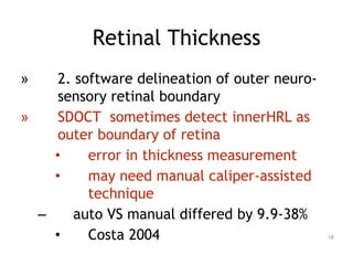
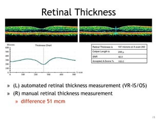
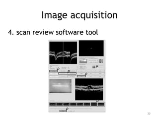
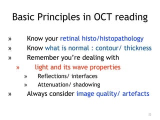
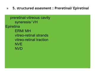
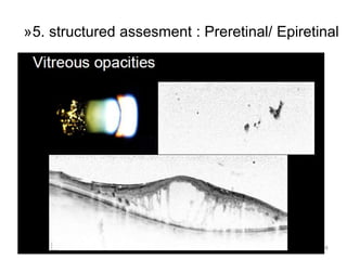
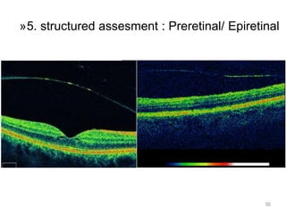
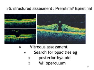
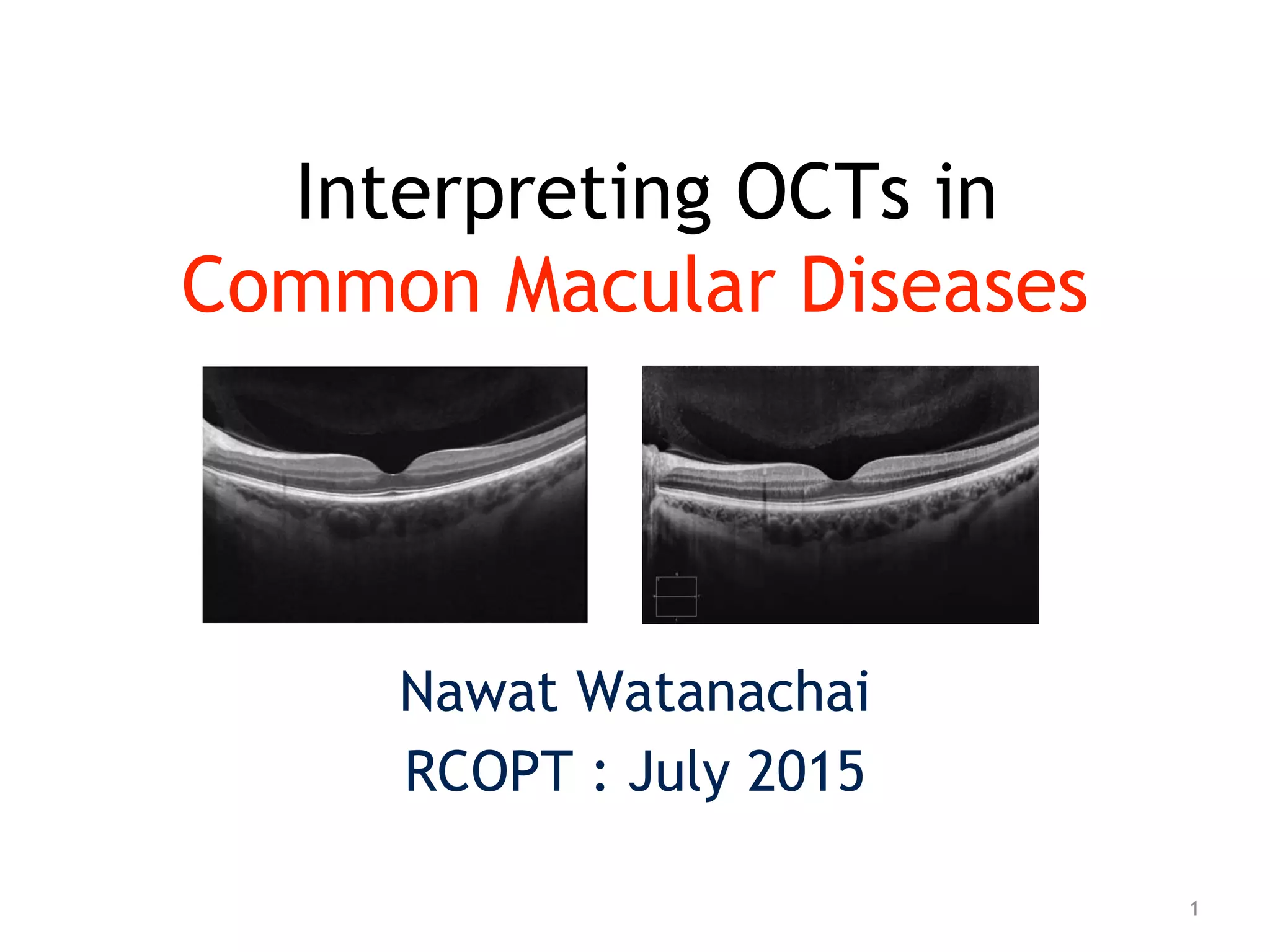
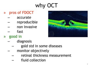
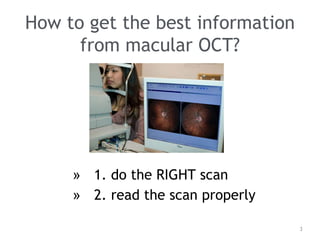
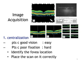
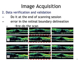
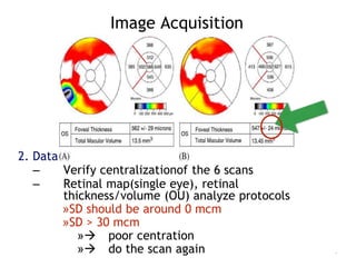
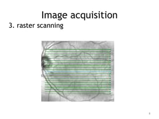
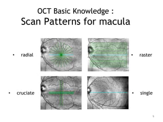
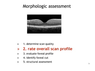
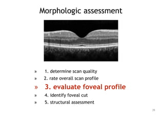
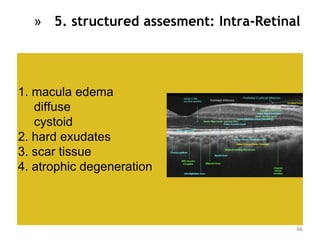
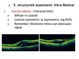
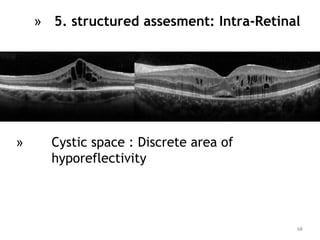
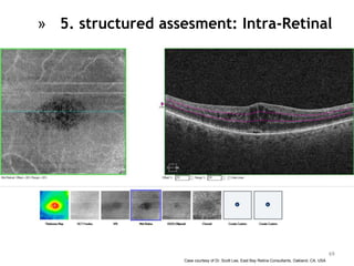
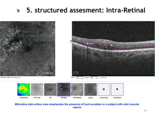
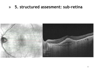
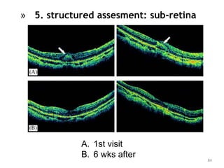
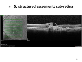
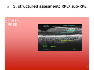
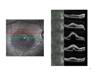
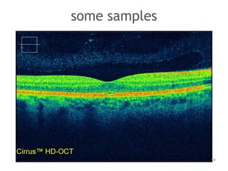
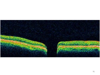
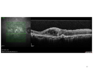
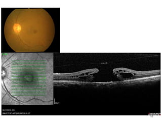
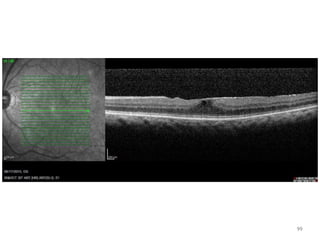
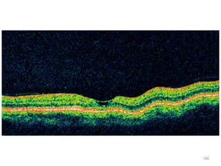
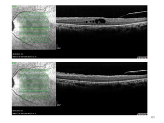
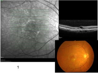
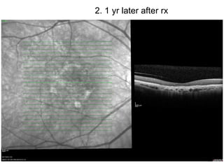
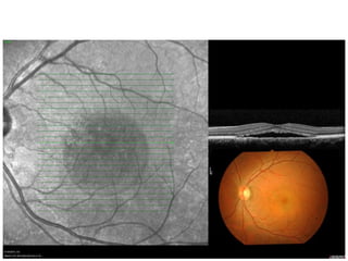
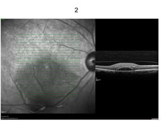
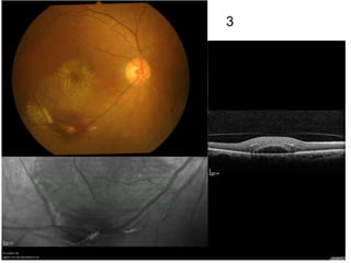
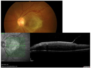
Interpreting OCTs in
Common Macular Diseases
Nawat Watanachai
RCOPT : July 2015
1
» pros of FDOCT
– accurate
– reproducible
– non invasive
– fast
» good in
– diagnosis
– gold std in some diseases
– monitor objectively
– retinal thickness measurement
– fluid collection
why OCT
2
How to get the best information
from macular OCT?
» 1. do the RIGHT scan
» 2. read the scan properly
3
How to get the best information
from macular OCT?
» 1. do the RIGHT scan
» take the best image
» minimise all possible
error
» 2. read the scan properly
4
Image
Acquisition
1. centralization
– pts c good vision : easy
– Pts c poor fixation : hard
• Identify the fovea location
• Place the scan on it correctly 5
Image Acquisition
2. Data verification and validation
– Do it at the end of scanning session
– error in the retinal boundary delineation
– re-do the scan
6
Image Acquisition
7
2. Data verification and validation
– Verify centralizationof the 6 scans
– Retinal map(single eye), retinal
thickness/volume (OU) analyze protocols
»SD should be around 0 mcm
»SD > 30 mcm
» poor centration
» do the scan again
Image acquisition
3. raster scanning
8
• radial
OCT Basic Knowledge :
Scan Patterns for macula
• raster
• cruciate • single
9
OCT Basic Knowledge :
radial line protocol
– 6X 6mm-long lines, 30’ apart
– Center at foveal center
10
3. raster scanning
– to minimize the chance of missing morphological
details
– 8 mm length scan
– best for vitreomacular adhesion
– May hit small lesions that missed on radial
protocol
11
OCT Basic Knowledge :
raster scan protocol
Image acquisition
A. foveal split B. lamellar hole
A’ FTMH B’ FTMH
12
OCT Basic Knowledge :
Retinal thickness map
13
color Thickness (microns)
White >470
red 350-470
Orange 320-350
Yellow 270-320
Green 210-270
blue 150-210
» from radial scan
Retinal Thickness
» 1. fundamental of OCT automatic retinal thickness
measurement
– algorithm (math. calculation)
– presumes 2 high reflective structures
• 1. VR surface
• 2. RPE-photoreceptor outer segment interface
– compares the shape of 1 a-scan to adjacent a-scans 14
OCT Basic Knowledge :
Retinal thickness map
15
» Depth 2 mm
» For thickness map
» Interpolation for thickness between sample point
16
Retinal Thickness
machine can be error
» 2. software delineation of outer neuro-sensory
retinal boundary
» HRL
– TDOCT : RPE-choriocapillaries reflective complex
– SDOCT : 2 lines
• IS/OS junction
• RPE-choriocapillaries reflective complex
Retinal Thickness
17
» 2. software delineation of outer neuro-
sensory retinal boundary
» SDOCT sometimes detect innerHRL as
outer boundary of retina
• error in thickness measurement
• may need manual caliper-assisted
technique
– auto VS manual differed by 9.9-38%
• Costa 2004
Retinal Thickness
18
Retinal Thickness
» (L) automated retinal thickness measurement (VR-IS/OS)
» (R) manual retinal thickness measurement
» difference 51 mcm
19
Image acquisition
4. scan review software tool
20
How to get the best information
from macular OCT?
» 1. do the RIGHT scan
» 2. read the scan properly
» systematic approach
21
Basic Principles in OCT reading
» Know your retinal histo/histopathology
» Know what is normal : contour/ thickness
» Remember you’re dealing with
» light and its wave properties
» Reflections/ interfaces
» Attenuation/ shadowing
» Always consider image quality/ artefacts
22
OCT Basic Knowledge :
Retinal layers in OCT
23
»High reflectivity : NFL/ IS-OS Junction/ RPE-choriocapillaris
»Intermediate reflectivity : plexiform layers
»Low reflectivity : nuclear layers/ photoreceptor outer segment/ vitreous
»Fovea
»Absence of inner retinal layer
»Increased thickness of the photoreceptor layer 24
OCT Basic Knowledge :
Retinal layers in OCT
»High reflectivity : NFL/ RPE/ choriocapillatis
»Intermediate reflectivity : plexiform layers
»Low reflectivity : nuclear layers/ photoreceptor outer segment/ vitreous
»Fovea
»Absence of inner retinal layer
»Increased thickness of the photoreceptor layer
25
OCT Basic Knowledge :
Retinal layers in OCT
»High reflectivity : NFL/ RPE/ choriocapillatis
»Intermediate reflectivity : plexiform layers
»Low reflectivity : nuclear layers/ photoreceptor outer segment/ vitreous
»Fovea
»Absence of inner retinal layer
»Increased thickness of the photoreceptor layer 26
OCT Basic Knowledge :
Retinal layers in OCT
• defining inner and outer HRL
• HRL - Highly Reflective Layer
• TD OCT : single line
• FD OCT : 2 lines 27
OCT Basic Knowledge :
Retinal layers in OCT
• defining inner and outer HRL
• FD OCT : not 1 line, but 2
• inner line : IS/OS junction
• outer line : RPE choriocapillaris complex
28
OCT Basic Knowledge :
Retinal layers in OCT
» IS/OS junction
» correlated with VA
» irregularities at the level of inner HRL after MH Sx
prevent VA improvement
» Uemoto 2002, Kitaya 2004, Villate 2005.
29
OCT Basic Knowledge :
Retinal layers in OCT
IS/OS junction : correlated with VA
RP
localization of missing photoreceptor
component
Jacobson 1998, 2000
cone-rod dystrophy, Best macular dystrophy
prediction of subret./ subRPE deposits
Aleman 2002, Pianta203
30
OCT Basic Knowledge :
Retinal layers in OCT
OCT Basic Knowledge :
Retinal layers in OCT
shadows
31
Let’s read
32
Morphologic assessment
» 1. determine scan quality
» 2. rate overall scan profile
» 3. evaluate foveal profile
» 4. identify foveal cut
» 5. structural assessment
33
Morphologic assessment
» 1. determine scan quality
» 2. rate overall acan profile
» 3. evaluate foveal profile
» 4. identify foveal cut
» 5. structural assessment
34
» 1. determine scan quality
» identify inner and outer retinal boundaries
» good signal to noise ratio
» rescan?
35
Morphologic assessment
» 1. determine scan quality
» 2. rate overall scan profile
» 3. evaluate foveal profile
» 4. identify foveal cut
» 5. structural assessment
36
» 2. rate overall scan profile
» normal over-all retinal profile
» slightly concave curvature
» abnormal
» exaggerated concavity and
convexity or retinal folds
» watch for artefact
37
» 1. retinal detachment
» RRD/ TRD/ ERD/ HRD
» 2. retinal thickening
» CSME/ CME/ CNV
» 3. RPED
» fibrous/ serous/ hemorrhage
38
» 2. rate overall scan profile
Morphologic assessment
» 1. determine scan quality
» 2. rate overall scan profile
» 3. evaluate foveal profile
» 4. identify foveal cut
» 5. structural assessment
39
» 3. evaluate foveal profile
» normal foveal profile
» slightly depression in the surface of
retina
40
Morphologic assessment
loss of foveal depression
 some problems
41
» 3. evaluate foveal profile
» deformations in the foveal profile
» VR surface
» ERM/ pseudohole
» MH/ Lamellar hole/ macular cyst
» Retina
42
Morphologic assessment
» 1. determine scan quality
» 2. rate overall acan profile
» 3. evaluate foveal profile
» 4. identify foveal cut
» 5. structural assessment
43
» 4. identify foveal cut
» do we need rescan?
44
back to
Image
Acquisition
1. centralization
• Identify the fovea location
• Place the scan on it correctly
– Centre line tool(OCT3 software) : right
click
45
Morphologic assessment
» 1. determine scan quality
» 2. rate overall acan profile
» 3. evaluate foveal profile
» 4. identify foveal cut
» 5. structural assessment
46
Morphologic assessment
» 5. structured assesment
» alteration of layers
» Systematic : antpost
47
» 5. structured assesment : Preretinal/ Epiretinal
preretinal-vitreous cavity
syneresis/ VH
Epiretina
ERM/ MH
vitreo-retinal strands
vitreo-retinal traction
NVE
NVD
48
49
»5. structured assesment : Preretinal/ Epiretinal
50
»5. structured assesment : Preretinal/ Epiretinal
» Vitreous assessment
» Search for opacities eg
» posterior hyaloid
» MH operculum
51
»5. structured assesment : Preretinal/ Epiretinal
52
»5. structured assesment : Preretinal/ Epiretinal
53
» consider looking for ERM/ PVD
traction in difficult DME cases
»5. structured assesment : Preretinal/ Epiretinal
DME: high prevalence of perifoveal PVD
Gaucher 2005
favorable macular remodeling in DME
after spontaneous PVD
Watanabe 2000, Yamagachi 2003
54
»5. structured assesment : Preretinal/ Epiretinal
A,B : PVD
C : VM traction
D. remodel after completion of PVD
55
»5. structured assesment : Preretinal/ Epiretinal
» Look for areas of abnormal VMT
» Identified areas where thin
hyperreflective band from the vit insert
into the retina
» Look for ERM
» Thin hyperreflective structures which
show multiple areas of attachment and
separation from the inner retinal surface
» may demonstrate free posterior hyaloid
face
56
»5. structured assesment : Preretinal/ Epiretinal
57
»5. structured assesment : Preretinal/ Epiretinal
VMT
58
»5. structured assesment : Preretinal/ Epiretinal
ERM
59
»5. structured assesment : Preretinal/ Epiretinal
ERM
» early MH
» oblique vitreofoveal tractional forces
» intrafoveal split
» Hee1995, Gaudric1999,
Haouchine2001, Tornambe2003 60
»5. structured assesment : Preretinal/ Epiretinal
61
»5. structured assesment :
Preretinal/ Epiretinal
» early MH
» late MH
62
»5. structured assesment
: Preretinal/ Epiretinal
» FTMH: typical configuration
» round cystic margins
» SRF
63
»5. structured assesment : Preretinal/ Epiretinal
64
»5. structured
assesment :
Preretinal/
Epiretinal
65
» 5. structured assesment: Intra-Retinal
1. macula edema
diffuse
cystoid
2. hard exudates
3. scar tissue
4. atrophic degeneration
66
» macula edema : Characteristics
» diffuse vs cystoid
» Central/symmetric vs asymmetric (eg RVO)
» Remember thickened retina can attenuate
signal
67
» 5. structured assesment: Intra-Retinal
» Cystic space : Discrete area of
hyporeflectivity
68
» 5. structured assesment: Intra-Retinal
Case courtesy of Dr. Scott Lee, East Bay Retina Consultants, Oakland, CA, USA
69
» 5. structured assesment: Intra-Retinal
Mid-retina slab enface view emphasizes the presence of hard exudates in a subject with mild macular
edema
70
» 5. structured assesment: Intra-Retinal
71
» 5. structured assesment: Intra-Retinal
72
» 5. structured assesment: Intra-Retinal
73
» 5. structured
assesment:
» Intra-Retinal
» Retinal thinning
» Increase transmission of light to the deeper layer
74
» 5. structured assesment: Intra-Retinal
75
» 5. structured assesment: Intra-Retinal
» Retinal thinning
» mostly found in late stage of many
diseases
IS/OS-Ellipsoid Enface Slab: Hydroxychloroquine toxicity example with classic bull’s eye
maculopathy
Case courtesy of Dr. Scott Lee, East Bay Retina Consultants, Oakland, CA, USA
76
» 5. structured assesment: Intra-Retinal
77
» 5. structured assesment: Intra-Retinal
thick/ thin/ edema
» 1. CNV
» 2. RPED
» 3. drusen
» 4. subretinal fibrosis
» 5. scar
» 6. RPE atrophy
» 7. SRF
78
» 5. structured assesment: sub-retina
» Separation between neural retina and RPE
» Generally hyporeflective
» Look for associated RPE level change eg
small PED in CSR
» Partial preservation of foveal depression
79
» 5. structured assesment: sub-retina
clear SRF
80
81
» 5. structured assesment: sub-retina
» Not hyporeflective space
» Presence of tissue eg typeII CNV,
subretinal fibrosis
» Hemorrhage or lipid
» Viscous SRF eg fibrinous CSR
» Correlate with color fundus photo/ FA/
ICG***
82
» 5. structured assesment: sub-retina
NOT so clear SRF?
83
» 5. structured assesment: sub-retina
84
» 5. structured assesment: sub-retina
A. 1st visit
B. 6 wks after
85
» 5. structured assesment: sub-retina
86
- drusen
- RPED
87
» 5. structured assesment: RPE/ sub-RPE
88
» 5. structured assesment: RPE/ sub-RPE
drusen
Small/ discrete low-lying areas of RPE elevation
c highly reflectivity
» RPED : area of hypo-reflectivity
underneath RPE elevation
89
» 5. structured assesment: RPE/ sub-RPE
» RPED
» Assess its size, contour and reflectivity
» Look for adjacent areas
» eg atrophy from RPE tear
90
» 5. structured assesment: RPE/ sub-RPE
Morphologic assessment : RPE elevation
» RPED : content
» serous/ hemorrhagic/ fibrovascular
91
92
93
some samples
94
95
96
97
98
99
100
101
102
1
103
2. 1 yr later after rx
104
105
106
107
108
1
109
2
110
3
111
112
the end

Nw2015 rcopt oct_retina22

  • 1.
    Interpreting OCTs in CommonMacular Diseases Nawat Watanachai RCOPT : July 2015 1
  • 2.
    » pros ofFDOCT – accurate – reproducible – non invasive – fast » good in – diagnosis – gold std in some diseases – monitor objectively – retinal thickness measurement – fluid collection why OCT 2
  • 3.
    How to getthe best information from macular OCT? » 1. do the RIGHT scan » 2. read the scan properly 3
  • 4.
    How to getthe best information from macular OCT? » 1. do the RIGHT scan » take the best image » minimise all possible error » 2. read the scan properly 4
  • 5.
    Image Acquisition 1. centralization – ptsc good vision : easy – Pts c poor fixation : hard • Identify the fovea location • Place the scan on it correctly 5
  • 6.
    Image Acquisition 2. Dataverification and validation – Do it at the end of scanning session – error in the retinal boundary delineation – re-do the scan 6
  • 7.
    Image Acquisition 7 2. Dataverification and validation – Verify centralizationof the 6 scans – Retinal map(single eye), retinal thickness/volume (OU) analyze protocols »SD should be around 0 mcm »SD > 30 mcm » poor centration » do the scan again
  • 8.
  • 9.
    • radial OCT BasicKnowledge : Scan Patterns for macula • raster • cruciate • single 9
  • 10.
    OCT Basic Knowledge: radial line protocol – 6X 6mm-long lines, 30’ apart – Center at foveal center 10
  • 11.
    3. raster scanning –to minimize the chance of missing morphological details – 8 mm length scan – best for vitreomacular adhesion – May hit small lesions that missed on radial protocol 11 OCT Basic Knowledge : raster scan protocol
  • 12.
    Image acquisition A. fovealsplit B. lamellar hole A’ FTMH B’ FTMH 12
  • 13.
    OCT Basic Knowledge: Retinal thickness map 13 color Thickness (microns) White >470 red 350-470 Orange 320-350 Yellow 270-320 Green 210-270 blue 150-210 » from radial scan
  • 14.
    Retinal Thickness » 1.fundamental of OCT automatic retinal thickness measurement – algorithm (math. calculation) – presumes 2 high reflective structures • 1. VR surface • 2. RPE-photoreceptor outer segment interface – compares the shape of 1 a-scan to adjacent a-scans 14
  • 15.
    OCT Basic Knowledge: Retinal thickness map 15 » Depth 2 mm » For thickness map » Interpolation for thickness between sample point
  • 16.
  • 17.
    » 2. softwaredelineation of outer neuro-sensory retinal boundary » HRL – TDOCT : RPE-choriocapillaries reflective complex – SDOCT : 2 lines • IS/OS junction • RPE-choriocapillaries reflective complex Retinal Thickness 17
  • 18.
    » 2. softwaredelineation of outer neuro- sensory retinal boundary » SDOCT sometimes detect innerHRL as outer boundary of retina • error in thickness measurement • may need manual caliper-assisted technique – auto VS manual differed by 9.9-38% • Costa 2004 Retinal Thickness 18
  • 19.
    Retinal Thickness » (L)automated retinal thickness measurement (VR-IS/OS) » (R) manual retinal thickness measurement » difference 51 mcm 19
  • 20.
    Image acquisition 4. scanreview software tool 20
  • 21.
    How to getthe best information from macular OCT? » 1. do the RIGHT scan » 2. read the scan properly » systematic approach 21
  • 22.
    Basic Principles inOCT reading » Know your retinal histo/histopathology » Know what is normal : contour/ thickness » Remember you’re dealing with » light and its wave properties » Reflections/ interfaces » Attenuation/ shadowing » Always consider image quality/ artefacts 22
  • 23.
    OCT Basic Knowledge: Retinal layers in OCT 23
  • 24.
    »High reflectivity :NFL/ IS-OS Junction/ RPE-choriocapillaris »Intermediate reflectivity : plexiform layers »Low reflectivity : nuclear layers/ photoreceptor outer segment/ vitreous »Fovea »Absence of inner retinal layer »Increased thickness of the photoreceptor layer 24 OCT Basic Knowledge : Retinal layers in OCT
  • 25.
    »High reflectivity :NFL/ RPE/ choriocapillatis »Intermediate reflectivity : plexiform layers »Low reflectivity : nuclear layers/ photoreceptor outer segment/ vitreous »Fovea »Absence of inner retinal layer »Increased thickness of the photoreceptor layer 25 OCT Basic Knowledge : Retinal layers in OCT
  • 26.
    »High reflectivity :NFL/ RPE/ choriocapillatis »Intermediate reflectivity : plexiform layers »Low reflectivity : nuclear layers/ photoreceptor outer segment/ vitreous »Fovea »Absence of inner retinal layer »Increased thickness of the photoreceptor layer 26 OCT Basic Knowledge : Retinal layers in OCT
  • 27.
    • defining innerand outer HRL • HRL - Highly Reflective Layer • TD OCT : single line • FD OCT : 2 lines 27 OCT Basic Knowledge : Retinal layers in OCT
  • 28.
    • defining innerand outer HRL • FD OCT : not 1 line, but 2 • inner line : IS/OS junction • outer line : RPE choriocapillaris complex 28 OCT Basic Knowledge : Retinal layers in OCT
  • 29.
    » IS/OS junction »correlated with VA » irregularities at the level of inner HRL after MH Sx prevent VA improvement » Uemoto 2002, Kitaya 2004, Villate 2005. 29 OCT Basic Knowledge : Retinal layers in OCT
  • 30.
    IS/OS junction :correlated with VA RP localization of missing photoreceptor component Jacobson 1998, 2000 cone-rod dystrophy, Best macular dystrophy prediction of subret./ subRPE deposits Aleman 2002, Pianta203 30 OCT Basic Knowledge : Retinal layers in OCT
  • 31.
    OCT Basic Knowledge: Retinal layers in OCT shadows 31
  • 32.
  • 33.
    Morphologic assessment » 1.determine scan quality » 2. rate overall scan profile » 3. evaluate foveal profile » 4. identify foveal cut » 5. structural assessment 33
  • 34.
    Morphologic assessment » 1.determine scan quality » 2. rate overall acan profile » 3. evaluate foveal profile » 4. identify foveal cut » 5. structural assessment 34
  • 35.
    » 1. determinescan quality » identify inner and outer retinal boundaries » good signal to noise ratio » rescan? 35
  • 36.
    Morphologic assessment » 1.determine scan quality » 2. rate overall scan profile » 3. evaluate foveal profile » 4. identify foveal cut » 5. structural assessment 36
  • 37.
    » 2. rateoverall scan profile » normal over-all retinal profile » slightly concave curvature » abnormal » exaggerated concavity and convexity or retinal folds » watch for artefact 37
  • 38.
    » 1. retinaldetachment » RRD/ TRD/ ERD/ HRD » 2. retinal thickening » CSME/ CME/ CNV » 3. RPED » fibrous/ serous/ hemorrhage 38 » 2. rate overall scan profile
  • 39.
    Morphologic assessment » 1.determine scan quality » 2. rate overall scan profile » 3. evaluate foveal profile » 4. identify foveal cut » 5. structural assessment 39
  • 40.
    » 3. evaluatefoveal profile » normal foveal profile » slightly depression in the surface of retina 40
  • 41.
    Morphologic assessment loss offoveal depression  some problems 41
  • 42.
    » 3. evaluatefoveal profile » deformations in the foveal profile » VR surface » ERM/ pseudohole » MH/ Lamellar hole/ macular cyst » Retina 42
  • 43.
    Morphologic assessment » 1.determine scan quality » 2. rate overall acan profile » 3. evaluate foveal profile » 4. identify foveal cut » 5. structural assessment 43
  • 44.
    » 4. identifyfoveal cut » do we need rescan? 44
  • 45.
    back to Image Acquisition 1. centralization •Identify the fovea location • Place the scan on it correctly – Centre line tool(OCT3 software) : right click 45
  • 46.
    Morphologic assessment » 1.determine scan quality » 2. rate overall acan profile » 3. evaluate foveal profile » 4. identify foveal cut » 5. structural assessment 46
  • 47.
    Morphologic assessment » 5.structured assesment » alteration of layers » Systematic : antpost 47
  • 48.
    » 5. structuredassesment : Preretinal/ Epiretinal preretinal-vitreous cavity syneresis/ VH Epiretina ERM/ MH vitreo-retinal strands vitreo-retinal traction NVE NVD 48
  • 49.
    49 »5. structured assesment: Preretinal/ Epiretinal
  • 50.
    50 »5. structured assesment: Preretinal/ Epiretinal
  • 51.
    » Vitreous assessment »Search for opacities eg » posterior hyaloid » MH operculum 51 »5. structured assesment : Preretinal/ Epiretinal
  • 52.
    52 »5. structured assesment: Preretinal/ Epiretinal
  • 53.
    53 » consider lookingfor ERM/ PVD traction in difficult DME cases »5. structured assesment : Preretinal/ Epiretinal
  • 54.
    DME: high prevalenceof perifoveal PVD Gaucher 2005 favorable macular remodeling in DME after spontaneous PVD Watanabe 2000, Yamagachi 2003 54 »5. structured assesment : Preretinal/ Epiretinal
  • 55.
    A,B : PVD C: VM traction D. remodel after completion of PVD 55 »5. structured assesment : Preretinal/ Epiretinal
  • 56.
    » Look forareas of abnormal VMT » Identified areas where thin hyperreflective band from the vit insert into the retina » Look for ERM » Thin hyperreflective structures which show multiple areas of attachment and separation from the inner retinal surface » may demonstrate free posterior hyaloid face 56 »5. structured assesment : Preretinal/ Epiretinal
  • 57.
    57 »5. structured assesment: Preretinal/ Epiretinal VMT
  • 58.
    58 »5. structured assesment: Preretinal/ Epiretinal ERM
  • 59.
    59 »5. structured assesment: Preretinal/ Epiretinal ERM
  • 60.
    » early MH »oblique vitreofoveal tractional forces » intrafoveal split » Hee1995, Gaudric1999, Haouchine2001, Tornambe2003 60 »5. structured assesment : Preretinal/ Epiretinal
  • 61.
    61 »5. structured assesment: Preretinal/ Epiretinal » early MH
  • 62.
    » late MH 62 »5.structured assesment : Preretinal/ Epiretinal
  • 63.
    » FTMH: typicalconfiguration » round cystic margins » SRF 63 »5. structured assesment : Preretinal/ Epiretinal
  • 64.
  • 65.
  • 66.
    » 5. structuredassesment: Intra-Retinal 1. macula edema diffuse cystoid 2. hard exudates 3. scar tissue 4. atrophic degeneration 66
  • 67.
    » macula edema: Characteristics » diffuse vs cystoid » Central/symmetric vs asymmetric (eg RVO) » Remember thickened retina can attenuate signal 67 » 5. structured assesment: Intra-Retinal
  • 68.
    » Cystic space: Discrete area of hyporeflectivity 68 » 5. structured assesment: Intra-Retinal
  • 69.
    Case courtesy ofDr. Scott Lee, East Bay Retina Consultants, Oakland, CA, USA 69 » 5. structured assesment: Intra-Retinal
  • 70.
    Mid-retina slab enfaceview emphasizes the presence of hard exudates in a subject with mild macular edema 70 » 5. structured assesment: Intra-Retinal
  • 71.
    71 » 5. structuredassesment: Intra-Retinal
  • 72.
    72 » 5. structuredassesment: Intra-Retinal
  • 73.
  • 74.
    » Retinal thinning »Increase transmission of light to the deeper layer 74 » 5. structured assesment: Intra-Retinal
  • 75.
    75 » 5. structuredassesment: Intra-Retinal » Retinal thinning » mostly found in late stage of many diseases
  • 76.
    IS/OS-Ellipsoid Enface Slab:Hydroxychloroquine toxicity example with classic bull’s eye maculopathy Case courtesy of Dr. Scott Lee, East Bay Retina Consultants, Oakland, CA, USA 76 » 5. structured assesment: Intra-Retinal
  • 77.
    77 » 5. structuredassesment: Intra-Retinal thick/ thin/ edema
  • 78.
    » 1. CNV »2. RPED » 3. drusen » 4. subretinal fibrosis » 5. scar » 6. RPE atrophy » 7. SRF 78 » 5. structured assesment: sub-retina
  • 79.
    » Separation betweenneural retina and RPE » Generally hyporeflective » Look for associated RPE level change eg small PED in CSR » Partial preservation of foveal depression 79 » 5. structured assesment: sub-retina clear SRF
  • 80.
  • 81.
    81 » 5. structuredassesment: sub-retina
  • 82.
    » Not hyporeflectivespace » Presence of tissue eg typeII CNV, subretinal fibrosis » Hemorrhage or lipid » Viscous SRF eg fibrinous CSR » Correlate with color fundus photo/ FA/ ICG*** 82 » 5. structured assesment: sub-retina NOT so clear SRF?
  • 83.
    83 » 5. structuredassesment: sub-retina
  • 84.
    84 » 5. structuredassesment: sub-retina A. 1st visit B. 6 wks after
  • 85.
    85 » 5. structuredassesment: sub-retina
  • 86.
  • 87.
    - drusen - RPED 87 »5. structured assesment: RPE/ sub-RPE
  • 88.
    88 » 5. structuredassesment: RPE/ sub-RPE drusen Small/ discrete low-lying areas of RPE elevation c highly reflectivity
  • 89.
    » RPED :area of hypo-reflectivity underneath RPE elevation 89 » 5. structured assesment: RPE/ sub-RPE
  • 90.
    » RPED » Assessits size, contour and reflectivity » Look for adjacent areas » eg atrophy from RPE tear 90 » 5. structured assesment: RPE/ sub-RPE
  • 91.
    Morphologic assessment :RPE elevation » RPED : content » serous/ hemorrhagic/ fibrovascular 91
  • 92.
  • 93.
  • 94.
  • 95.
  • 96.
  • 97.
  • 98.
  • 99.
  • 100.
  • 101.
  • 102.
  • 103.
    103 2. 1 yrlater after rx
  • 104.
  • 105.
  • 106.
  • 107.
  • 108.
  • 109.
  • 110.
  • 111.
  • 112.

Editor's Notes

  • #10 New Smart HD Scans deliver targeted visualizations of critical anatomy HD 21 Line – more scans covering a larger area to better assess the retina HD Radial – 12 radial lines with the fovea as the comm